References

Ahmad I. Anterior dental aesthetics: dental perspective. Br Dent J. 2005; 199:(3)135-141
Ahmad I. Anterior dental aesthetics: gingival perspective. Br Dent J. 2005; 199:(4)195-202
Ahmad I. Anterior dental aesthetics: dentofacial perspective. Br Dent J. 2005; 199:(2)81-88
Ahmad I. Anterior dental aesthetics: facial perspective. Br Dent J. 2005; 199:(1)15-21
Ahmad I. Anterior dental aesthetics: historical perspective. Br Dent J. 2005; 198:(12)737-742
Magne P, Gallucci GO, Belser UC. Anatomic crown width/length ratios of unworn and worn maxillary teeth in white subjects. J Prosthet Dent. 2003; 89:(5)453-461
Qualtrough AJ, Burke FJ. A look at dental esthetics. Quintessence Int. 1994; 25:(1)7-14
Liebler M, Devigus A, Randall RC, Burke FJ, Pallesen U, Cerutti A Ethics of esthetic dentistry. Quintessence Int. 2004; 35:(6)456-465
Brisman AS. Esthetics: a comparison of dentists' and patients' concepts. J Am Dent Assoc. 1980; 100:(3)345-352
Morley J, Eubank J. Macroesthetic elements of smile design. J Am Dent Assoc. 2001; 132:(1)39-45
Gillen RJ, Schwartz RS, Hilton TJ, Evans DB. An analysis of selected normative tooth proportions. Int J Prosthodont. 1994; 7:(5)410-417
Smales RJ. Effects of enamel-bonding, type of restoration, patient age and operator on the longevity of an anterior composite resin. Am J Dent. 1991; 4:(3)130-133
Redman CD, Hemmings KW, Good JA. The survival and clinical performance of resin-based composite restorations used to treat localised anterior tooth wear. Br Dent J. 2003; 194:(10)566-572
Horn HR. Porcelain laminate veneers bonded to etched enamel. Dent Clin North Am. 1983; 27:(4)671-684
Burke FJ, Lucarotti PS. Ten-year outcome of porcelain laminate veneers placed within the general dental services in England and Wales. J Dent. 2009; 37:(1)31-38
Friedman MJ. A 15-year review of porcelain veneer failure – a clinician's observations. Compend Contin Educ Dent. 1998; 19:(6)625-632
Rouse JS. Full veneer versus traditional veneer preparation: a discussion of interproximal extension. J Prosthet Dent. 1997; 78:(6)545-549
Dumfahrt H, Schaffer H. Porcelain laminate veneers. A retrospective evaluation after 1 to 10 years of service: Part II – Clinical results. Int J Prosthodont. 2000; 13:(1)9-18
Garber D. Porcelain laminate veneers: ten years later. Part I: Tooth preparation. J Esthet Dent. 1993; 5:(2)56-62
Saunders WP, Saunders EM. Prevalence of periradicular periodontitis associated with crowned teeth in an adult Scottish subpopulation. Br Dent J. 1998; 185:(3)137-140
Davies SJ, Gray RJ, Qualtrough AJ. Management of tooth surface loss. Br Dent J. 2002; 192:(1)11-6
Capp NJ. Occlusion and splint therapy. Br Dent J. 1999; 186:(5)217-222
Carrier DD. A laboratory technique for custom incisal guidance. J Prosthet Dent. 2001; 86:(5)551-552

Aesthetic treatment related to clinical need – an illustrated case report

From Volume 42, Issue 3, April 2015 | Pages 282-290

Authors

Paul Worskett

Dentist, Amblecote Dental Care, Brierley Hill

Articles by Paul Worskett

Abstract

Aesthetic treatment may be patient driven and usually by a ‘want’, rather than a ‘need’. This paper describes the management of a patient who presented with aesthetic wants and clinical needs, both of which were caused as a result of unsuccessful aesthetic treatment which the patient had received previously. The diagnostic process, discussion of the treatment plan and clinical procedures, which produced a satisfactory result, are described and illustrated.

Clinical Relevance: This case demonstrates that aesthetic treatment may commit the patient to future dental treatment needs, particularly if treatment provided is poorly planned and carried out to an unsatisfactory standard.

Article

The clinical need for treatment is based upon the principle that, whenever treatment is proposed for a patient, the treatment is necessary to secure and maintain the oral health of that patient. A patient may seek out treatment to solve a clinical problem that he/she may be aware of, such as pain, trauma or disease. In such cases, there is a clinical need for treatment, unrelated to aesthetics, and treatment objectives are based on:

  • Establishing oral health;
  • Restoring oral function;
  • Providing occlusal stability;
  • Achieving an acceptable aesthetic result;
  • Maintaining a stable oral environment in the long term.
  • In some aesthetic cases, the primary reason for attendance may be to improve the appearance of the patient's mouth and teeth. This may require treatment in an otherwise healthy and stable mouth and, in so doing, any treatment provided is unnecessary from a clinical need point of view. However, there is no reason for the objectives of treatment to deviate from the list presented above.

    In an ideal world, any treatment provided for purely aesthetic reasons would not compromise the long-term viability of the teeth or oral health. Unfortunately, we do not live in an ideal world and purely aesthetic treatment carries the risk that, even if carried out to an exceptionally high standard, the teeth may be compromised as a result of intrusive treatment.

    This case describes a patient whose primary reason for attendance was the improvement of her dental appearance. However, it became clear that there was also a clinical need for treatment, which was necessary as a result of aesthetic treatment she had received previously that had been poorly executed and resulted in compromised oral health. Therefore, there was a clinical need to provide aesthetic treatment.

    The patient

    History and objectives

    The patient was a 29-year-old female whose occupation at a bank involved contact with the public. She had received orthodontic treatment in the past and had not attended a dentist for four years, following the placement of porcelain veneers to her six maxillary anterior teeth. The veneers were, reportedly, placed to eliminate gaps between the teeth and the patient had apparently been informed that veneers were the only option.

    Her medical history was unremarkable; she was a non-smoker and social alcohol drinker (5 units per week).

    She reported that she was unhappy with her dental appearance. She did not like the ‘look’ of the veneers and didn't like her ‘gummy smile’. She also complained of ‘bleeding gums’ and ‘bad breath’. She indicated that some of the veneers had chipped or cracked over time and had been repaired.

    She was apprehensive about dental treatment, but wanted to have a healthy mouth and eventually wanted the veneers replacing to improve her appearance.

    Basic examination

    Extra-oral examination

    There were no signs of any pathology to the soft issues, muscles, lymph nodes or temporomandibular joints (TMJ) (Figure 1).

    Figure 1. Extra–oral full facial photograph.

    Intra-oral examination

    Soft tissues

    There was no underlying pathology of the soft tissues.

    Teeth present and existing restorations

    The tooth chart of the pre-existing state of the teeth was recorded (Figure 2). Porcelain veneers were present on the six maxillary anterior teeth. All the restorations in the posterior teeth were amalgam. The existing restorations and teeth were satisfactory except that:

    Figure 2. Tooth chart – Base chart.
  • Caries was noted in the maxillary left first molar (UL6) distally, with breakdown of amalgam mesially;
  • There was a vertical fracture running through the veneer of the maxillary right lateral incisor;
  • The incisal edge of the veneer in the maxillary left central incisor had been repaired, presumably as a result of fracture to the incisal edge;
  • The incisal tip of the veneer in the maxillary left canine had fractured and had been repaired;
  • There was staining around the veneers due to poor marginal fit.
  • Periodontium

    Calculus deposits were noted. The BPE chart was recorded as:


    2 2 2
    3 2 3
  • There was general bleeding on probing, especially around the veneers;
  • There were significant overhangs at the gingival margins of the veneers, contributing to poor oral hygiene and consequent gingivitis.
  • Occlusal examination

    Occlusal function was normal and guidance was noted on the maxillary right lateral incisor and maxillary left central incisor in the respective lateral excursions.

    Aesthetic examination

    A full aesthetic analysis revealed numerous discrepancies that are summarized in Figure 3 (A–F) and Figure 3 (g) and (h) reveal a vertical fracture through the veneer at UR2 (3g) and incisal edge fracture to the veneer at UL1 and a short gingival margin discrepancy at UL2 and UL3 (3h).’

    Figure 3. (A) High lip line revealing a large gingival display. (B) Gingival level of lateral incisors higher than central incisors and higher on the left than the right. (C) Occlusal level canted down to the right, emphasized by larger right central incisor. Both central incisors too square. (D) General tooth form too bulbous and out of proportion. Overall, asymmetrical and unbalanced with midline discrepancy. (E) Complete vertical fracture through veneer at UR2. (F) Incisal edge fracture (repaired) to veneer at UL1.
    Figure 3. (g, h) Close up photographs of lateral incisors.

    Special investigations

    Radiographs

    Radiographic examination confirmed the absence of any periapical pathology, although calculus was noted around the veneer margins interdentally (Figures 4 a–c).

    Figure 4. (a, b, c) Periapical radiographs of maxillary anterior teeth.

    Vitality tests

    The vitality of the upper incisors and canines was positively established with ethyl chloride.

    Articulated study casts and diagnostic wax-ups

    Diagnostic records were taken in order to construct and mount study casts on a semi-adjustable articulator.

    A duplicate maxillary cast was made on which diagnostic wax-ups were prescribed. The aesthetic prescription was defined following analysis of the deficiencies and discussion with the patient. A prescription form was used to communicate the prescription to the laboratory (Figure 5). Guidance was directed onto the maxillary canines in both lateral excursions.

    Figure 5. Wax–up prescription form – copy of actual document.

    Diagnosis and problem list

    As a result of the investigations, the following problems were diagnosed:

  • Generalized gingivitis;
  • Calculus deposits;
  • Halitosis;
  • Caries in UL6 distally;
  • Acute dental anxiety;
  • Chipped and cracked veneers with overhanging margins resulting from a poorly designed occlusal scheme;
  • Poor aesthetics of maxillary anterior veneers:
  • – Gingival aesthetics;
  • Excessive gingival display;
  • – Macro aesthetics;
  • Midline;
  • Smile line;
  • – Micro aesthetics;
  • Tooth form and asymmetry;
  • Tooth position;
  • Tooth proportion.
  • Treatment plan

    There was existing pathology related to the periodontal health in the form of gingivitis, and intensive oral hygiene instruction, coupled with detailed scaling and debridement, would address this. The caries at UL6 would be restored with a direct composite resin restoration to achieve an aesthetic result in accordance with the patient's requirements. This would be carried out initially to establish oral health and compliance of the patient.

    Given that porcelain restorations were already in place on the maxillary anterior teeth, and these were unsatisfactory, there was no alternative but to replace these restorations. Porcelain was selected as the material, although the design and occlusal scheme would be modified to overcome the errors in the existing design, which had resulted in failure.

    As a consequence of the acute anxiety problem, it was agreed that treatment would be staged over a period of time to allow the patient to build up confidence. The treatment plan was therefore structured into phases.

    Phase 1: address periodontal and caries pathology and achieve stability

    Designed to address problems 1, 2, 3, 4 and 5, this would consist of:

  • Initially, two hygienist visits for scaling and intensive oral hygiene instruction;
  • Placement of composite resin restoration in UL6 with Enamel Plus HFO (Micerium), carried out under intra-venous sedation (Figure 6).
  • Figure 6. (a, b) Pre- and post-operative photographs of UL6.

    Phase 2: aesthetic treatment planning

    Following successful completion of phase 1, phase 2 would address problems 6 and 7 and would be carried out under sedation, addressing problem 5.

    In order to achieve the desired result, the following plan was proposed:

  • Crown lengthening to improve the lip line, correct the gingival height discrepancies and reduce the amount of gingival display;
  • Remove the existing veneers and replace with acrylic prototype veneers to assess aesthetics, function and stability;
  • Following a period of evaluation and, provided the prototypes perform satisfactorily in every respect, convert to porcelain restorations.
  • Phase 3: maintain ongoing health and stability. This would consist of regular examinations and hygiene visits.

    The treatment plan and waxed-up model were presented to the patient and the implications of treatment discussed. Subsequently, a written treatment plan was sent to the patient and written consent obtained.

    Treatment sequence for phase 2: treatment to maxillary anterior teeth

    In preparation for the crown lengthening, polycarbonate surgical guides were fabricated which reproduced the gingival level of the waxed-up model (Figure 7). The crown lengthening was carried out using an Ellman Radiolase 3 (Ellman International NY, USA) electro-surgery unit. The gingival margins of the maxillary central incisors were raised by between 2 and 2.5 mm and the maxillary right lateral incisor by 1–1.5 mm.

    Figure 7. Crown lengthening guide in situ.

    Following early resolution of the area, persistent inflammation was noted around some of the margins, which were still overhanging and rough, and were causing plaque retention. The areas were hand scaled, during which the veneer on UL1 became dislodged. Following discussion with the patient, it was agreed that the existing veneers would all be removed to allow better plaque control.

    The remaining existing veneers were removed and the teeth prepared for new veneers. The preparations were extended to include any cavities, which had previously been filled. Clearance was established by using sliced, laboratory constructed putty matrices formed over the waxed-up model, and a flexible clearance guide (Kerr Corporation, CA, USA). Prototypes were formed using preVISION CB (Heraeus Kulzer Inc, NY, USA), the morphology duplicated from the wax-ups with the aid of a putty matrix (Figure 8).

    Figure 8. Provisional restorations immediately after placement.

    At subsequent appointments, minor modifications and refinements were made to the morphology of the prototypes until the appearance and function was satisfactory. Final impressions were taken using Extrude Wash Low Viscosity (Kerr Corporation, CA, USA) and Flexitime Monophase (Heraeus Kulzer Inc, NY, USA) in a double mix technique. An alginate impression was recorded of the prototypes in situ, which the laboratory could then cast up to enable them to copy the morphology of the teeth. The laboratory was instructed to copy the occlusion from the prototypes by constructing a custom incisal guidance table. The patient attended the laboratory for shade-taking.

    The patient attended for the fitting of the final restorations. Following removal of the prototypes, the preparations were cleaned using an air abrasion unit (PrepStart, Danville Materials, CA, USA) and final cementation was with Rely-x veneer cement (3M ESPE MN, USA) under rubber dam isolation.

    Regular visits were planned for re-examination and hygiene as part of the treatment plan for maintenance. At a recent visit, some four years following treatment completion, it was noted that the oral health, function, occlusal stability and aesthetics had remained intact and there was no evidence of the re-occurrence of the failures that had affected the original restorations (Figure 9).

    Figure 9. Final photographs: (a) close-up retracted; (b) close-up smile; (c) full face.

    Discussion

    Aesthetic considerations

    There are a number of factors that contribute to dental aesthetics, and it is beyond the scope or intention of this article to discuss these at length. The reader is referred to standard texts on the subject for further information and a useful reference is A Clinical Guide to Anterior Dental Aesthetics,1,2,3,4,5 a series of articles, which have been collated into a book for publication.

    In order to achieve the patient's objectives in this case, the following changes were planned.

    Crown-lengthening surgery

    This presented the opportunity to:

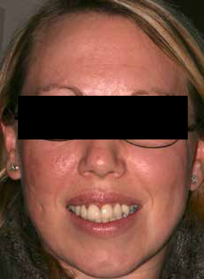
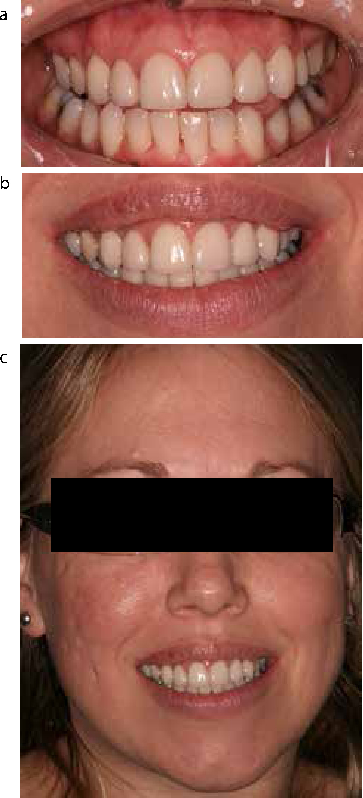
  • Reduce the excessive gingival display;
  • Correct the gingival level discrepancies between the lateral incisors;
  • Enable the central incisors to be proportioned more correctly;
  • Central incisors form the focal point of the smile and the width/height ratio should be approximately 0.75–0.8 for unworn maxillary incisors.6 Raising the gingival level allowed for the height of the central incisors to be increased and modified to approximate this proportion, thus reducing their ‘squareness’.
  • Replacement veneers

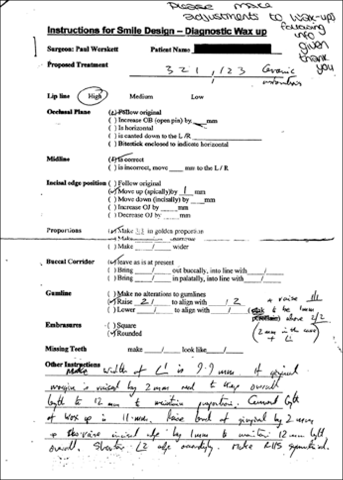
    The design for the new veneers was prescribed to the laboratory by using a ‘diagnostic wax-up prescription form’ which the author uses in his practice. This directs the technician to construct the wax-ups in line with the clinician's requirements, rather than the technician's assumptions. As a result, the veneers were designed to achieve symmetry, balance and harmony, with consideration for golden proportion in line with generally accepted principles of anterior dental aesthetics.7,8,9,10,11

    Clinical considerations

    It is unfortunate that the patient had already been committed to porcelain veneers, as these are irreversible restorations. With the benefit of hindsight, other treatment alternatives might have been considered which may have provided a more conservative solution. These might have included orthodontics, to improve the tooth positions and close spaces, if the natural teeth were otherwise satisfactory in appearance and condition; and composite resin restorations, which can achieve satisfactory short to medium term results and have documented satisfactory performance. Success rates for composite resin restorations have been quoted at 50% survival at 8–9 years,12 although this may be as low as 6 years in toothwear cases.13 However, composite resin restorations are not necessarily invasive and avoid the tooth destruction associated with veneer preparation. Unfortunately, there was no other option in this case but to replace the failed porcelain restorations with new porcelain veneers.

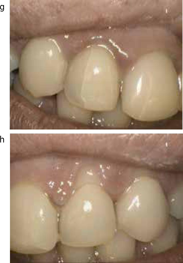
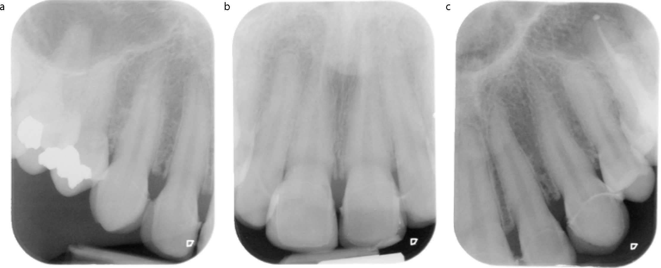
    The use of porcelain laminate veneers were first described in 1983 as a method of restoring teeth14 and has become a common treatment modality over the years as a means of improving the appearance of the anterior teeth. In England and Wales, porcelain veneers placed under NHS regulations achieved a 53% survival rate over 10 years.15 When planned well and executed with care and precision, treatment results can be very satisfactory and the longevity can also be acceptable, although the chances of success are improved when preparation margins do not extend beyond enamel.16, 17 When the finishing margin is in dentine, the success of veneers is reduced, with an increased risk of fracture, poorer marginal integrity and greater discoloration.18 However, the success rate for veneers may be more than 90% over 10 years and as much as 93% over 15 years.16,18,19 Unfortunately, even when performed to high standards, irreversible damage is inflicted on the dental hard tissues, which can only compromise the long-term survival of the teeth. When performed poorly, and with little regard for occlusal function and health of the dental tissues, significant damage can result. The risk of irreversible pulp damage due to tooth preparation may be around 20%.20 Furthermore, the restorations themselves may fail. A significant reason for failure of porcelain veneers is fracture of the porcelain.16 In this particular case, different types of porcelain fracture were noted. At UR2, a longitudinal fracture indicated a static fracture. This may be due to occlusal loading, cyclical fatigue or polymerization shrinkage.16 Incisal edge fractures are an example of a cohesive fracture, which in this case was likely due to excessive occlusal loading.16 In some patients, bruxism may be a cause of fracture and tooth surface loss and, in such cases, consideration should be given to protecting restorations from occlusal dysfunction, where possible.21 This can be achieved with an acrylic occlusal stabilization splint, which will also achieve muscle relaxation and reduce the tendency for bruxism.22,23

    In this particular case, the use of diagnostic wax-ups enabled the design of an occlusal scheme whereby the occlusion of the anterior teeth was protected from interference in lateral excursion by directing the guidance away from the incisal edges of the anterior maxillary incisors in excursive movements. The provisional restorations were designed with the same morphology to allow a period of evaluation in the mouth. Following successful review, the successful occlusal scheme could then be copied into the final porcelain restorations by making a study cast of the provisional restorations and creating a custom incisal guidance table so that the laboratory could duplicate the occlusal scheme.23

    Poorly planned and executed treatment not only compromises the patient's oral health, but can also cause dissatisfaction and unhappiness for the patient. To add further insult, the treatment also brings with it financial compromise due not only to the cost of the original treatment, but also the costs involved in corrective treatment. Therefore, before embarking on elective treatment, such as aesthetic treatment which is not clinically necessary, it is imperative that patients are made fully aware of the significance and consequences of any proposed treatment and the possible long-term implications with respect to further treatment need and the costs which would be incurred, both financial and clinical. Only when this has been done can the patient give full and complete informed consent to the treatment, as required by the General Dental Council,24 safe in the knowledge that they understand the risks that they are accepting by embarking on the treatment. For the ethical practitioner, this is an essential approach to patient care, not only as a professional duty of care, but also to reduce the possibility of a potentially damaging litigation claim.

    Conclusion

    This case demonstrates that, if poorly planned and executed, aesthetic dentistry is arguably worse than doing no treatment at all. Certainly, the patient was left dissatisfied and her oral health was compromised as a result of the treatment she had received previously.

    The current treatment has provided the patient with an aesthetic result, with which she is happy. The occlusion has been stabilized and the long-term relationship with the patient has been established to maintain oral health in the future. However, the longevity of the teeth have been compromised as a consequence of the previous treatment and, indeed, the retreatment. There will be a clinical need in the future to maintain the aesthetic treatment of the past.