Chadwick SM, Banks P, Wright JL. The use of myofunctional appliances in the UK: a survey of British orthodontists. Dent Update. 1998; 25:(7)302-308
Samuels RH. A review of orthodontic face-bow injuries and safety equipment. Am J Orthod Dentofacial Orthop. 1996; 110:(3)269-272
Advice Sheet 8: The Use of Headgear and Facebows.: Development and Standards Committee of the British Orthodontic Society; 2006
Yang CS, Lu CK, Lee FL, Hsu WM, Lee YF, Lee SM. Treatment and outcome of traumatic endophthalmitis in open globe injury with retained intraocular foreign body. Ophthalmologica. 2010; 224:(2)79-85
Singh P, Grammati S, Kirschen R. Orthodontic retention patterns in the United Kingdom. J Orthod. 2009; 36:(2)115-121
Shah AA, Sandler PJ, Murray AM. How to place a lower bonded retainer. J Orthod. 2005; 32:(3)206-210
Advice Sheet 9: Management of Inhaled or Ingested Foreign Bodies.: Development and Standards Committee of the British Orthodontic Society; 2003
Emergencies in orthodontics part 2: management of removable appliances, functional appliances and other adjuncts to orthodontic treatment Paul Dowsing Alison Murray Jonathan Sandler Dental Update 2024 42:3, 707-709.
In the second of two papers, management of orthodontic emergencies involving appliances other than fixed appliances will be detailed. Problems relating to removable appliances, as well as other orthodontic adjuncts, will be discussed. Unfortunately, orthodontic appliance breakage does occur, despite the clinicians giving clear and concise instructions to the patients and their parents at fitting. If general dental practitioners have a practical knowledge of how to diagnose problems and to provide appropriate advice or timely ‘emergency’ treatment, this will significantly reduce the inconvenience for all parties concerned. It should also ensure that treatment progresses in the most efficient and comfortable manner for their patients. In specific situations the early, accurate identification of the problem and instigation of its appropriate management can avoid more serious consequences.
Clinical Relevance: Appropriate handling of an orthodontic ‘emergency’ by the dentist can, on many occasions, provide immediate relief to the patient. This will, in turn, allow treatment to continue in the right direction, thus allowing more efficient and effective use of valuable resources.
Article
In the first paper, general problems encountered in orthodontics and those specific to fixed appliances were described in detail. In this second paper, problems associated with the wearing of removable appliances, functional appliances, retainers and other orthodontic auxiliaries will be discussed. The aim is to remind general practitioners about the various components of these appliances, as well as to provide useful and practical advice should the patient attend his/her surgery for an unexpected ‘emergency’ visit. By managing the problem appropriately, the inconvenience to the patient can be minimized. There are some orthodontic problems which are common to all appliance types, as well as others that are of a more specific nature and both types will be described.
Removable appliances
The widespread use of single-arch removable appliances is now on the decrease, but there are still a significant number of practitioners who prescribe them in specific circumstances. General problems with removable appliances include:
Initial difficulties with speech;
Temporary excessive production of saliva; and
Initial general discomfort.
These symptoms will soon pass once the patient has become used to the appliance, therefore every patient should be encouraged to persevere. If any of these problems persist for more than a few days, then it is more than likely that the patient is not wearing the appliance for a sufficient amount of time to get used to it. Patients should be encouraged to wear appliances as directed by their orthodontist and also informed that, only if they do so will the initial feelings of discomfort subside. They also need to be reminded that failure to follow the instructions will almost certainly compromise the treatment outcome.
The basic design of a removable appliance has not changed significantly, though each is customized in its particular components (Figure 1). Knowledge and familiarity with the most common components will ensure that the practitioner is in the best possible position to deal with any complications that may arise.
Figure 1. Components of a removable appliance: (a) retentive; (b) baseplate; (c) active.
Fractured retentive components
Loose, non-retentive appliances are a common cause of emergency visits and can be avoided by careful design and adjustment of the retention components of the appliance, ensuring that sufficient clasps are prescribed at the outset. Satisfactory retention of a removable appliance will add to the patient's confidence and his/her enthusiasm to wear the appliance, which will therefore maximize the chance of a successful outcome.
It is not uncommon for an Adams clasp to fracture (Figure 2a). If there are a number of other Adams clasps providing retention, often the only treatment required is the removal of the fractured clasp and smoothing of any cut wires. Another option, if there are fewer retentive components, is to remove the fractured bridge of the Adams clasp (Figure 2b) leaving the arrowheads engaging the undercut for retention (Figure 2c). The majority of clasp fractures occur due to work hardened wire suffering fatigue. The likelihood of clasp fracture increases greatly when the patient clicks the appliance ‘in and out’. The patient and his/her parents must be informed how harmful this irritating habit is to the appliance and how the inevitable clasp fracture will undoubtedly delay the progress of treatment.
Figure 2.
(a) Fractured Adams clasp. (b) Wire work trimmed to remove fractured section. (c) Single arrowhead of Adams clasp used for retention.
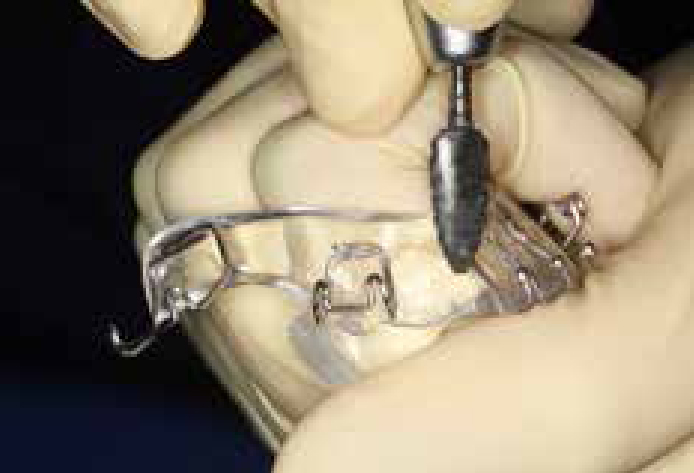
Active components
Appliances are susceptible to damage around areas containing active components such as springs or expansion screws (Figure 3). If a spring is significantly distorted, then there may be no option but to replace it, if further tooth movement is required. Unfortunately, with significant damage, there is little that can be usefully done by the general dental practitioner apart from removing sharp ends or loose wires and referring the patient back to his/her orthodontist. If the patient using an expansion screw has left the appliance out of the mouth for a day or two, even after a temporary repair, the appliance can be extremely difficult to seat fully, as some relapse of the expansion will have occurred. The dentist can attempt to turn the screw back, in quarter-turn increments, until the patient can comfortably, fully insert the appliance. The patient should then be referred back to the orthodontist, with a note detailing the exact adjustments that had to be made to the screw, to allow re-insertion. This information will then allow the orthodontist to issue appropriate instructions.
Figure 3. Irreversible damage to an active component of a removable type of appliance.
Baseplate
A common problem patients often present with is fracture of small areas of the acrylic. If this is fairly minimal, and doesn't affect the design or the integrity of the appliance, then smoothing of the rough edges is all that is required to prevent soft tissue trauma. If the damage is more severe, a new impression is usually necessary for the appliance to be repaired by a technician. Referral back to the orthodontist is therefore recommended.
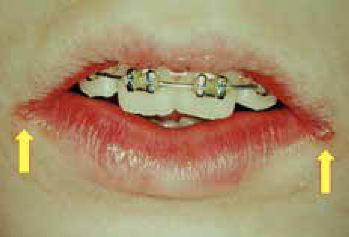
Problems related to appliance hygiene, such as Candida albicans infection causing inflammation to the palatal tissues on the fitting surface of the appliance (Figure 4), are not uncommon. Alternatively, infection may manifest as angular cheilitis with cracks appearing at the corners of the mouth (Figure 5). Measures instituted by the general dental practitioner initially involve instruction to achieve meticulous appliance hygiene, which may include recommendation of a proprietary brace cleaner. If rapid resolution does not occur, antifungal medication, such as Miconazole applied to the affected area, may be required.
Figure 4.
(a, b) Candida infection associated with baseplate of upper removable appliance.Figure 5. Angular cheilitis at corners of mouth.
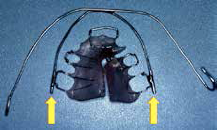
Functional appliances
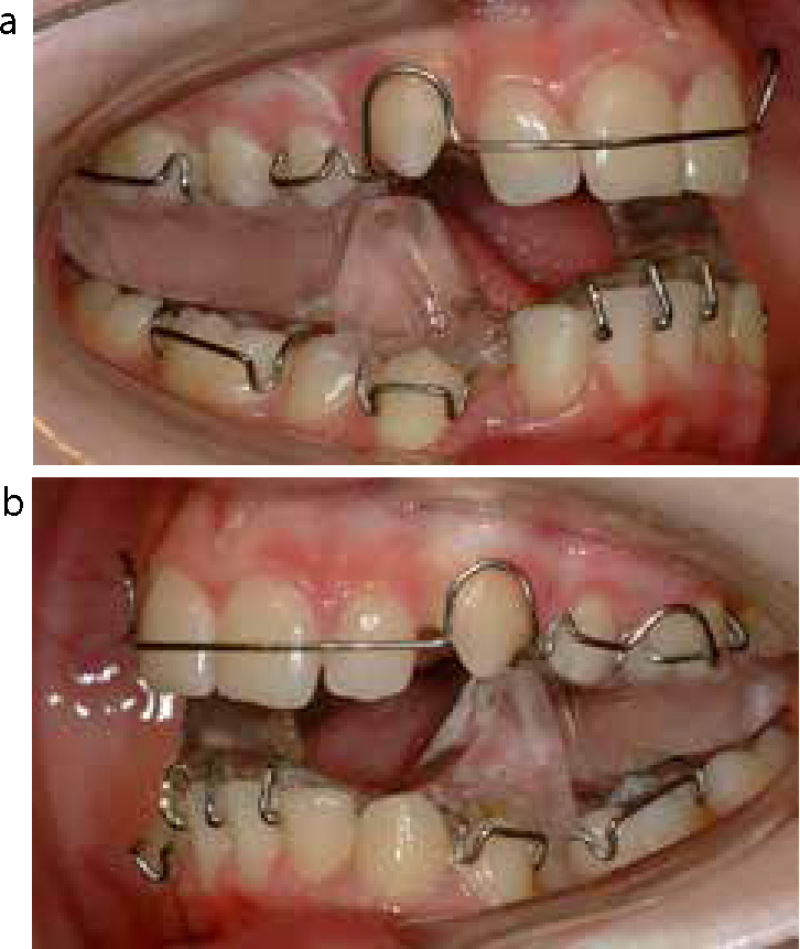
The most commonly used functional appliance in the United Kingdom is the twin block1 (Figure 6a, b). General problems are very similar to those encountered with single-arch removable appliances, such as initial discomfort, excessive saliva production and speech interference, and they should be dealt with as described previously.
Figure 6.
(a, b) Twin block appliance.
After initial fit of the twin block appliance, the muscles of the jaw, as well as the teeth themselves, will ache for the first couple of days of wear. The patient needs to be informed that this is to be expected and reassurance should be given that these symptoms will pass. Another common initial problem is difficulty in speaking for the first day or two. As the twin block appliance is recommended as a full-time appliance, patients should also be advised that speech will rapidly improve and, with time and perseverance, they will learn to speak intelligibly with their teeth together.
Ulceration in the lingual sulcus caused by the lower lingual flanges is not uncommon, often in the lower canine/premolar area. This can be readily dealt with by the GDP by appropriate trimming of the acrylic, just in the area of the inflamed mucosa or ulcer (Figure 7). If the twin block is fractured (most commonly the lower block in the midine area), new working impressions and a new bite registration are often required. The patient will almost certainly need to return to his/her orthodontist for this.
Figure 7. Trimming of acrylic, usually lingually, in lower canine premolar area.
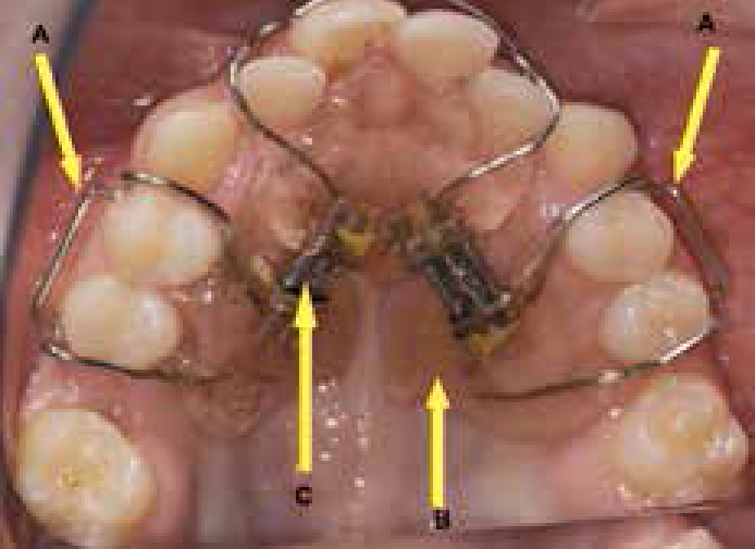
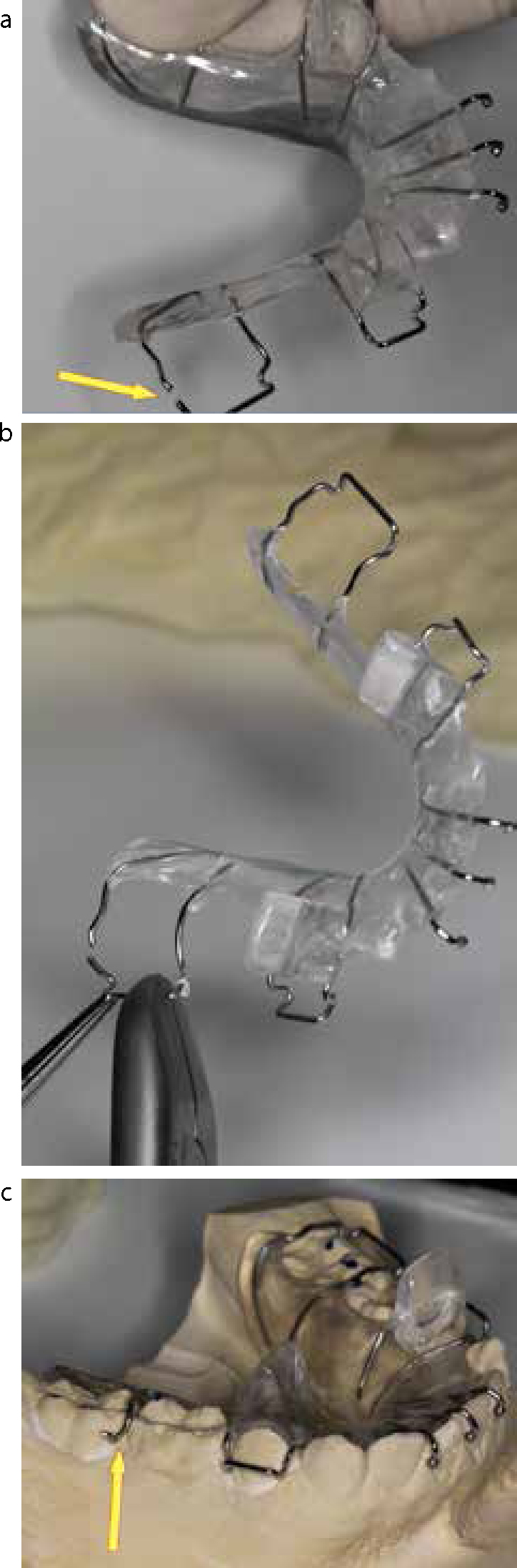
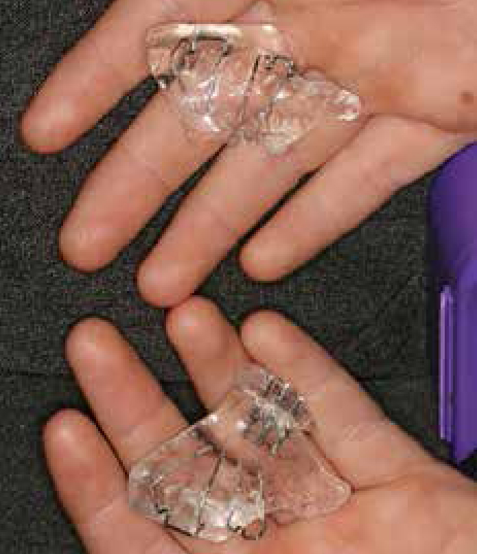
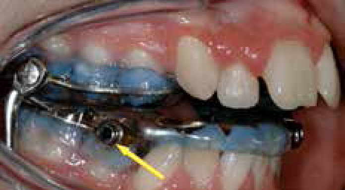
Fixed functional appliance
Although the use of these types of appliances is fairly uncommon, the general practitioner should be aware of their existence. The ‘Class 2 corrector’ appliance is usually attached to the bands placed on the upper molar teeth and to either bands or brackets in the lower arch, depending on the particular appliance. The components of these complicated appliances are prone to breakage (Figure 8) and their repair is not at all straightforward. The general dental practitioner may secure or remove any loose components, which might provide immediate relief from discomfort for the patient, and then return both the appliance components and the patient to the orthodontist as soon as possible.
Figure 8. Example of a popular fixed/functional appliance – Herbst appliance.
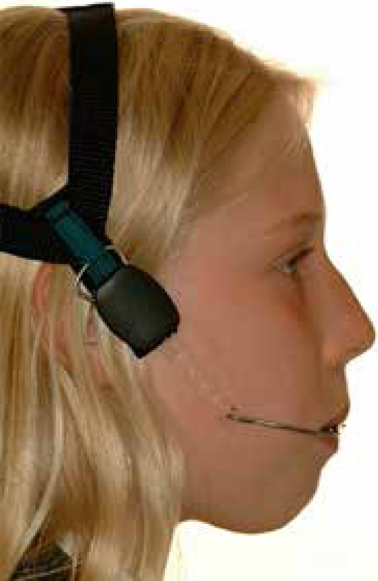
Headgear
Headgear usually consists of an external headgear cap connected to a facebow that transfers force from the back of the head to the dentition (Figure 9). This method of supplementing anchorage is not without risk. Ocular injuries from headgear have been reported in the past,2 usually caused by the inner arms of the facebow (Figure 10). At least two independent safety mechanisms are now recommended in all headgear patients, to prevent recoil injuries following accidental disengagement.3 If there is any evidence whatsoever that the facebow has a tendency to come out of the headgear tubes, the headgear wear should be immediately stopped. The patient must be referred back to see his/her orthodontist as soon as possible.
Figure 9. Headgear and facebow being worn.Figure 10. Facebow inner arms can be potential source of eye damage
Should an ocular injury be suspected, then immediate referral of the patient to the local hospital accident and emergency unit for an ophthalmic opinion is indicated. Any undue delay may well compromise the possibility of a successful restoration of vision.4
Retainers
Removable retainers
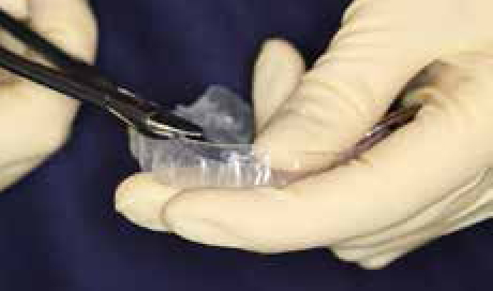
The removable type, vacuum-formed retainers are now commonplace.5 Problems caused by these retainers include, occasionally, trauma on insertion, particularly around the gingival margins. It is a very simple matter for the general dental practitioner, or even the patient, to trim the prominent flange back with a pair of sharp scissors then smooth the cut ends with an emery board (Figure 11). Retainers can also wear down or fracture, in which case replacement will be necessary. Most orthodontists advise the patients that, at some time in the future, their own dentist may need to take impressions to replace lost or broken retainers, albeit on a private basis.
Figure 11. Trimming of a vacuum-formed retainer with sharp scissors or crown shears.
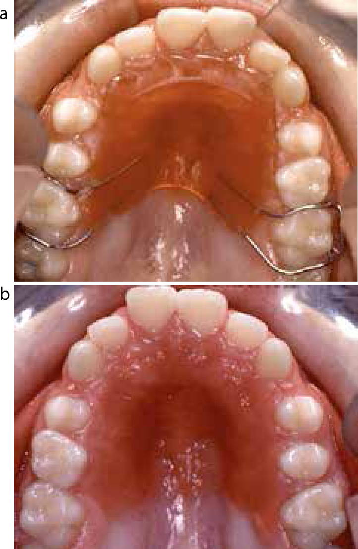
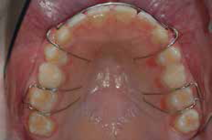
Hawley retainers suffer from all the problems of removable appliances and therefore have exactly the same solutions. Additional problems specific to the Hawley type of retainer may include distortion of the labial bow, which will need to be carefully readapted to the upper labial segment teeth to minimize the chance of relapse (Figure 12). Anything more than very minor distortion will need the appliance to be sent back to the laboratory, or to the treating orthodontist for attention.
Figure 12. Hawley retainer with labial bow.
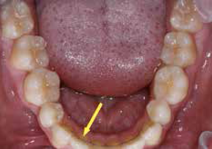
Bonded retainers
Bonded retainers are used these days in a significant number of cases. A multistrand wire is attached to the individual teeth using composite cement and these retainers are designed to stay in place for many years. Problems occurring with these retainers can include the fracture of the wire in between the teeth or the retainer becoming either partially or fully detached from the teeth. If just one composite pad has become dislodged from its tooth (Figure 13), it is usually a fairly simple matter to remove the remnants of composite with a burr, clean the lingual tooth surface and re-bond another pad of composite using an acid etch technique.6 This can provide an easy and quick solution, as long as there has been no distortion of the retainer wire or movement of the associated tooth. If the retainer wire has actually fractured or distorted, the loose or sharp ends should be cut and smoothed and the patient redirected to his/her original orthodontist for any necessary further treatment, as there is always the possibility that relapse may occur.
Figure 13. Composite off one tooth only, no relapse yet seen.
Other potential orthodontic problems
Possible inhalation or ingestion of an orthodontic component
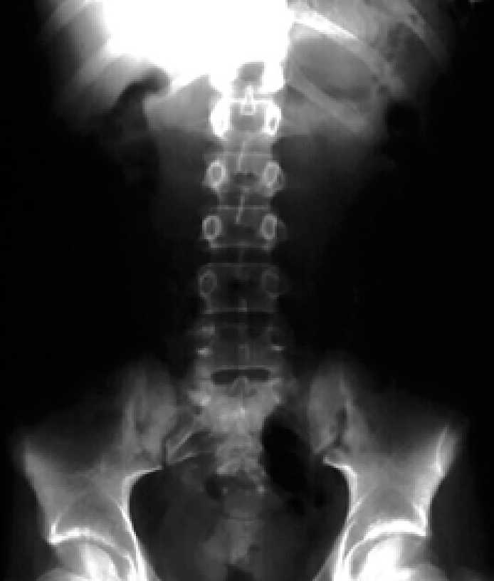
Comprehensive guidelines for the management of inhaled or ingested foreign bodies have been produced by the British Orthodontic Society7 and these are an invaluable guide that should be available in every dental practice. Common sense should dictate the most immediate and appropriate action, especially in the first few seconds when it is suspected that a problem may have occurred.
If the loose or detached object is still visible in the mouth or oropharynx, an attempt should be made to remove it if at all possible. If this is not possible then the patient should be encouraged to cough up the foreign body. If the airway appears compromised then an ambulance should be called, and if possible an attempt made to try and remove the obstructive cause, if this can be done safely. If there is a suspicion or concern that the component has been inhaled or swallowed, the patient should be referred immediately to the local hospital for an appropriate radiographic examination (Figure 14), and further management as appropriate. Ideally, a similar orthodontic component to the one inhaled or swallowed should also be sent with the patient; this will help the radiologist enormously to identify this particular foreign body. Advice should also be sought from the attending physician in the accident and emergency department as to the need for further observation or care, and the incident fully documented in the patient's notes.
Figure 14. Abdominal x-ray taken to locate foreign body.
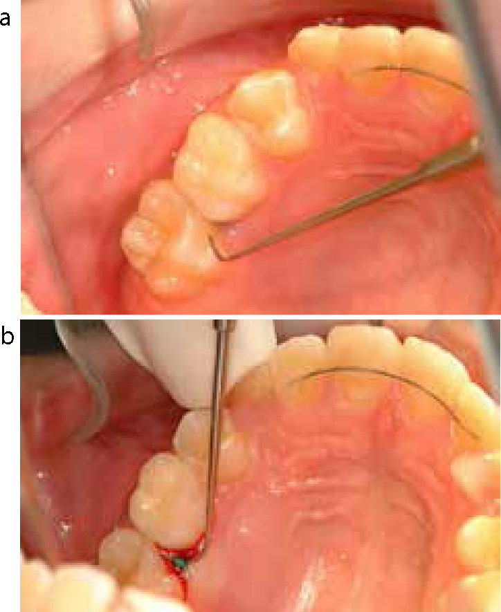
Orthodontic separator subgingivally
All patients will experience varying levels of pain or discomfort following the placement of separating elastics, as often this is the most uncomfortable part of the whole orthodontic treatment, and they should certainly be forewarned about this. Prophylactic analgesia is probably advisable and the patients should continue to take analgesics, strictly following the prescribed instructions, for as long as they feel the need. Care must always be taken to ensure the number and the site of separator placement is recorded in the notes, as well as the site from which they are removed at the next visit. Each separator placed should always be accounted for, and if any are missing the patient should be asked whether they remember losing them. If unsure, it is always advisable to probe the area gently with the aid of the air syringe. Occasionally, a separator may be hidden subgingivally (Figure 15a, b). If this hidden separator is inadvertently ignored, and an orthodontic band is just placed, this could lead to significant periodontal problems, infection and possible ultimate tooth loss.
Figure 15.
(a) Orthodontic separator not immediately visible but dark shadow mesially. (b) Gentle probing subgingivally reveals hidden separator.
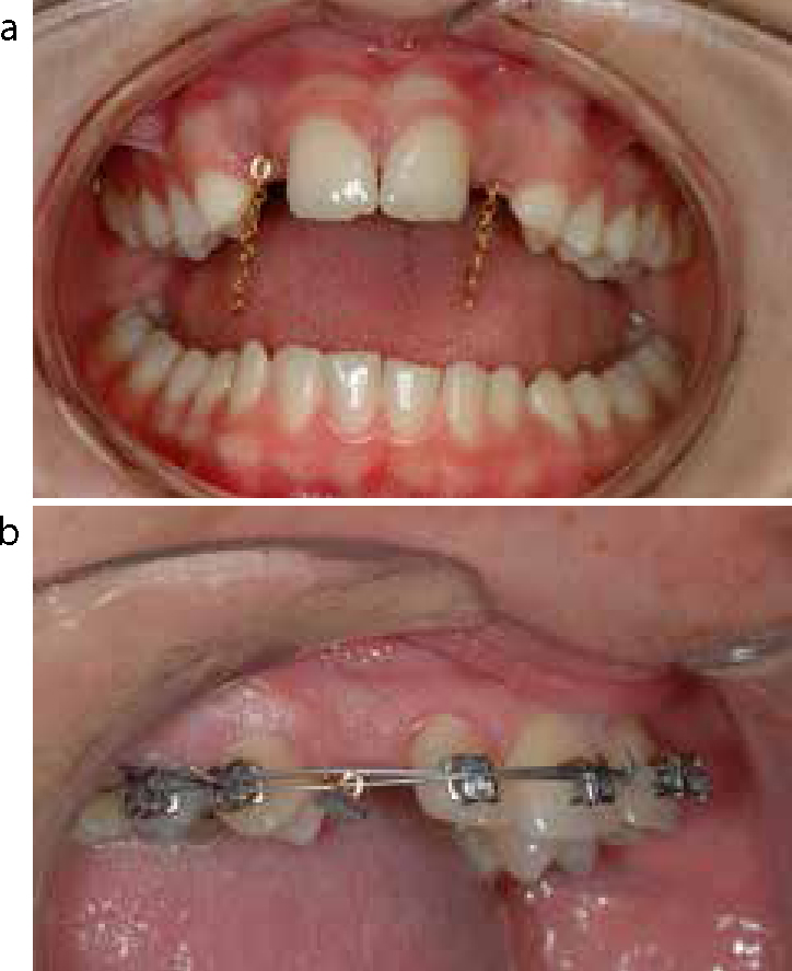
Debonding of gold chain attached to impacted tooth
Following a closed surgical procedure to expose and bond an unerupted impacted tooth, a gold chain is attached to its crown to allow subsequent orthodontic traction. This free end of the chain is usually sutured into the sulcus, however, it can sometimes become loose and hang down in the patient's mouth (Figure 16a). If this is irritating the patient, the loose part of the chain can easily be re-attached either to the brace itself or re-bonded onto an adjacent tooth. Alternatively, the chain can be carefully shortened taking care to ensure that there are still a sufficient number of links protruding to allow subsequent orthodontic traction. This ‘high risk’ solution is probably best left to the treating orthodontist.
Figure 16.
(a) Loose gold chains can be bonded to adjacent teeth or shortened. (b) Bond failure between unerupted tooth and pad of gold chain.
Occasionally, the gold chain may become loose due to the failure of the bond to the enamel of the unerupted tooth itself (Figure 16b). In these circumstances, all that can be done is to remove the detached part of the chain, and the patient must be informed a further surgical procedure will probably be required in the near future to re-bond the chain to the crown.
Summary
Most patients undergoing orthodontic treatment will only have a few problems during their period of appliance therapy. Orthodontics is a specialist branch of dental treatment, but it is important that all general dental practitioners have an understanding of the problems with all common types of appliances. The main objective of an emergency visit is primarily to get the patient out of discomfort or pain, but also to ensure continuation of progress with treatment. The help of the general dental practitioner throughout treatment is invaluable to achieving this end.
The problems listed in these two papers are by no means exhaustive, but they hopefully identify the majority of day-to-day problems that may be encountered by general dental practitioners. The papers offer simple and straightforward, practical solutions. In the first instance, an attempt should always be made by the patient to get in touch with his/her orthodontic provider, but if for some reason this is not possible, then the GDP will often still be able to ‘save the day’!