Snauwaert K, Duyck J, van Steenberghe D, Quirynen M, Naert I Time dependent failure rate and marginal bone loss of implant supported prostheses: a 15-year follow-up study. Clin Oral Invest. 2000; 4:13-20
Buser D, Weber HP, Donath K, Fiorellini JP, Paquette DW, Williams RC Soft tissue reactions to non-submerged unloaded titanium implants in beagle dogs. J Periodont. 1992; 63:225-235
Weber HP, Buser D, Donath K, Fiorellini JP, Doppalapudi V, Paquette DW, Williams RC Comparison of healed tissues adjacent to submerged and non-submerged unloaded titanium dental implants. A histometric study in beagle dogs. Clin Oral Implants Res. 1996; 7:11-19
Cochran DL The scientific basis for and clinical experiences with Straumann implants including the ITI Dental Implant System: a consensus report. Clin Oral Implants Res. 2000; 11:33-58
Abrahamsson I, Berglundh T, Glantz PO, Lindhe J The mucosal attachment at different abutments. An experimental study in dogs. J Clin Periodont. 1998; 25:721-727
Marsh PD Dental plaque: biological significance of a biofilm and community life-style. J Clin Periodont. 2005; 32:7-15
Mombelli A, Lang NP The diagnosis and treatment of peri-implantitis. Periodontology 2000. 1998; 17:63-76
Renvert S, Roos-Jansåker AM, Claffey N Non-surgical treatment of peri-implant mucositis and peri-implantitis: a literature review. J Clin Periodont. 2008; 35:305-315
Jovanovic S The management of peri-implant breakdown around functioning osseointegrated dental implants. J Periodont. 1993; 64:1176-1183
Luterbacher S, Mayfield L, Brägger U, Lang NP Diagnostic characteristics of clinical and microbiological tests for monitoring periodontal and peri-implant mucosal tissue conditions during supportive periodontal therapy (SPT). Clin Oral Implants Res. 2000; 11:521-529
Lang NP, Berglundh T Peri-implant diseases: where are we now? Consensus of the Seventh European Workshop on Periodontology. J Clin Periodont. 2011; 38:178-181
Ericsson I, Berglundh T, Marinello C, Liljenberg B, Lindhe J Long-standing plaque and gingivitis at implants and teeth in the dog. Clin Oral Implants Res. 1992; 3:99-103
Berglundh T, Zitzmann NU, Donati M Are peri-implantitis lesions different from periodontitis lesions?. J Clin Periodont. 2011; 38:188-202
Klinge B, Hultin M, Berglundh T Peri-implantitis. Dent Clinic N Am. 2005; 49:661-676
Lindhe J, Berglundh T, Ericsson I, Liljenberg B, Marinello C Experimental breakdown of peri-implant and periodontal tissues. A study in the beagle dog. Clin Oral Implants Res. 1992; 3:9-16
Lindhe J, Meyle J Peri-implant diseases: Consensus Report of the Sixth European Workshop on Periodontology. J Clin Periodont. 2008; 35:282-285
Heitz-Mayfield LJ, Huynh-Ba G History of treated periodontitis and smoking as risks for implant therapy. Int J Oral Maxillofac Implants. 2009; 24:(Suppl)39-68
Renvert S, Persson GR Periodontitis as a potential risk factor for peri-implantitis. J Clin Periodont. 2009; 36:9-14
Dentino A, Lee S, Mailhot J, Hefti AF Principles of periodontology. Periodontolology 2000. 2013; 61:16-53
Quirynen M, Listgarten MA Distribution of bacterial morphotypes around natural teeth and titanium implants ad modum Brånemark. Clin Oral Implants Res. 1990; 1:8-12
Heydenrijk K, Meijer HJ, van der Reijden WA, Raghoebar GM, Vissink A, Stegenge B Microbiota around root-form endosseous implants: a review of the literature. Int J Oral Maxillofac Implants. 2002; 17:829-838
Hultin M, Gustafsson A, Hallström H, Johansson LA, Ekfeldt A, Klinge B Microbiological findings and host response in patients with peri-implantitis. Clin Oral Implants Res. 2002; 13:349-358
Eke PI, Braswell LD, Fritz ME Microbiota associated with experimental peri-implantitis and periodontitis in adult Macaca mulatta monkeys. J Periodont. 1998; 69:190-194
Mombelli A, Müller N, Cionca N The epidemiology of peri-implantitis. Clin Oral Implants Res. 2012; 23:67-76
Klinge B, Meyle J Peri-implant tissue destruction. The Third EAO Consensus Conference 2012. Clin Oral Implants Res. 2012; 23:108-110
Zittzman NU, Berglundh T Definition and prevalence of peri-implant diseases. J Clin Periodont. 2008; 35:286-291
Atieh MA, Alsabeeha NH, Faggion CM, Duncan WJ The frequency of peri-implant diseases: a systematic review and meta-analysis. J Periodont. 2012;
Roos-Jansåker AM Long-time follow up of implant therapy and treatment of peri-implantitis. Swed Dent J. 2007; 188:(Suppl)7-66
Lang NP, Berglundh T, Heitz-Mayfield LJ, Pjetursson BE, Salvi GE, Sanz M Consensus statements and recommended clinical procedures regarding implant survival and complications. Int J Oral Maxillofac Implants. 2004; 19:(Suppl)150-154
Mombelli A, Mühle T, Brägger U, Lang NP, Bürgin WB Comparison of periodontal and peri-implant probing by depth-force pattern analysis. Clin Oral Implants Res. 1997; 8:448-454
Gerber JA, Tan WC, Balmer TE, Salvi GE, Lang NP Bleeding on probing and pocket probing depth in relation to probing pressure and mucosal health around oral implants. Clin Oral Implants Res. 2009; 20:75-78
Garnick JJ, Silverstein L Periodontal probing: probe tip diameter. J Periodontol. 2000; 71:96-103
Claffey N, Polyzois I, Ziaka P An overview of nonsurgical and surgical therapy. Periodontology 2000. 2004; 36:35-44
Serino G, Turri A, Lang NP Probing at implants with peri-implantitis and its relation to clinical peri-implant bone loss. Clin Oral Implants Res. 2013; 24:91-95
Lang NP, Wetzel AC, Stich H, Caffesse RG Histologic probe penetration in healthy and inflamed peri-implant tissues. Clin Oral Implants Res. 1994; 5:191-201
Schou S, Holmstrup P, Stoltze K, Hjorting-Hansen E, Fiehn NE, Skovgaard LT Probing around implants and teeth with healthy or inflamed peri-implant mucosa/gingiva. A histologic comparison in cynomolgus monkeys (Macaca fascicularis). Clin Oral Implants Res. 2002; 13:113-126
Etter TH, Håkanson I, Lang NP, Trejo PM, Caffesse RG Healing after standardized clinical probing of the peri-implant soft tissue seal: a histomorphometric study in dogs. Clin Oral Implants Res. 2002; 13:571-580
Serino G, Turri A, Lang NP Probing at implants with peri-implantitis and its relation to clinical peri-implant bone loss. Clin Oral Implants Res. 2013b; 24:91-95
Christensen MM, Joss A, Lang NP Reproducibility of automated periodontal probing around teeth and osseointegrated oral implants. Clin Oral Implants Res. 1997; 8:455-464
Luterbacher S, Mayfield L, Brägger U, Lang NP Diagnostic characteristics of clinical and microbiological tests for monitoring periodontal and peri-implant mucosal tissue conditions during supportive periodontal therapy (SPT). Clin Oral Implants Res. 2000; 11:521-529
Haffajee AD, Socransky SS, Lindhe J, Kent RL, Okamoto H, Yoneyama T Clinical risk indicators for periodontal attachment loss. J Clin Periodont. 1991; 18:117-125
Jepsen S, Ruhling A, Jepsen K, Ohlenbusch B, Albers HK Progressive peri-implantitis. Incidence and prediction of peri-implant attachment loss. Clin Oral Implants Res. 1996; 7::133-142
Boynueğri D, Nemli SK, Kasko YA Significance of keratinized mucosa around dental implants: a prospective comparative study. Clin Oral Implants Res. 2013; 24:928-933
Bouri A, Bissada N, Al-Zahrani MS, Faddoul F, Nouneh I Width of keratinized gingiva and the health status of the supporting tissues around dental implants. Int J Oral Maxillofac Implants. 2008; 23:323-326
Chung DM, Oh TJ, Shotwell JL, Misch CE, Wang HL Significance of keratinized mucosa in maintenance of dental implants with different surfaces. J Periodontol. 2006; 77:1410-1420
Kim BS, Kim YK, Yun PY, Yi YJ, Lee HJ, Kim SG, Son JS Evaluation of peri-implant tissue response according to the presence of keratinized mucosa. Oral Surg Oral Med Oral Pathol Oral Radiol Endodont. 2009; 107:24-28
Atassi F Peri-implant probing: positives and negatives. Implant Dent. 2002; 11:356-362
Sewerin IP Errors in radiographic assessment of marginal bone height around osseointegrated implants. Scand J Dent Res. 1990; 98:428-433
Fransson CSweden: Department of Periodontology, Institute of Odontology Sahlgrenska, Academy University of Gothenburg; 2009
Schwartz F, Herten M, Sager M, Bieling K, Sculean A, Becker J Comparison of naturally occurring and ligature-induced peri-implantitis bone defects in humans and dogs. Clin Oral Implants Res. 2007; 18:161-170
Dörtbudak O, Haas R, Bernhart T, Mailath-Pokorny G Lethal photosensitization for decontamination of implant surfaces in the treatment of peri-implantitis. Clin Oral Implants Res. 2001; 12:104-108
Leonhard Å Five-year clinical, microbiological, and radiological outcome following treatment of peri-implantitis in man. J Periodont. 2003; 74:1415-1422
Máximo MB, de Mendonça AC, Renata Santos V, Figueiredo LC, Feres M, Duarte PM Short-term clinical and microbiological evaluations of peri-implant diseases before and after mechanical anti-infective therapies. Clin Oral Implants Res. 2009; 20:99-108
Renvert S, Polyzois I, Maguire R Re-osseointegration on previously contaminated surfaces: a systematic review. Clin Oral Implants Res. 2009; 20:216-227
Lee J-H, Kwon Y-H, Herr Y, Shin S-II, Chung J-H Effect of erbium-doped: yttrium, aluminium and garnet laser irradiation on the surface microstructure and roughness of sand-blasted, large grit, acid-etched implants. J Periodont Implant Sci. 2011; 41:135-142
Heitz-Mayfield LJ, Salvi GE, Mombelli A, Faddy M, Lang NP Anti-infective surgical therapy of peri-implantitis. A 12-month prospective clinical study. Clin Oral Implants Res. 2012; 23:205-210
Claffey N, Clarke E, Polzois I, Renvert S Surgical treatment of peri-implantitis. J Clin Periodont. 2008; 35:316-332
Porras R, Anderson GB, Caffesse R, Narendran S, Trejo PM Clinical response to 2 different therapeutic regimens to treat peri-implant mucositis. J Periodont. 2002; 73:1118-1125
Ji YJ, Tang ZH, Wang R, Cao J, Cao CF, Jin LJ Effect of glycine powder air-polishing as an adjunct in the treatment of peri-implant mucositis: a pilot clinical trial. Clin Oral Implants Res. 2014; 25:683-689
Karring ES, Stavropoulos A, Ellegaarg B, Karring T Treatment of peri-implantitis by the Vector system. Clin Oral Implants Res. 2005; 16:288-293
Schwartz F, Sculean A, Rothamel D, Schwenzer K, Georg T, Becker J Clinical evaluation of an Er:YAG laser for nonsurgical treatment of peri-implantitis: a pilot study. Clin Oral Implants Res. 2005; 16:44-52
Renvert S, Lessem J, Dahlén G, Lindahl C, Svensson M Topical minocycline microspheres versus topical chlorhexidine gel as an adjunct to mechanical debridement of incipient peri-implant infections: a randomized clinical trial. J Clin Periodont. 2006; 33:362-369
Kreisler M, Götz H, Duschner H Effect of Nd:YAG, Ho:YAG, Er:YAG, CO2, and GaAIAs laser irradiation on surface properties of endosseous dental implants. Int J Oral Maxillofac Implants. 2002; 17:202-211
Renvert S, Lessem J, Dahlén G, Renvert H, Lindahl C Mechanical and repeated antimicrobial therapy using a local drug delivery system in the treatment of peri-implantitis: a randomized clinical trial. J Periodont. 79:836-844
Rimondini L, Farè S, Brambilla E, Felloni A, Consonni C, Brossa F, Carrassi A The effect of surface roughness on early in vivo plaque colonization on titanium. J Periodont. 1997; 68:556-562
Thierbach R, Eger T Clinical outcome of a nonsurgical and surgical treatment protocol in different types of peri-implantitis: a case series. Quintessence Int. 2013; 44::137-148
Schou S, Berglundh T, Lang NP Surgical treatment of peri-implantitis. Int J Oral Maxillofac Implants. 2004; 19:140-149
Hayek RR, Araújo NS, Gioso MA, Ferreira J, Baptista-Sobrinho CA, Yamada AM, Ribeiro MS Comparative study between the effects of photodynamic therapy and conventional therapy on microbial reduction in ligature-induced peri-implantitis in dogs. J Periodont. 2005; 76:1275-1281
Serino G, Turri A Outcome of surgical treatment of peri-implantitis: results from a 2-year prospective clinical study in humans. Clin Oral Implants Res. 2011; 22:1214-1220
Romeo E, Lops D, Chiapasco M, Ghisolfi M, Vogel G Therapy of peri-implantitis with resective surgery. A 3-year clinical trial on rough screw-shaped oral implants. Part II: radiographic outcome. Clin Oral Implants Res. 2007; 18:179-187
Sharon E, Shapira L, Wilensky A, Abu-Hatoum R, Smidt A Efficiency and thermal changes during implantoplasty in relation to bur type. Clin Implant Dent Relat Res. 2013; 15:292-296
Hitti RA, Kerns DG Guided bone regeneration in the oral cavity: a review. Open Pathol J. 2011; 5:33-45
Nyman S, Lindhe J, Karring T, Rylander H New attachment following surgical treatment of human periodontal disease. J Clin Periodont. 1982; 9:290-296
Gottlow J, Nyman S, Lindhe J, Karring T, Wennstrom J New attachment formation in the human periodontium by guided tissue regeneration. Case reports. J Clin Periodont. 1986; 13:604-616
Dahlin C, Sennerby L, Lekholm U, Linde A, Nyman S Generation of new bone around titanium implants using a membrane technique: an experimental study in rabbits. Int J Oral Maxillofac Implants. 1989; 4:19-25
Jovanovic SA, Spiekermann H, Richter EJ Bone regeneration around titanium dental implants in dehisced defect sites: a clinical study. Int J Maxillofac Implants. 1992; 7:233-245
Zybutz MD, Laurell L, Rapoport DA, Persson GR Treatment of intrabony defects with resorbable materials, non-resorbable materials and flap debridement. J Clin Periodont. 2000; 27:169-178
Weltman R, Trejo PM, Morrison E, Caffesse R Assessment of guided tissue regeneration procedures in intrabony defects with bioabsorbable and non-resorbable barriers. J Periodont. 1997; 68:582-590
Khoury F, Buchmann R Surgical therapy of peri-implant disease: a 3-year follow-up study of cases treated with 3 different techniques of bone regeneration. J Periodont. 2001; 72:1498-1508
Wikesjö UM, Qahash M, Thomson RC, Cook AD, Rohrer MD, Wozney JM, Hardwick WR Space-providing expanded polytetrafluoroethylene devices define alveolar augmentation at dental implants induced by recombinant human bone morphogenetic protein 2 in an absorbable collagen sponge carrier. Clin Implant Dent Relat Res. 2003; 5:112-123
Nguyen-Hieu T, Borghetti A, Aboudharam G Peri-implantitis: from diagnosis to therapeutics. J Investig Clin Dent. 2012; 3:79-94
Hürzeler MB, Quiñones CR, Morrison EC, Caffesse RG Treatment of peri-implantitis using guided bone regeneration and bone grafts, alone or in combination, in beagle dogs. Part 1: Clinical findings and histologic observations. Int J Oral Maxillofac Implants. 1995; 10:474-484
Hürzeler MB, Quiñones CR, Schüpback P, Morrison EC, Caffesse RG Treatment of peri-implantitis using guided bone regeneration and bone grafts, alone or in combination, in beagle dogs. Part 2: Histologic findings. Int J Oral Maxillofac Implants. 1997; 12:168-175
Machado MA, Stefani CM, Sallum EA, Sallum AW, Tramontina VA, Nociti Júnior FH Treatment of ligature-induced peri-implantitis defects by regenerative procedures: a clinical study in dogs. J Oral Sci. 1999; 41:181-185
Schwartz F, Boeling K, Latz T, Nuesry E, Becker J Healing of intrabony peri-implantitis defects following application of a nanocrystalline hydroxyapatite (Ostim) or a bovine-derived xenograft (Bio-Oss) in combination with a collagen membrane (Bio-Gide). A case series. J Clin Periodont. 2006; 33:491-499
Schwarz F, Sculean A, Bieling K, Ferrari D, Rothamel D, Becker J Two-year clinical results following treatment of peri-implantitis lesions using a nanocrystalline hydroxyapatite or a natural bone mineral in combination with a collagen membrane. J Clin Periodont. 2008; 35:80-87
Renvert S, Polyzois I, Claffey N Surgical therapy for the control of peri-implantitis. Clin Oral Implants Res. 2012; 6:(Suppl)84-94
Oh TJ, Yoon J, Misch CE, Wang HL The causes of early implant bone loss: myth or science?. J Periodont. 2002; 73:322-333
Steigenga JT, Al-Shammari KF, Nociti FH, Misch CE, Wang HL Dental implant design and its relationship to long-term implant success. Implant Dent. 2003; 12:306-317
Adell R, Lekholm U, Rockler B, Brånemark PI, Lindhe J, Eriksson B, Sbordone L Marginal tissue reaction at osseointegrated titanium fixtures (1). A 3-year longitudinal prospective study. Int J Oral Maxillofac Surg. 1986; 15:39-52
The use of dental implants in replacing missing teeth is proven to be a valid treatment with a high success rate. To achieve the best treatment outcome in all implant systems, the implant has to be able to integrate with the surrounding tissue. However, dental implants are affected by peri-implant diseases and may fail as a result. As the number of implants placed continues to increase, the prevalence of peri-implant disease will also increase. This requires preventive measures to inhibit the development of the disease and stop its progression.
Clinical Relevance: Understanding how to maintain healthy peri-implant tissue as well as diagnosis and treatment of disease are vital for every dentist and dental student.
Article
Dental implants provide an alternative and a predictable treatment option for replacement of missing teeth with a high success rate.1,2 However, dental treatment using oral implants may fail as a result of biological complications such as peri-implant mucositis and peri-implantitis.1,2,3,4,5 When such complications occur and are not treated properly, this may lead to the loss of the affected implant. Thus, following implant placement, an effective check-up protocol and supportive therapy to maintain peri-implant tissue health is of paramount importance. This article provides an overview of peri-implant diseases, their diagnosis and treatments which are vital for every dental care professional.
Implant-soft tissue interface
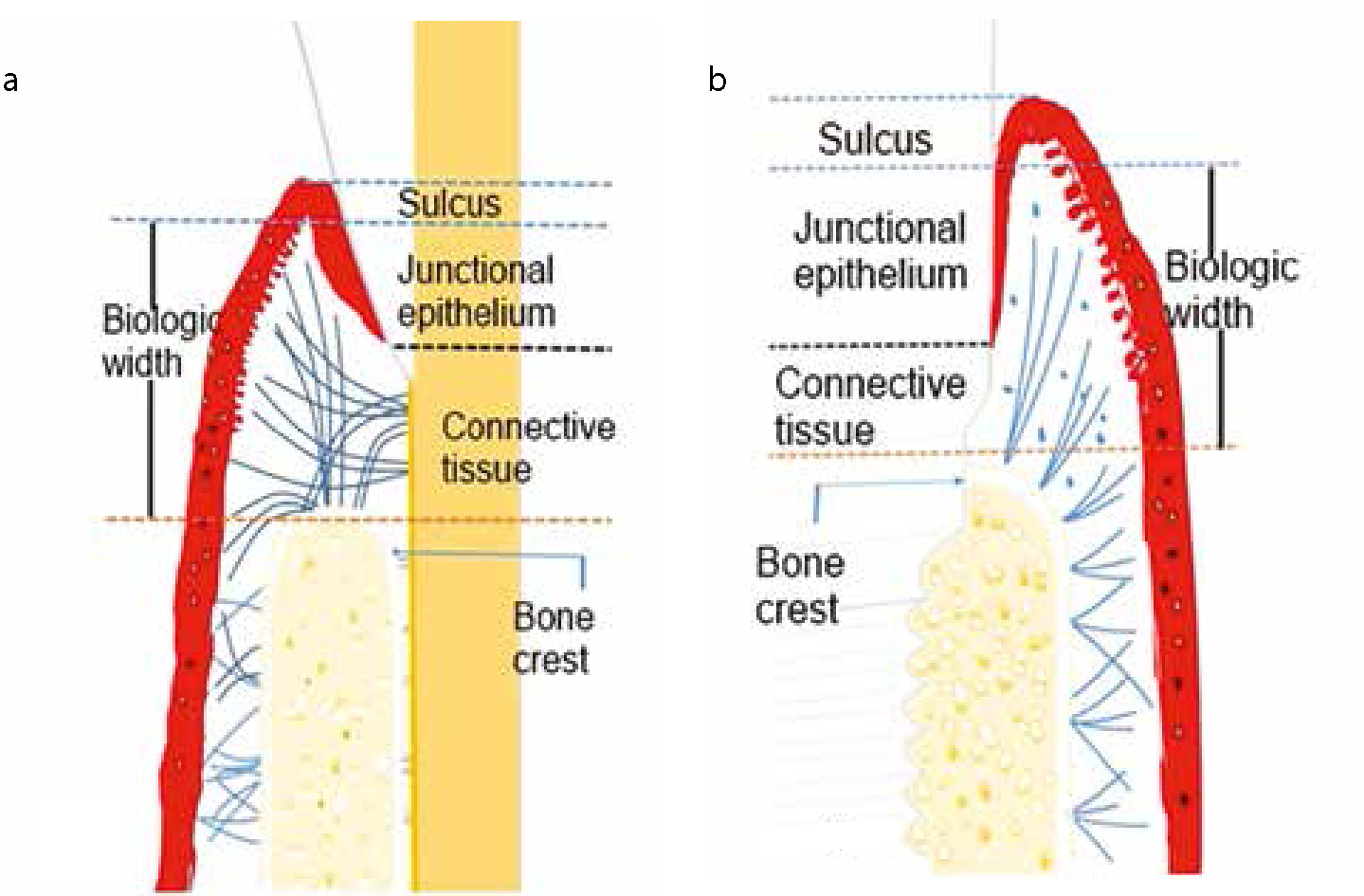
Several studies have reported that the soft tissue that surrounds an implant has similar features to the soft tissue that surrounds teeth (Figure 1 and Table 1).2,3,4,5 Therefore, the peri-implant soft tissue consists of a junctional epithelium which is attached to the implant and/or abutment surface through a hemi-desmosomal attachment and a basal lamina. However, the basal lamina is less evident around an implant than teeth. Apical to the junctional epithelium and coronal to the crest of alveolar bone, there is an area of connective tissue which includes a dense circular avascular zone of fibres that are surrounded by a loose vascular connective tissue.5 Collagen fibres arising from the crest of alveolar bone are oriented parallel to the implant surface/abutment towards the oral epithelium. Unlike connective tissue that is attached to the root surface, connective tissue that is located close to implant surface is not attached to it. Horizontal fibres, originating from the periosteum and the alveolar crest to the oral epithelium, were also found. The junctional epithelium and connective tissue are collectively known as the biologic width. This biologic width is comparable to those found around teeth. The apical extension of the peri-implant epithelium may be varied according to the implant placement technique; submerged and non-submerged. A study in beagle dogs4 reported that the apical extension of the peri-implant junctional epithelium was significantly smaller and the attachment level significantly higher around non-submerged, one-stage implants than submerged implants with second-stage trans-mucosal abutments.4
Figure 1. A schematic representation of a comparison between: (a) periodontal and (b) peri-implant tissues.
Parameters
Teeth
Implants
Gingival/mucosal sulcus depth
Shallow (on average 0.69 mm).
Dependent on implant type and prosthetic component length. In general, it is deeper than around the teeth.
Relationship of oral epithelium with the junctional epithelium
A well-keratinized oral epithelium joins the sulcular epithelium.
A well-keratinized oral epithelium joins the sulcular epithelium.
Location of crestal bone
1–2 mm apical to cemento-enamel junction.
Dependent on implant design; ranges from 0.5–2.5 mm from implant shoulder or the first thread.
Biologic width
Junctional epithelium approx. 0.97 mm long and a connective tissue attachment of 1.07 mm in crono-apical direction.
Junctional epithelium about 2 mm long and connective tissue about l–1.5 mm.
Connective tissue attachment
Collagen fibres inserted into alveolar bone and cementum.
Collagen fibres arise from the crestal bone and run parallel to the implant surface.
Collagen content and fibroblast density
The gingiva contains less collagen content and more fibroblast density than the peri-implant mucosa.
The peri-implant mucosa contains more collagen and less fibroblast density than the gingiva.
The occlusal thickness perception
Teeth-to-teeth contact is about 20 µm.
Implants with opposing teeth is about 48 µm.
Supporting mechanisms
As a result of the presence of periodontal ligament and its viscoelastic properties, when teeth are loaded, they move within the socket with lateral movements that range from 56–108 μm and apical movements which range from 25 to 100 μm.
No periodontal ligament but an intimate implant–alveolar bone contact present: when implants are loaded, they move laterally approximately 10–50 μm and 3–5 μm apically due to bone deformation.
Materials from which the abutment was made affect the location and the quality of the attachment that occurs between the peri-implant mucosa and the abutment.6 For instance, when abutments are made of gold or porcelain, no proper attachment is formed at the abutment level, but soft tissue recession and bone resorption occurs, consequently the abutment-implant junction is exposed and the mucosal barrier is located on the implant body. On the other hand, when the abutments were made of commercial pure titanium or highly sintered aluminum based ceramic (Al2O3), proper mucosal attachment was found on the abutment surface.6 These findings may be due to differences in adhesive properties or to the corrosion resistance of the materials.
It seems that the implant-soft tissue interface makes a protective seal between the oral environment and the bone, which plays a vital role in the success of the implants. It is important to mention that the peri-implant tissue is more likely to resemble a scar tissue with less vascularity and more collagen fibres in comparison with the soft tissue around teeth.5
Peri-implant diseases
Biological complications that are reported to affect the peri-implant tissue may be caused by the inflammatory response of this tissue to bacteria that colonize the implant surface and form a biofilm.7,8 It occurs when the balance between the bacterial load and host defence is shifted in favour of the bacteria. The response of the peri-implant tissue to such bacterial insult may be limited to the soft tissues, or it may also extend to affect peri-implant crestal bone and lead subsequently to its resorption. This inflammatory response occurs in a similar manner to that which is seen in periodontal tissue and periodontal disease.
The diseases that affect the peri-implant tissue are collectively known as peri-implant diseases. They are classified as peri-implant mucositis or peri-implantitis. Mucositis represents an inflammation of the peri-implant mucosa, but is not associated with bone resorption.9 On the other hand, peri-implantitis is considered when peri-implant soft tissue inflammation is associated with bone resorption.10,11,12 The Seventh European Workshop on Periodontology (2011)13 specified an objective diagnostic criterion for peri-implant diseases as follows:
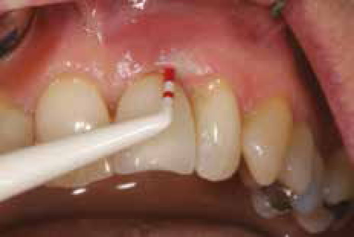
Mucositis, when there is bleeding on gentle probing and peri-implantitis, when the bleeding is associated with crestal bone resorption with or without an increase in PPD and suppuration (Figure 2).
Figure 2. Probing using a plastic probe to avoid damaging the implant surface.
In peri-implant mucositis, the inflammatory response resembles the response in gingivae when exposed to pathogenic bacteria which leads to gingivitis. Thus, peri-implant mucositis is not essentially different from gingivitis. For instance, after three months of plaque accumulation in beagle dogs, the histological examination of the gingiva and the peri-implant mucosa revealed that both tissues, which were obtained from gingivitis and mucositis sites, contained inflammatory cell infiltrates (ICIs), but the peri-implant mucositis had more ICIs than that found in gingivitis.14 In peri-implantitis, the response to bacterial insult may differ from periodontitis, both in the extent and the composition of cells in addition to the progression rate.14 Furthermore, histopathological features of peri-implantitis and periodontitis in humans are not closely identical and some variations may be present.15 A review of several studies,16 which were carried out on human biopsy material, indicated that the ICIs were found to be more pronounced and located more apically in peri-implantitis than in periodontitis. Neutrophils and macrophages were present in more numbers in peri-implantitis lesions than in periodontitis.15 In animal studies, plaque formation is observed following ligature placement, which results in the loss of supporting tissues and large ICIs around both implants and teeth.17 However, after ligature removal, the ICTs extended to the bone crest in peri-implant tissues, whereas a band of connective tissue capsule separated the ICIs from bone in the periodontia.15
Several risk indicators are identified to be associated with the presence of peri-implant diseases. These factors include poor oral hygiene, history of periodontitis, smoking, type 1 diabetes, genetic traits and excessive alcohol consumption18,19,20,21,22 (Table 2).
History of periodontitisGeneticsAcquired factors, ie diabetesEnvironmental factors such as smoking, alcohol consumption and stress
Microbiota associated with peri-implant diseases
The sub-mucosal part of the abutment is colonized by the microbiota which closely resemble those found on the neighbouring teeth in partially edentulous patients. It was proposed that teeth are the source of the bacterial biofilm on implant surfaces in the same individual.16,23,24 The literature indicates that the microbiota associated with chronic periodontitis are also found on surfaces of implants that have failed due to peri-implantitis.24,25
The microbiota that are most commonly found to colonize the surfaces of failed implants include: Gram-negative anaerobic bacteria, such as Fusobacteria, Spirochetes, Bacteroides forsythus and Aggregatibacter actinomycetemcomitans and ‘black-pigmented bacteria’ like Prevotella intermedia, Prevotella nigrescens and Porphyromonas gingivalis.24,25
There is increasing evidence which shows that bacteria are the main cause of the peri-implant disease but its severity is influenced by other environmental factors as well as the individual's genetic make-up.24 A study carried out on adult monkeys by Eke et al26 indicated that the microbiota associated with progression of experimentally induced peri-implantitis and periodontitis, which occurred simultaneously in partially edentulous mouths, are similar. However, while peri-implantitis sites contain a greater number of Spirochetes, the periodontitis sites harbour more Actinomyces.26
Prevalence of peri-implant diseases
The precise prevalence rate of peri-implant diseases is difficult to estimate due to many factors, such as a lack of a standardized definition of each disease entity, different limits for bone loss, which indicate the presence of the disease, and the variability in the other clinical parameters used in diagnosis of the disease.27,28 However, peri-implant mucositis was reported to occur in about 80% of subjects and in 50% of implant sites, whereas peri-implantitis occurs in about 28% to 56% of patients and in 12% to 43% of implant sites.29 In a recently published systematic review and meta-analysis study, Atieh et al30 reported that the frequency of peri-implant mucositis in the patients and on the implants was at 63.4% and 30.7%, respectively. The figures for peri-implantitis were about 19% for the patients and 9.6% for the implant sites. These figures indicate that mucositis is more common than peri-implantitis when patients or implant sites were considered. There is a general consensus that during 5–10 years after implant placement, about 20% of patients and 10% of implants were estimated to be affected by peri-implantitis.27,28 It has been assumed that 9–14 years after implant placement peri-implantitis is a common clinical finding and smoking and periodontal disease are risk factors in the development of the disease.31
Diagnosis of peri-implant diseases: mucositis and peri-implantitis
Clinical parameters that are used in the diagnosis of periodontal diseases are also used for diagnosis of the peri-implant diseases. These parameters are:
The quality of peri-implant mucosa;
Plaque accumulation;
Bleeding on probing (BoP);
Probing pocket depth (PPD);
Clinical attachment level (CAL); and
Width of peri-implant keratinized mucosa.
Peri-implant mucosa should be carefully examined for detection of redness, hyperplasia or recession.2 Monitoring for presence or absence of suppuration, which indicates the association of peri-implant disease with deep lesion and progressive bone loss, should also be considered. Implant mobility, which may indicate a complete implant failure or fracture, should also be included in the clinical examination. Radiograph evaluation is also important in the diagnoses. However, these evaluating measures are not without drawbacks that should be considered when clinical and radiographic examinations are made.
Peri-implant probing
The periodontal probe is used in examination of the pre-implant tissue in a similar manner to that used for periodontal examination (Figure 2). It is commonly used for evaluating the gingival health status and the connective tissue attachment level around teeth as well as around implants. Thus, in order to avoid tissue trauma, the use of a gentle probing force of 0.2 to 0.25N is considered suitable for determination of PPD and CAL.32,33 However, according to Gerber et al34 a force of 0.15 N might represent the threshold that is to be used during implant probing to avoid a false positive BoP. It appears that probing around implants has a higher sensitivity compared with probing around teeth.
There is strong evidence which indicates that the probe tip position within the gingival sulcus is affected by periodontal and peri-implant tissue health conditions. For instance, in inflamed conditions the probe tip was found to penetrate the junctional epithelium and reach the connective tissue (closer to bone), whereas in healthy tissue the probe will stop short of the base of the junctional epithelium.35,36,37,38,39 In healthy sites around implants and teeth, the distance between the probe tip and the alveolar bone was found to range from 0.5 to 1.5mm. Furthermore, even in the presence of minor marginal inflammation, the probe penetrates deeper around implants than around teeth.39 It was also reported that the PPD was consistently greater at implants with peri-implantitis than at implants with mild mucositis or healthy mucosa.38 However, in an animal study, Etter et al40 reported that probing of the healthy peri-implant mucosal tissues caused a separation of the junctional epithelium from the implant surface but not the connective tissue. The probe tip was located at the most coronal level of the supra-crestal connective tissue as determined histologically. The epithelial adaptation was found to return to its original position within five days after the peri-implant soft tissue probing.40 This may indicate the delicate nature and fragility of the peri-implant tissue compared with the periodontal tissue.
It is important to emphasize that other factors, such as diameter of the probe tip and angulation of the probe, as well as presence of restoration, could affect the measurement accuracy of the PPD and CAL. The accuracy could also be affected by the angulation and position of the implant. The PPD may also vary according to the technique by which the implant is placed, to implant systems and position of implant shoulder in relation to crestal bone level.
Several clinical studies reported a deeper PPD in implants with radiographic peri-implant bone loss when compared to implants without such bone loss.33,37 It is important to indicate here that the clinical measurement in these studies was carried out without the removal of the prosthesis, which may affect the measurements. A weak correlation between PPD before the removal of implant-retained prosthesis and the amount of bone loss evaluated during surgery was reported.41 On the other hand, a high correlation was reported when the prosthesis was removed, as better access to probing was achieved.41 Furthermore, removal of the restoration may also be required for debridement and decontamination, when sub-mucosal access is inadequate as a result of the presence of the restoration or due to design or position of the implant.24 Thus, to obtain consistent PPD and CAL measurements and to achieve a proper treatment outcome, removal of the restoration is of vital importance in certain clinical situations.
It may be concluded that PPD is an important indicator of bone loss at implant sites if proper access is provided41 and an increasing PPD with time is usually an indicator of crestal bone loss. Monitoring PPD is important and its increase when associated with BoP is an indication of a risk for peri-implant disease progression. Thus, probing should be carried out after attachment of restorative component to the implant body and this probing measurement is taken as a baseline record from which the peri-implant tissue status is monitored. It should be pointed out that the literature indicates that the reproducibility and sensitivity of PPD measurements, in general, is low.42 Therefore, all of these deficiencies in probing should be considered when peri-implant tissue probing is carried out and interpreted.
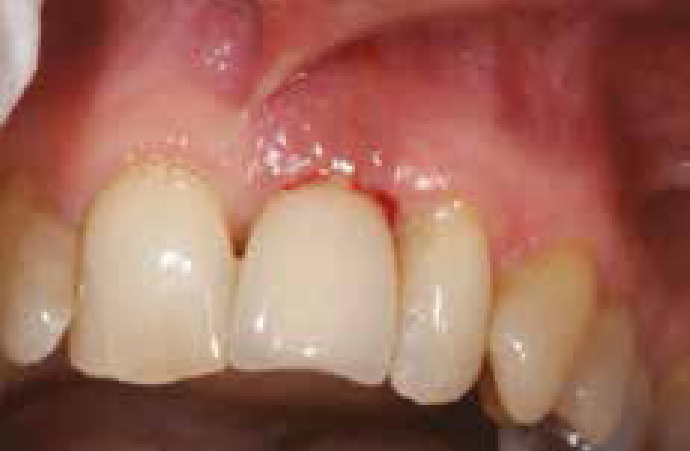
During the probing procedure, bleeding areas are also observed and recorded (Figure 3). It indicates mucosal inflammation.12,43 However, in the evaluation of periodontal disease, BoP does not have a good predictable value. Nevertheless, the absence of BoP is considered a reliable indicator of stable and healthy periodontal tissue.44,45 When BoP is considered around the implants, it seems to have a better diagnostic accuracy than around the teeth.12 It has been reported that BoP occurred in about 91% of peri-implantitis sites.38 Thus, it is a valuable diagnostic parameter. However, smoking may affect peri-implant mucosa as it has a suppressive effect on its BoP, which may lead to an improper diagnosis.
Figure 3. Bleeding on probing with suppuration.
Keratinized mucosa
It has been suggested that the presence of keratinized mucosa around implants may be beneficial in maintaining the mucosal health as it facilitates plaque control and enhances its removal. The importance of keratinized mucosa around dental implants was evaluated by Boynueğri and co-workers46 in 15 edentulous patients with implant-retained complete overdentures. The results of this study indicated that less plaque accumulation and less mucosal inflammation were associated with the presence of a minimum of 2 mm band of keratinized mucosa when compared with implants with no keratinized mucosa. This is in agreement with the results reported by Bouri and co-workers,47 who found a higher mean of gingival and plaque scores, as well as radiographic bone loss around implants with a decreased keratinized mucosa (<2 mm), than implants with more keratinized mucosa. Inversely, according to Chung and co-workers,48 the absence of adequate keratinized mucosa was not found to be associated with higher plaque accumulation and mucositis.48 It has also been reported that inadequate peri-implant keratinized mucosa was not found to affect the implant hygiene or peri-implant soft tissue health status adversely. Nevertheless, the susceptibility to gingival recession and the peri-implant crestal bone loss may increase.49 Even though no conclusive recommendation can be made, healthy peri-implant mucosa can still be seen in the presence and absence of keratinized peri-implant mucosa. There is, however, a general consensus that the presence and preservation of peri-implant keratinized mucosa is important for the health of the mucosa32 and for long-lasting maintenance and management of tissue.32,49
Radiographic evaluation
After implant placement a radiograph is needed. This radiograph is usually used to confirm the position of the implant and may also be used in comparing the peri-implant bone level with this bone level in the future. However, after placement of the implant, a 3-month healing period should be given and peri-implant bone level recorded radiographically, as the osseointegration of peri-implant bone is probably now achieved. This radiograph is used as a baseline to monitor the bone level.24 The radiograph should also be standardized in order to minimize distortion and to obtain a comparable view which is important to reach an accurate interpretation.
Patient clinical assessment is required first and a radiograph is carried out only if it is a necessity and is used to confirm the clinical finding and for an estimation of the peri-implant bone level and to detect any existing pathology. Nevertheless, a repeat exposure of the patient to unnecessary radiation is not ethically sound and adherence to what is known as the ALARA (As Low As Reasonably Achievable) principal is essential.
An intra-oral radiograph may underestimate the bone level and defect depth around implants when compared to direct bone measurements during surgical exposures. It has also been estimated that about 30% reduction in calcified structures is essential before it can be seen radiographically.50
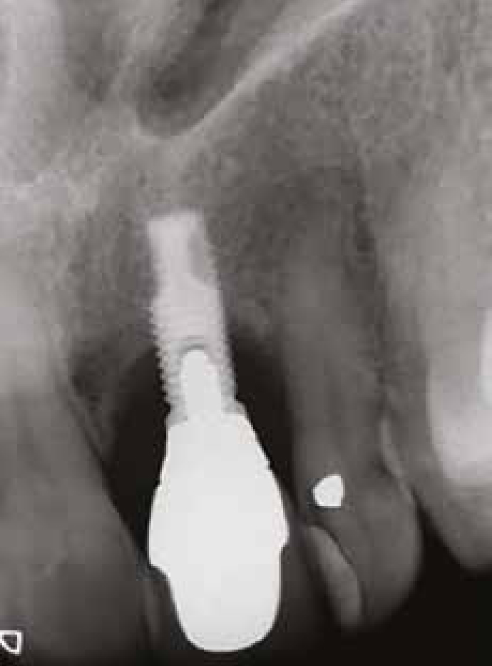
The evaluation of the peri-implant bone level around an implant is also influenced by the angulation of the central X-ray beam to the long axis of the implant body. Thus, a precise parallelism between implant body axes and film plane is important to obtain valid results.51 The central beam is then directed at a right angle to the film. In this case, implant threads can clearly be seen with no overlapping. Figure 4 shows a radiograph of an affected implant.
Figure 4. Radiographic representation of peri-implantitis lesion for the same patient as in Figures 2 and 3.
Radiographically, the distortion of buccal and lingual bone margins may lead to an overestimation of peri-implant bone heights.51 The degree of overestimation is also affected by the bucco-lingual position of the implant body, as well as the width of the alveolar bone ridge.51 Thus, the wider the ridge, the less accurate bone level readings will be. It has been concluded that the width of the alveolar ridge and the amount of bone loss may negatively affect the accuracy and precision of the intra-oral radiograph evaluation.52
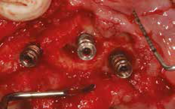
We should bear in mind that radiography is 2-dimensional imaging of a 3-dimensional object. Furthermore, radiographic assessments of marginal bone loss are highly specific and are limited to the mesial and distal aspects of the implants.37 Therefore, changes in bone level on the labial and lingual aspect cannot be radiographically detected as the implant material will absorb the radiation and, consequently, the bone level will not be seen. In a clinical study carried out by Serino et al,37 only 66% of the implants presented with circumferential bone loss (Figure 5). While 34% had a tendency to have more bone loss buccally, radiographs were poor in detecting such loss. In another study examining 40 implants undergoing surgical treatment for peri-implantitis, only 55% of the implants had circumferential bone loss.53
Figure 5. Circumferential bone defect around implant.
It is worth mentioning that a 3-dimensional image may be obtained by use of conventional or cone-beam computed tomography, which provide a more accurate representation, but its availability and expense may preclude its use in diagnosis of peri-implant disease.
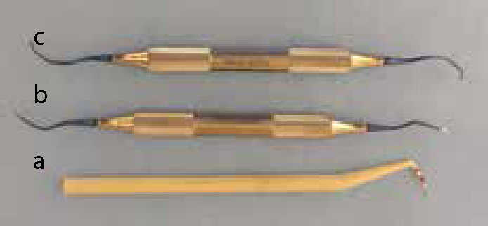
It may be concluded that diagnosis of peri-implant diseases requires the use of probing techniques to identify the presence or absence of pocketing, bleeding, as well as CAL, all of which may indicate the presence of peri-implant disease. Radiographs are also required to detect and estimate peri-implant bone level. Diagnostic information should be obtained for all implant patients once placement and healing of the implant is complete, to allow for longitudinal monitoring of peri-implant conditions. Monitoring the health status of the treated sites requires regular check-ups, and enhancement of the patient's oral hygiene, to keep plaque accumulation on the implant body and/or abutment surface as low as it is achievable. Furthermore, clinical examination and peri-implant disease therapy require instruments that are designed and made from materials which are unlikely to damage the implant surface during the examination and treatment. The instruments used for clinical examination and implant surface debridement are displayed in Figure 6.
Figure 6. The instruments used for clinical examination and treatment: a plastic probe used to avoid scratching and damaging the implant surface (a); titanium implant curettes (Titan®) used for implant surface debridement (b and c).
A number of methods have been suggested for cleaning and decontamination of implant surfaces.54,55,56,57,58,59 These methods may be categorized into three classes as follows:
Mechanical techniques such as the use of titanium-coated Gracey curettes, carbon fibre curettes, plastic-coated ultrasonic scalers, or air-powder abrasives;
Use of chemicals such as chlorhexidine, citric acid, hydrogen peroxide, tetracycline, or stannous fluoride; and
Laser-based treatments.
However, no single surface cleaning technique seems to be superior.60
Treatment of peri-implant diseases
The principal step in any treatment option for peri-implant diseases consists of debridement of the tissue defect and decontamination of the affected implant surface. Largely, the treatment may be divided into two categories: 1. Those targeting peri-implant mucositis; and 2. Those used in treatment of peri-implantitis (Table 3).
Clinical Parameters
Clinical Diagnosis
Treatment Protocols
PPD (Shallow)No plaqueNo BoP
Healthy peri-implant tissue
No treatment is needed, just regular check-ups and enhancement of oral hygiene
PPD (Shallow)Plaque is presentBoP is present
Mucositis
A. Mechanical debridement and polishing using a rubber cup and non–abrasive paste and regular check-ups and enhancement of oral hygiene
PPD ≤5 mm
Mucositis
B. Treatment includes treatment A with antiseptic cleaning
PPD >5 mm associated with bone loss of up to 2 mm
Peri-implantitis
C. Same as treatment B in addition to the use of local or systemic antibiotic
PPD >5 mm associated with bone loss >2 mm
Severe peri-implantitis
D. Same as treatment C combined with surgery (access flap, resective method or regenerative technique)
While the treatment of mucositis is non-surgical, the treatment of peri-implantitis may be surgical or non-surgical, depending on several factors such as the severity of the disease, aesthetic requirement and the presence/absence of neighbouring teeth.
Treatment of peri-implant mucositis
The aim of the treatment is to decrease and disturb the peri-implant microbial community in order to stop further tissue destruction and to change the microbial composition to those associated with healthy tissue. In general, treatment of peri-implant mucositis consists of mechanical debridement, oral hygiene education and regular check-ups. The outcome of such treatment could be enhanced when an antimicrobial mouthrinse is used as an adjunctive.10
Mechanical debridement, as well as mechanical debridement supplemented with chlorhexidine, resulted in a reduction of plaque, inflammation and PPD, as well as a gain in CAL, and were effective in suppressing or eradicating the pathogenic bacteria which is often associated with peri-implant inflammation.61 It appears that such a treatment option is effective and could lead to the reduction in peri-implant mucosal inflammation. Furthermore, a positive effect was reported when antimicrobial mouthrinses were used as an adjunctive to the mechanical intervention.18,57,62 Thus, it is correct to say that treatment of mucositis is identical to that used in treatment of gingivitis.
Treatment of peri-implantitis
Similar to the objectives of the mucositis treatment, the goal of peri-implantitis treatment is to stop further peri-implant tissue destruction and to establish healthy peri-implant soft and hard tissues. Secondary goals, in certain situations, are to obtain bone fill of the defective bone and re-osseointegration of exposed implant surfaces. Several treatment options have been suggested for treatment of peri-implantitis.52,53,54,55,56,57,58,62,63,64,65,66,67,68,69,70 These options are based on several factors, such as degree and extent of the disease and morphology of bone defect. They can be divided into two main categories as follows:
The non-surgical approach: mechanical debridement or laser applications, either alone or combined with antiseptic or antibiotic agents;
The surgical approach: open flap access with or without resective or regenerative techniques.
Non-surgical approach
This approach involves mechanical debridement of the peri-implant tissues and the affected implant surface without raising a flap. In general, this method is not always effective, particularly when the peri-implantitis is associated with a deep PPD.10,24 Furthermore, the outcome of non-surgical treatment of peri-implantitis was not predictable and mechanical debridement alone may be efficient when the PPD is shallow,32 but not enough when the PPDs are deep and with exposed implant threads.63 However, the use of mechanical debridement alone was not found to achieve considerable re-osseointegration,57 which is an important treatment outcome.
It would seem rational to combine the mechanical debridement with chemical antiseptic agents.64,65,66 In a human study66 of oral implants with an initial to moderate peri-implantitis, the use of carbon curettes combined with chlorhexidine antiseptic was compared with the use of air-abrasive devices alone. Both treatment measures led to comparable but limited reductions in PPD and in CAL gains at 6 months.66 However, the result of the study indicated that the first treatment regimen was less effective in decreasing BoP than the second one. It was found that, in peri-implant lesions with a PPD ≥ 5 mm, the combination of mechanical debridement and antiseptic therapy may provide an improvement in clinical parameters. However, residual defects remain after the therapy, which may require additional treatment. According to Renvert et al65 the addition of antiseptic therapy to mechanical debridement does not provide adjunctive benefits in shallow peri-implant lesions (mean PPD <4 mm). Therefore, it may be concluded that the use of antiseptic agents as an adjuvant to mechanical debridement is needed in deep PPD. However, surgical intervention may also be required.
It was demonstrated2,65 that the adjunctive use of a topical antibiotic, minocycline microspheres, to mechanical debridement tends to have greater benefits than those achieved through the use of an antiseptic, ie chlorhexidine and mechanical debridement together. In the study conducted by Renvert and co-workers,65 where 32 subjects had peri-implant lesions with a PPD ≥4 mm with BoP and/or exudate, the adjunctive use of chlorhexidine resulted in limited reduction of BoP, but the adjunctive use of minocycline led to improvements in both BoP and PPDs. The observed improvements in bleeding scores and PPDs obtained by the adjunctive use of minocycline were maintained during a period of 12 months. In another study conducted by Renvert et al,67 using the same treatment protocols as used in the previous study, the treatment was repeated three times: baseline, one and three months. Follow-up examinations were carried out at 10 days and at 1, 3, 6, 9 and 12 months. The results of this study indicated that the use of minocycline microspheres as an adjunctive is beneficial in the treatment, but the treatment may have to be repeated. This raises the question as to whether peri-implantitis with deep PPD can be adequately treated non-surgically by a combination of mechanical debridement and a local antibiotic, and whether the positive effect of this treatment can be maintained for a longer period of time. Hence, well-controlled randomized clinical studies are required.
Lasers are used currently in dentistry and their use has extended to the treatment of peri-implant diseases. For instance, Thierbach and Eger69 divided 28 patients with peri-implantitis into two categories: those with peri-implantitis with pus formation (Figure 3) and those without. Both groups were treated using the same protocol initially after microbiologic diagnosis. All patients were treated at baseline with full-mouth scaling and root planing. Two months later, full-mouth scaling and root debridement and antimicrobial photodynamic therapy were applied. The study indicated that the presence of pus influences the clinical outcome of the treatment of peri-implantitis. According to this investigation, non-surgical treatment of peri-implantitis may be effective when the peri-implantitis lesion has no pus formation but not when pus is present. Nevertheless, when the lesion is associated with pus formation, it can be successfully treated with a supplementary access flap surgery following an additional observation time of 3 months.
It should be emphasized here that, although the use of lasers for implant surface de-contamination is a promising treatment method, their use may be associated with implant surface changes that may negatively affect the treatment result. Furthermore, laser irradiation may raise the temperature beyond the degree which the peri-implant tissue can tolerate and leads to its damage.58,66 Likewise, not all laser types are suitable as implant surface decontaminators. For instance, YAG and Ho:YAG were not found to be appropriate as implant surface decontaminators, irrespective of the power output.58,66 Additionally, the application of laser results in an increase in implant surface roughness that may encourage more plaque accumulation and complicates its removal.68 Thus, the proper laser type, irradiation angle and energy intensity and application time that is suitable for each implant surface should be recognized and used.58
Surgical approach
Surgical treatment of peri-implantitis lesions can be performed after the acute infection has resolved and proper oral hygiene has been established.2,32,70 The primary objective of surgical treatment in peri-implantitis is to get direct access to the affected site and the implant surface, which facilitates decontamination of the implant surface and eliminates granulation tissue. The selected surgical protocol is also dictated by the degree and morphology of the peri-implant bony defect. The amount and quality of the remaining peri-implant soft tissue may play a role in surgical protocol selection.2 The surgical approach can be categorized as follows:
Access surgery (an open flap technique);
Resective surgery with or without implant surface modification (implanto-plasty);
Regenerative approach (use of grafting materials with or without membrane).
Access surgery (an open flap technique): This approach consists of raising a flap, removal of granulation tissue and debridement/decontamination of implant surfaces.55,56,59 This method provides a direct vision and access to the affected peri-implant tissue and to the unexposed implant surface. The effect of this treatment was evaluated clinically, microbiologically and radiographically at 6 months, 1 year and 5 years after surgical exposure and implant surface decontamination using curettes and sterile saline.55 Systemic antibiotics were also given according to a susceptibility test of target bacteria. Six implants of the 26 studied demonstrated bone gain while seven implants were lost and four continued to lose more bone. Nine implants revealed unchanged peri-implant bone levels. This study indicated that the treatment protocol used may be useful in controlling peri-implantitis lesions but with a low predictable value. However, the number of implants used in the study may preclude from drawing a valid conclusion and further well-controlled studies are important. Heitz-Mayfield et al59 studied moderate to advanced peri-implantitis lesions in 36 implants in 24 partially dentate patients. They were treated with open flap surgery and implant surface decontamination using titanium-coated Gracey curettes or carbon fibre curettes and rubbing of the implant surface with gauze soaked in sterile saline. Adjunctive systemic antibiotics (amoxicillin and metronidazole) were also prescribed. Clinical parameters such as BoP, PPD and suppuration were evaluated at 3, 6 and 12 months and the treatment outcomes were analysed during these intervals. Forty seven percent of the affected implants had complete resolution of inflammation (BoP negative) and stable crestal bone levels or bone gain, which was seen in 92% of implants. It was concluded that the conducted treatment protocol was effective and it was possible to maintain the three month positive treatment outcomes for at least one year post-operatively if a strict oral hygiene protocol was followed.
The use of an access flap for debridement of the implant surface combined with chlorhexidine irrigation was compared with non-surgical scaling before photodynamic therapy was implemented for microbial reduction in ligature-induced peri-implantitis in dogs.71 This investigation reported that several bacterial species, that are believed to be associated with peri-implantitis, were significantly reduced by both treatments. However, photodynamic therapy is a non-invasive approach which may be an advantage over surgical access.71
It may be concluded that the open flap technique permits direct access into the affected site, which may enable the clinician to evaluate the bony defect and to debride the implant surface and also eliminate the granulation tissues effectively. In addition, this approach may be necessary when the PPD is deep or when the angulation of the implant body is in an inconvenient position that may preclude effective treatment.
Resective surgery (with or without implant surface modification): Resective surgery is used in treatment of periodontal diseases to correct soft and hard tissue defects around natural teeth. It is based on periodontal pocket elimination, such as apical repositioning flap and gingivectomy or pocket reduction to make the periodontal tissue more accessible for cleaning, thus facilitating the patient's oral hygiene. Resective surgery may also involve an osseous resection/re-contouring which means the removal of defective/correction of supporting bone. This resective treatment option is extended for use in treatment of peri-implantitis lesions. In the oral implant, it is carried out in the treatment of peri-implant disease in order to reduce PPD. It may involve correction of the peri-implant soft tissue when the bone loss is mainly of the horizontal type. Resective surgery combined with apical repositioning flap may be performed in the treatment of peri-implantitis that results in a two- or one-walled bony defect or horizontal bone resorption. However, the resective approach should be carried out cautiously as the amount of removed peri-implant tissues may compromise the aesthetic and/or mechanical results of the implant and the neighbouring teeth.
In a clinical study of 31 subjects with clinical signs of peri-implantitis, 42% of the implants still had the disease after two years of treatment with bone re-contouring and pocket elimination and plaque control before and following the surgery.72 Seventy four percent of the implants with an initial bone loss of 2–4 mm as measured radiographically showed no sign of peri-implantitis during the 2-year period in comparison with 40% of the implants which had a greater bone loss (≥ 5 mm). These findings reflected the effects of the severity of initial peri-implant bone loss on the treatment outcome and on the stability of the achieved improvement.
A combination of resective surgery and modification of the implant surface (implanto-plasty) has been proposed in treatment of peri-implantitis.73,74 The implanto-plasty technique involves the use of rotary instruments to smooth the exposed implant surfaces in order to reduce formation of plaque on the implant surface and to facilitate its removal.73 The use of proper burs with coolant is of paramount importance for efficient cutting to reduce clinical time and to avoid excess heat generation that can damage the peri-implant tissue.74 Romeo and colleagues73 studied peri-implantitis lesions in 19 patients with 38 implants. The treatment involved systemic antibiotics (Amoxicillin for 8 days) with full-mouth disinfection. Nine patients were treated with resective surgery only and ten with resective surgery and implanto-plasty. Marginal bone levels were measured radiographically at 1, 2 and 3 years after surgery. The resective method combined with implanto-plasty was found to be more effective than the resective method alone. Consequently, it is not unreasonable to conclude that resective surgery coupled with implanto-plasty could have a positive influence on the survival rates of rough-surface implants affected by peri-implantitis, as well as on clinical peri-implant parameters such as BoP, PPD and suppuration.73
It is important to remember that the resective approach is indicated when the regenerative treatment is not the treatment of choice.32 For instance, in horizontal bone loss where the regenerative approach may not be a rational option. However, this surgical approach may be used in a non-aesthetic zone as tissue recession is most likely to follow.
Regenerative approach (use of grafting materials with or without membrane): In general, the regenerative approach consists of flap elevation, mechanical root debridement and placement of a graft material, either alone or combined with a membrane. The membrane protects the graft material and provides a confined space for formation of the desired tissues. When the membrane is used without a graft material, the surgical method is known as guided tissue regeneration (GTR). Guided tissue regeneration (GTR) was initially used for treatment of periodontal disease to hinder epithelial and connective tissue migration into the surgical area but allowing cells from periodontal ligament (PL) and bone to repopulate the root surface after its debridement,75,76,77,78 therefore new cementum, PL and alveolar bone are regenerated. The GTR concept is based on the hypothesis that the cell type which has the potential to colonize the exposed root surface at the periodontal healing site will dictate the nature of the resultant attachment or repair.75 Thus, if epithelial cells reach the surgical site and repopulate the exposed root surface, a long junctional epithelium will form. However, if the perivascular cells of the alveolar bone, as well as cells from PL, repopulate the surgical site, new bone, cementum and PL will be created.76,77,78 When the main goal of the surgical process is to regenerate new bone, the procedure is known as guided bone regeneration (GBR). This technique has been further applied to encourage bone formation on exposed surfaces of titanium implants in animal79 and human studies.80
The membrane used can be made of resorbable material, such as collagen membrane, for example (Bio-Gide®), or non-resorbable, such as expanded polytetrafluoroethylene (ePTFE). By the use of resorbable membrane the second surgical intervention needed to remove the membrane is avoided. However, resorbable membranes may be associated with more signs of post-surgical inflammation than non-resorbable ones.81 Studies comparing the use of resorbable and non-resorbable membranes indicated that both types had a comparable clinical effectiveness.81,82 However, complications such as membrane exposure is a real problem which can have a detrimental impact on the treatment outcome.83,84 The exposed membranes should be removed immediately because, if it is left in situ, the amount of bone gain will be negatively affected.
In treatment of peri-implantitis, the use of membrane is usually combined with graft materials. The objective here is to regenerate new bone, reduce PPD, gain of CAL and re-osseointegration of the decontaminated implant surface.85 The use of grafting material with or without a membrane has been used extensively in the treatment of peri-implantitis, with a short-term follow-up with a promising outcome. As the exposure of the membrane is a problem that adversely affects the treatment outcomes, the use of graft material alone appears to be an effective alternative to the use of membrane and the combination of graft materials and the membrane is not always an ideal treatment option.2 Nevertheless, Hürzeler et al86,87 reported no significant difference in bone fill between the use of membrane alone and when the membrane was used with a graft material. However, according to these two studies, the combination of membrane and graft materials led to a greater amount of re-osseointegration than debridement alone (with an air-powder abrasive), or when debridement combined with membrane or graft materials. It is important to note that the findings of Hürzeler et al86,87 were based on animal studies (seven beagle dogs) in which the peri-implantitis was induced by ligature applications for 3 months.86,87
In another study in dogs,88 no statistically significant differences in bone fill in the treatment of ligature-induced peri-implantitis were found when the use of membrane, bone graft, or a combination of the two methods, were compared. The three treatment options performed better and gained more bone fill than debridement alone. Accordingly, it was concluded that the three treatment techniques can improve the bone fill in defects caused by experimentally induced peri-implantitis in dogs.88
The clinical study by Schwartz et al89 demonstrated that treatment of moderate peri-implantitis with either nano-crystalline hydroxyapatite (Ostim®) or Bio-Oss®, in combination with the use of collagen membrane, provided clinically significant improvements in clinical parameters, such as PPD and CAL gains following 6 months of non-submerged healing. The two year results of the same clinical study90 once more demonstrated that both treatment modalities were effective in providing clinically significant reductions of PPD and gains in CAL. However, the combination use of Bio-Oss® and collagen membrane seemed to correlate with greater improvements in those clinical parameters and hence were associated with greater predictability and enhanced healing outcome.
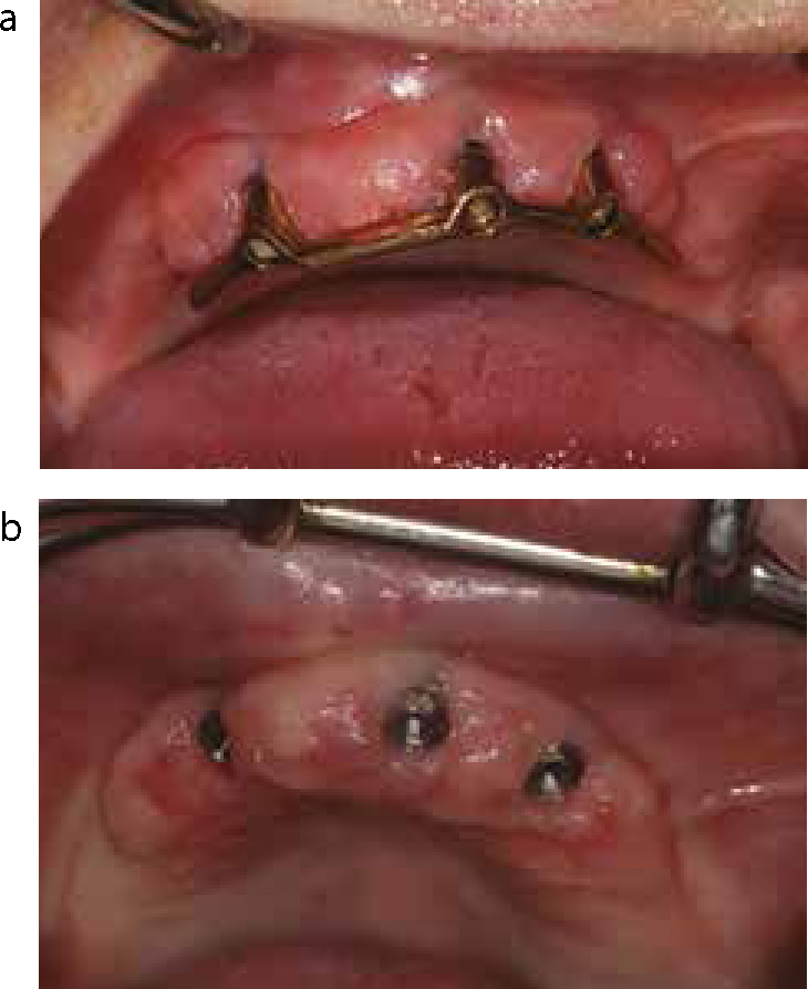
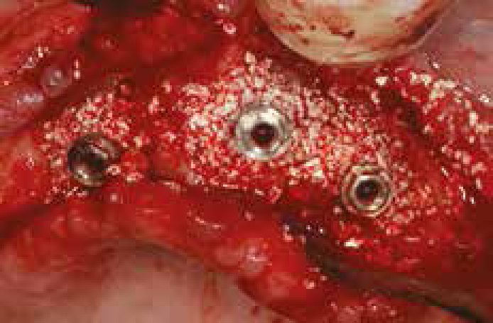
It may be concluded that the routine use of the membrane should be judgementally implemented83 and the use of the membrane with graft material may not always be required, as there is no strong evidence to support its advantage.91 However, the use of graft material without a membrane may be considered when the graft material can be retained within the bony defect, such as in a circumferential bone defect, ie a 4-wall bony defect (Figures 7 and 8). The use of the membrane may be employed when the bony defect morphology does not retain the graft material, ie a 2-wall bony defect. However, long-term, well-controlled randomized clinical studies are required.85 A summary of different treatment options for peri-implant disease, which is suggested by Lang and colleagues,32 is displayed in Table 3.
Figure 7. Peri-implant mucosal enlargement associated with peri-implantitis. Before removal of the bar (a) and after removal of the bar (b). Note deposition of calculus on the implant surface.Figure 8. The circumferential (4-walled) defect of the patient in Figures 5 and 7 treated with graft material (Bio-Oss®).
In general, it may be proposed that each peri-implantitis case should be evaluated clinically and radiographically in a similar manner, as in the treatment of periodontal disease. Oral hygiene should be observed and regular check-ups should be scheduled. In addition, a decision on how to handle the case can be made and which treatment modality should be considered and carried out. Moreover, the following points may be suggested as a general approach:
Peri-implantitis lesions with shallow PPDs may be treated non-surgically with further follow-up and oral hygiene enhancement;
Deep PPDs may be treated surgically with an access flap which aids further examination. This evaluation may dictate the resective approach or regenerative method, depending on many factors, such as the severity of the disease, degree and type of bony defect, aesthetic requirements and neighbouring teeth.
Following any treatment protocol, a follow-up period is essential in order to monitor oral hygiene and help stop and/or discover in time any further deterioration.
Other causes of peri-implant crestal bone loss
As peri-implantitis is associated with the peri-implant crestal bone loss, other factors that may cause such bone loss should be considered. Distinction between crestal bone loss as a result of the peri-implantitis or that due to other causes is an important starting point in stopping and treating such bone loss. It has been reported that an average of one millimetre of marginal bone loss is commonly seen after the first year of function.92,93 This may be followed by an annual loss of less than 0.1 mm thereafter.94 However, the exact cause of this bone loss is still debatable. Currently available literature indicates that the reformation of a ‘biologic width’ around dental implants, presence of a rough/smooth interface, and a micro-gap at implant-abutment interface and occlusal overload are the most likely causes of early peri-implant crestal bone loss. Nevertheless, other contributing factors, such as surgical trauma, may also have a role to play in the process of early implant bone loss.92,93 It is important to mention that occlusal overload does not cause peri-implant disease, but can accelerate its effect when the disease is already present.19 The factors that may contribute to, or cause, peri-implant crestal bone loss are displayed in Table 4.
Bone remodelling after implant placement and an annual loss of <0.1 mm.
Reformation of a ‘biologic width’.
Presence of a rough/smooth interface.
Presence of a micro-gap at implant-abutment interface.
Surgical trauma.
Occlusal overload.
A ‘stress shielding’ phenomenon.
Incomplete removal of luting cement.
Conclusion
Management of mucositis involves mechanical debridement and polishing of the restoration-abutment surface in addition to oral hygiene education and follow-up. The effect of mechanical debridement could be improved when it is combined with application of antiseptic agents. Management of peri-implantitis with several surgical approaches have been described. Unfortunately, there is no general agreement on the best method of treatment. Thus, due to lack of sufficient evidence, a recommendation to implement a specific protocol in the treatment of peri-implantitis is inappropriate. Nevertheless, the first step in any treatment of peri-implant disease is sub-gingival debridement and implant surface decontamination with the use of antimicrobial agents and oral hygiene reinforcement. This approach will control inflammation and prepare the site for more complex surgery, if required. Following peri-implantitis therapy, tissue recession and consequently exposure of the implant surface is common. Thus, the patient should be made aware of these complications before the prospective therapy is carried out as exposure of the rough implant surface may facilitate plaque accumulation that could be difficult to remove and will lead to further inflammation and endanger the implant. Furthermore, removal of part of the peri-implant bone may jeopardize aesthetics and change the mechanical support of the affected implant. It may also negatively affect tissue support of the neighbouring teeth. Thus, careful investigation, which should include clinical and radiographic examinations, is required before any treatment option is carried out.