
Article
The main aims of healthcare provision are generally to eradicate disease, improve health, prevent further disease and improve function. These ideals are seemingly in conflict with the cosmetic dental epidemic currently facing dentistry. Although some may argue that the provision of elective cosmetic treatment can improve well-being, this is often at the cost of the removal of sound tooth tissue. One aspect that may not be entirely apparent initially is damage to the adjacent periodontium.
Patients may complain of rotated, instanding, crowded or labially placed teeth with the desire to have a ‘perfectly’ straight alignment. For some dentists, the worrying apparent alternative to orthodontics, or simply accepting the tooth position, is the elective preparation of teeth to ‘align’ incisal edges and the bucco-palatal profile of the sound teeth. What follows is a critique of this ‘supposedly cosmetic’ technique.
The elective preparation of sound otherwise intact tooth tissue
When crowns and veneers are electively utilized to improve cosmetic appearance the co-morbidities are significant. One problem that requires specific attention is pulpal damage (Figure 1). Felton and colleagues, as long ago as 1987, recognized that, when crowning teeth, the greater the amount of tooth removed, the greater the likelihood of pulpal death.1 Where teeth in normal alignment are prepared for veneers or crowns, the vulnerable perimeter of the pulps may be encroached upon, depending on individual anatomy and the skill set of the operator. Unfortunately, these risks are magnified greatly where teeth are malaligned, especially when positioned towards the labial aspect (Figure 2). Somewhat futile attempts to overprepare teeth, not only to create space for the ceramic, but also to mask position, can be so aggressive that there will be a high probability of pulpal damage.
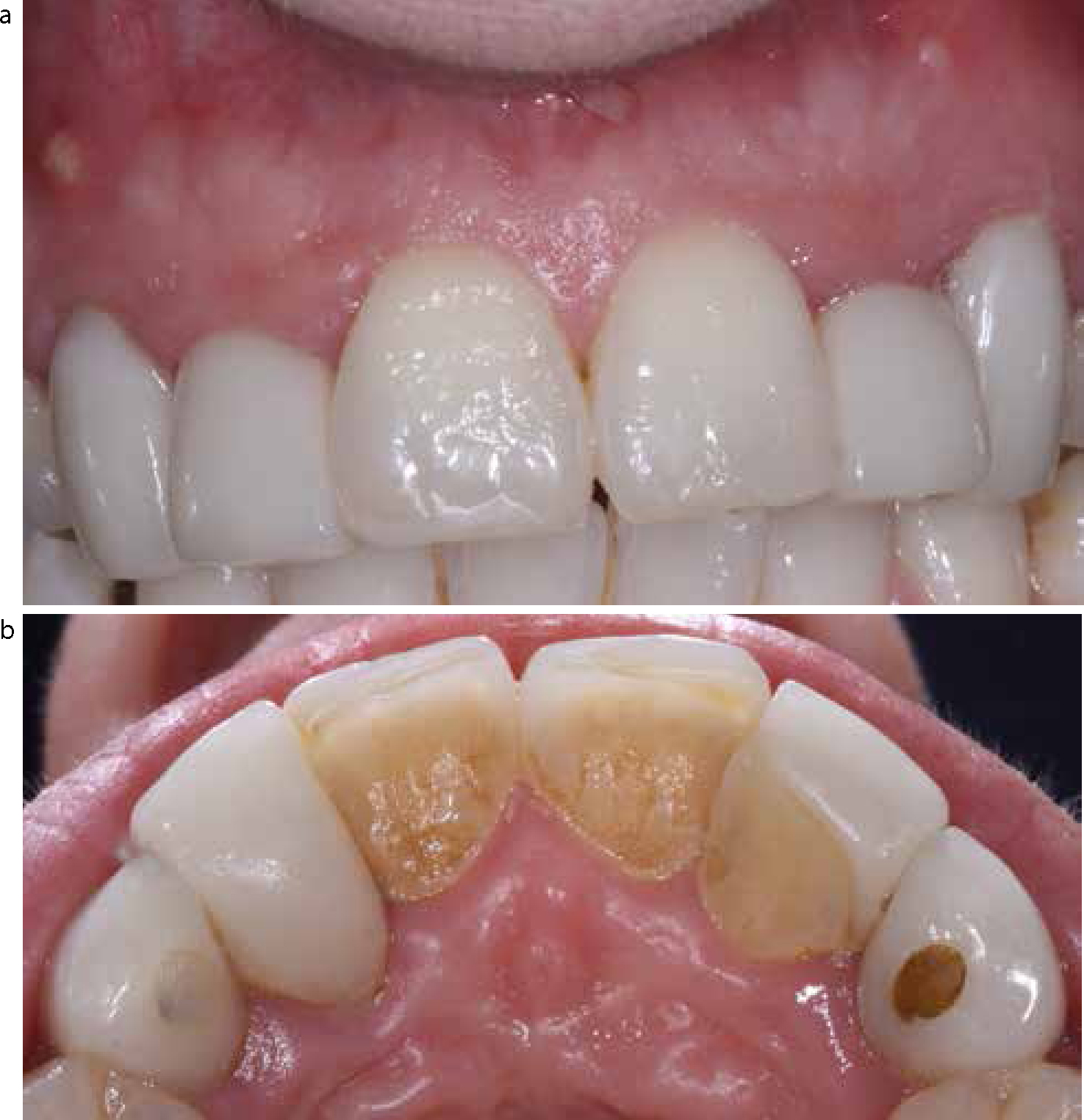

Unfortunately, where multiple veneer and crown units are provided in this way the contact zones become elongated and wide (Figure 1b and 2d). Owing to the extent of such contacts, restoration seating at cementation may be difficult to achieve. Another challenge that these contacts provide is the hindrance of interproximal cleaning. As a consequence, caries may remain undetected as direct visualization of the restoration/tooth interface is difficult.
Periodontal disease
Where teeth are instanding and labially cantilevered restorations are placed, these may become associated with periodontal disease development (Figures 1b and 2d). Such overcontouring creates a significant ‘shelf interface’ resulting in an ideal niche for pathogenic biofilm development. The repercussions of this can result in localized papillary inflammation or recession defects around the instanding tooth.
Treatment alternatives
Patients have been shown to re-consider treatment options when they have been informed of the risks associated with a procedure.2 Ethically and legally, this aspect assumes greater significance when the treatments are elective and invasive.3 Given these potential morbidities, the alternative of no treatment may often be the most sensible biologic option.
Where patients are keen on improving aesthetic appearance without significant tissue loss, the utilization of minimally invasive techniques have been outlined extensively.4 These techniques aim to optimize appearance by way of improving shade by bleaching and utilize direct composite to improve alignment and form without tissue removal.4 A properly informed patient is likely to weigh up the relative improvements to be gained by this non-invasive approach, against the risks associated with destructive veneer preparations, and probably consider the ‘least destructive’ as the best option.
Conclusion
The cosmetic dentistry epidemic, resulting in elective damage to teeth, seems to be widespread and increasing. The informed and evidence-based clinician has to understand the repercussions of elective preparation of sound teeth and recognize, in advance, the complications they can bring. Ignorance of these probable problems is no defence.