
Article
The applications for dental photography are continually growing. While digital single-lens reflex (DSLR) cameras may be used to provide outstanding clinical images, the use of smartphone cameras is becoming increasingly popular (in common with all types of photography).
Regardless of equipment, the main dental photography challenges relate to the limited amount of available light within the oral cavity, particularly when photographing posterior teeth.
Progressive developments in Smile-Line technology (St-Imier, Switzerland) are designed to overcome these exposure difficulties. The original Smite-Lite device (Optident, Ilkley, Yorkshire) was designed as a natural light source (colour temperature 5500K) to optimize shade-matching and to enable calibration of camera phones against a grey scale.
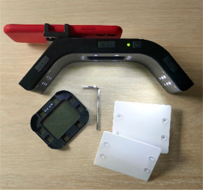
The latest Smile-Line innovation is Mobile Dental Photography (MDP) (Optident, Ilkley, Yorkshire), which is a continuous light source that is designed to provide a versatile range of lighting options, enabling higher-quality smartphone images and providing a stable platform that fits most mobile phones (width range 55–85 mm). The camera phone is easily positioned so that the lens is in the centre of the MDP unit and is secured using the hex key provided (Figure 1).2
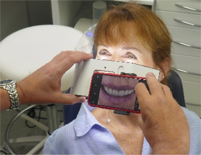
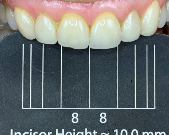
Familiarity with camera phone technology makes the MDP very easy to use. For most intra-oral photographs, the camera is held 10–12 cm from the subject and image framing, magnification and focusing are considerably simplified by the optimized lighting conditions (Figure 2).
The MDP's central and lateral light sources may be used individually or in combination, and each light array may be adjusted to four different levels of intensity. The versatile system also includes two light diffusers (which may be magnetically attached to the lateral lights) and a polarizing filter, which attaches to the centre of the main unit. These accessories may also be used, individually or at the same time, to further modify lighting conditions or to create artistic photographic effects. The MDP requires no batteries and is mains charged.
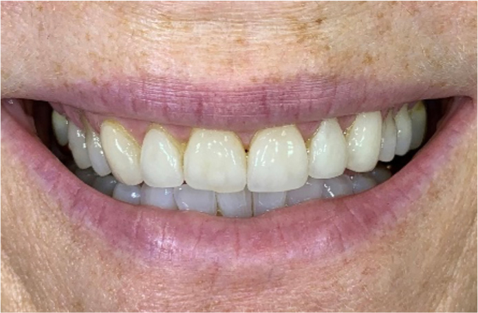
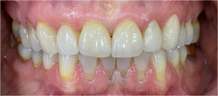
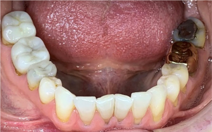
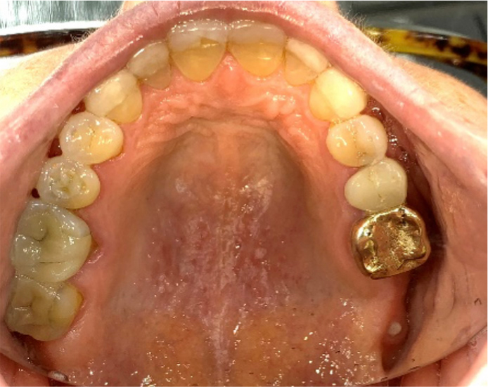
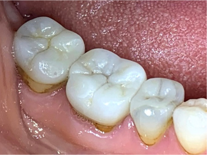
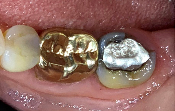
Learning to use an MDP is very easy and quickly enables an introduction to dental photography. Figures 3–11 are examples of MDP images used to convey a range of diagnostic information taken with an iPhone (Apple Inc, Cupertino, California).
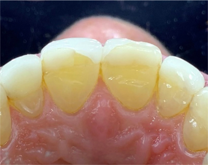
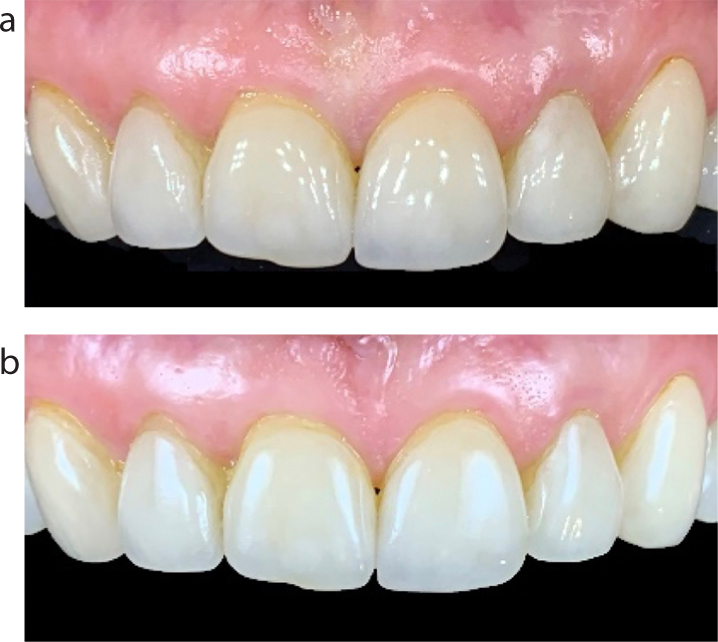
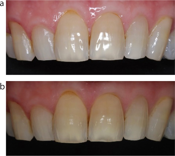
The MDP's polarizing filter may be used to reduce the amount of reflected light. This facilitates analysis of the optical properties of anterior teeth, eg translucency/opacity and diagnosis of internal dentine anatomy. When taking polarized images, it is recommended to use the central light at full power with the lateral lights turned off (Figure 12).
NB: When considering the use of camera phone technology for dental photography, detailed understanding is required with regard to consent, quality control, data protection, image storage and reporting (see ‘Dental photography: a practical guide’, this issue).1