Burke FJT. Amalgam to tooth-coloured materials – implications for clinical practice and dental education: governmental restrictions and amalgam-usage survey results. J Dent. 2004; 32:343-350
Dilley DC, Vann WF, Oldenburg TR, Crisp RM. Time required for placement of composite versus amalgam restorations. J Dent Child. 1990; 57:177-181
Burke FJT, Siddons C, Phipps S, Bardha J, Crisp RJ, Dopheide B. Clinical performance of reinforced glass ionomer restorations placed in UK dental practices. Br Dent J. 2007; 203
Frencken JE, Makoni F, Sithole WD. Atraumatic restorative treatment and glass ionomer sealants in a school oral health programme in Zimbabwe. Evaluation after 1 year. Caries Res. 1996; 30:428-433
Wilson AD, Kent BE. A new translucent cement for dentistry. The glass ionomer cement. Br Dent J. 1972; 132:133-135
Eastbourne, UK: Dental Practice Board;
Combe EC, Burke FJT, Douglas WH.Chicago: Kluwer Academic Publishers; 1999
Manhart J, Chen HY, Hamm G, Hickel R. Review of the clinical survival of direct and indirect restorations in posterior teeth of the permanent dentition. Oper Dent. 2004; 29:481-508
Sidhu SK, Watson TF. Resin modified glass ionomer materials. American Journal of Dentistry. Am J Dent. 1995; 8:59-67
Dentsply De Trey GmbH Professional Research.Germany: Dentsply De Trey; 1998
Magni E, Zhang L, Hickel R, Bussu M, Polimenti A, Ferrari M. SEM and microleakage evaluation of the marginal integrity of two types of Class V restorations with or without the use of a light-curable coating material and of polishing. J Dent. 2008; 36:885-891
Randall RC, Wilson NHF. Glass ionomer restoratives: a systematic review of a secondary caries treatment effect. J Dent Res. 1999; 78:628-637
Deligeorgi V, Mjör IA, Wilson NHF. An overview of reasons for the placement and replacement of restorations. Prim Dent Care. 2001; 8:5-11
Mickenautsch S. How well are GIC product labels related to current systematic review research?. Dent Update. 2011; 38:634-644
Wiegand A, Buchalla W, Attin T. Review of fluoride-releasing restorative materials – fluoride release and uptake, antibacterial activity and influence on caries activity. Dent Mater. 2007; 23:343-362
Burke FJT, Bardha JS. A retrospective practice-based clinical evaluation of Fuji IX aged over 5 years placed in loadbearing cavities. Br Dent J. 2013; 215
Friedl K, Hiller K-A, Friedl K-H Clinical performance of a new glass ionomer based restorative system: a retrospective cohort study. Dent Mater. 2011; 27:1031-1037
Scholtanus JD, Huysmans M-C DMJM Clinical failure of a highly viscous glass-ionomer material over a six year period: a retrospective study. J Dent. 2007; 35:156-162
Basso M, Heiss MA. Permanent restorations with glass ionomer cements: clinical evaluation on 319 cases. J Dent Res. 2013; 92:(Spec Iss A)
Burke FJT, Mackenzie L, Sands P. Dental materials – what goes where? Class I and II cavities. Dent Update. 2013; 40:260-274
Dental materials – what goes where? the current status of glass ionomer as a material for loadbearing restorations in posterior teeth FJT Burke Dental Update 2024 40:10, 707-709.
Authors
FJTBurke
Primary Dental Care Research Group, University Birmingham School of Dentistry, St Chad's Queensway, Birmingham B4 6NN, UK
Glass ionomer materials have been available for 40 years, but have not been indicated for loadbearing restorations, other than when used in the ART concept. However, there is anecdotal evidence that dentists are using the reinforced versions of this material in posterior teeth, possibly as a result of demands from patients to provide them with tooth-coloured restorations in posterior teeth at a lower cost than resin composite. This paper reviews the existing literature on reinforced glass ionomer restorations in posterior teeth, concluding that, under certain circumstances (which are not fully elucidated) these materials may provide reasonable service. However, the patient receiving such restorations should be made aware of the minimal amount of evidence for the success of these restorations and the potential need for the restorations to be re-surfaced in due course.
Clinical Relevance: Reinforced glass ionomer restorations may provide patients with tooth-coloured restorations in posterior teeth, but their survival may not equate to restorations in resin composite in loadbearing situations.
Article
Tooth-coloured restorations in posterior teeth
The demand for tooth-coloured restorations in posterior teeth is increasing worldwide,1 with the principal alternative to amalgam being considered to be resin composite. However, resin composite restorations in loadbearing situations in posterior teeth take longer to place than amalgam, with one study indicating that they take 2.5 times as long2 while, in another, they took only 35% longer, with the three composite steps of acid-etch, wash/dry, and light cure accounting for 86% of the mean time differences for the two materials.3 Since a dentist's time is the most expensive component in a restoration, resin composite restorations are more expensive than amalgam: patients have therefore sought cheaper alternatives. Accordingly, there is some evidence of dentists offering patients a glass ionomer material for restoration of posterior teeth.4 These materials may be placed in bulk, resulting in a saving in time when compared with the time-consuming incremental build-up required for posterior composite restorations. This, in turn, results in a restoration which is less expensive than the equivalent posterior composite. Such a technique has the advantage that the initial fee to the patient will be less than that for a composite (insofar as it could be considered that a glass ionomer restoration in a posterior tooth may be placed in a similar time as placing amalgam) and, even if the restoration later requires re-surfacing (vide infra), the fee to the patient will be spread over a number of years. However, the glass ionomer restoration in a loadbearing situation, while costing less to place initially, may not be cost-effective if its survival is less than an equivalent resin composite restoration.
It is therefore the aim of this paper to assess the clinical performance of glass ionomer materials in loadbearing situations in posterior teeth. It is not the intention of this paper to assess glass ionomer restorations placed using the Atraumatic Restorative Treatment (ART) technique,5 as these are placed under different conditions from those pertaining in general dental practice worldwide (and are often assessed in a different manner from scientific evaluations of resin composite), or so-called ‘sandwich restorations’ in which the cavity is filled with glass ionomer cement covered by a layer, or layers, of resin composite, given that research into such restorations essentially tests the performance of the composite rather than the glass ionomer.
Summary of glass ionomer as a restorative material
Glass ionomer cements were developed in the early 1970s. These materials comprised a fluoro-alumino-silicate (FAS) glass, mixed with a polyacrylic acid.6 Their popularity increased through the 1980s and, by 2000, these materials were used in the placement of circa 1.7 million restorations in the NHS in England and Wales, mainly in Class V non-loadbearing cavities.7
Principal advantages of conventional glass ionomer materials included:8
Their good compressive strength;
Their reliable adhesion to tooth substance (which, in turn, reduces the need for the clinician to cut sound tooth substance to create retention for the restoration); and
Release of fluoride.
Disadvantages of conventional materials included:8,9
Poor tensile/flexural strengths;
Suboptimal wear resistance (which may be considered to preclude the use of these materials in loadbearing cavities);
Initial sensitivity to moisture;
High solubility in dilute organic acids (such as may be found in plaque); and
Poor aesthetics, as a result of their opacity.
As a result of these disadvantages, variants of glass ionomer materials have been developed, becoming available as luting cements and as resin-modified glass ionomers. In the latter, the typical glass ionomer features are maintained, but a resin component is added to allow the materials to be command cured, while at the same time enhancing the material's physical properties – producing enhanced fluoride release, improved adhesion to dentine and reducing the material's solubility.10 The most recently developed generation of glass ionomer materials have been termed fast-setting, high-strength or reinforced glass ionomers. This group includes Fuji IX (GC, Tokyo, Japan), Chemflex (Dentsply, Weybridge, UK) and Ketac-Molar Easymix (3M ESPE, Seefeld, Germany). Manufacturers claim improved early physical properties and resistance to dissolution over conventional glass ionomers,11 this improvement being due to a reduction in the size of the glass particles in the matrix, allowing a faster speed of reaction between the glass and the polyacrylic acid. These materials are stiffer when mixed and have been termed ‘packable’ as a result. Manufacturers have considered that a reinforced glass ionomer material may be suitable as long-term temporary restoration of Class I and II cavities in permanent teeth,11 or permanent small Class I restorations, notwithstanding its suggested use in Class III and V cavities, Class I and II cavities in primary teeth, fissure fillings, core build-ups and Atraumatic Restorative Treatment (ART) technique (Ketac Molar Quick Product Brochure12). The manufacturers of Fuji IX GP suggest that this material is suitable for Class I, II and V restorations in permanent and primary teeth (GC Fuji IX and Fuji IX GP Fast company literature13), although some literature specifies ‘under conditions of minimal loading’.
It could be considered that further research (by way of one or more randomized controlled trials) is needed to determine the survival of reinforced glass ionomer materials accurately in Class I and II cavities and to ascertain the clinical situations which affect survival. Most recently, GC have developed Fuji IX into EQUIA, this being Fuji IX with a resin-based coating, G-Coat, this having been demonstrated to be effective in reducing microleakage.14
Reinforced glass ionomer materials may be considered to have the following properties:
Smaller particle size which leads to faster reaction;
Higher loading which brings improved physical properties;
Exhibit plastic features – can be condensed and packed;
Still a need for improved wear resistance;
Typical glass ionomer features.
The relevance of fluoride release from glass ionomer materials
The release of fluoride from glass ionomer materials was originally considered to inhibit the progress of caries around the restoration, although the literature on this is, by no means, equivocal.15 The relevant literature is presented below.
Randall RC, Wilson NHF. J Dent Res 1999; 78: 628–63715
While there is no doubt that glass ionomer materials release fluoride through their setting reaction, there is little evidence that this inhibits caries after the first week following placement. Randall and Wilson provided a review,15 which examined the research on cariostasis in respect of glass ionomer restorations. Exclusion/inclusion criteria were developed by the authors and 52 papers were identified. While no paper fulfilled the strict inclusion criteria, 28 papers were eventually identified and included. Of these, only one demonstrated a caries inhibitory effect of glass ionomer, with the authors concluding that there was no conclusive evidence for or against inhibition of secondary caries by the glass ionomer restoratives reviewed.
Deligeorgi V et al. Prim Dent Care 2001; 8: 5–1116
The results of this overview of 10 cross-sectional studies, which included 2137 glass ionomer restorations, have indicated that secondary caries is the reason for failure of between 17% and 40% of glass ionomer restorations. Additionally, it was considered that the benefit of fluoride release from glass ionomer restorations in respect of cariostasis is not clear.
This paper demonstrated a caries inhibitory effect for glass ionomer in laboratory caries models. In this work, 251 papers from PubMed from 1980 to 2004 were reviewed. Materials' fluoride release was related to their matrices, setting mechanisms and fluoride content, with the authors concluding that these materials may act as a fluoride reservoir and may increase the fluoride level in saliva, plaque and dental hard tissues and that glass ionomers and compomers may show cariostatic properties in vitro. The authors also concluded that it is not proven by prospective studies whether the incidence of secondary caries is significantly reduced by the fluoride release of restorative materials.
However, it is the author's view that there is a perception among many dentists that the fluoride release from glass ionomer restorations (which is high at placement but decreases after the first week) inhibits secondary caries. This is not well supported by the literature, although results from in vitro testing are generally positive.
Performance of glass ionomer materials in loadbearing situations in posterior teeth
From the foregoing, it may be considered that the properties of conventional glass ionomer materials (such as Chemfill or Ketac-Fil) is insufficient for their use in loadbearing situations in posterior teeth. However, the physical properties of reinforced glass ionomers are superior to conventional glass ionomers, but there is not a large volume of evidence on the performance of reinforced glass ionomer materials in the long term, when placed in loadbearing situations in cavities in posterior teeth.
Reinforced glass ionomer materials have been, and are, used for the Atraumatic Restorative Treatment (ART), in which the removal of decay is by hand instruments, after which the cavity is filled with a glass ionomer cement.5 It is considered that, while there is a reasonable volume of clinical work on the success or otherwise of this treatment, it does not readily stand comparison with clinical studies in which the failure of restorations is measured using recognized scientific criteria, such as USPHS. These studies therefore do not form part of this review.
A review of the relevant available literature has produced the following working peer reviewed publications.
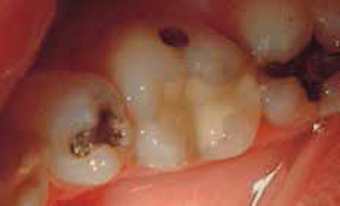
The principal author became aware of three dental practitioners who either placed reinforced glass ionomer restorations in posterior teeth, or who had purchased dental practices in which their predecessor had placed such restorations. Accordingly, a practice-based retrospective clinical evaluation of 169 Fuji IX restorations was carried out, with the results indicating high rates of success. This paper, while indicating that Fuji IX restorations had good potential for survival for two years, cannot be rated as high quality evidence, because the practitioners, following training, assessed their own restorations, with the attendant potential for bias. Selected restorations from the restorations in this study are presented in Figures 1 to 3.
Figure 1. Disto-occlusal reinforced glass ionomer restoration in lower first molar tooth at 2 years.Figure 2. Disto-occlusal reinforced glass ionomer restoration in second premolar tooth at 2 years. (Courtesy of Drs Siddons, Phipps and Bardha.) The restoration appears somewhat flat but, in the absence of a baseline photograph, it is not possible to determine whether wear has occurred or not.Figure 3. Reinforced glass ionomer restorations in first and second molar teeth at 2 years. (Courtesy of Drs Siddons, Phipps and Bardha.)
The practitioners involved in the original project (described above) were contacted in early 2007 and asked if they would be prepared to extend the project to five years. All three responded, but one had retired from practice, and a second had relocated and was uncertain as to whether he could identify the patients involved in the original project. The third practitioner agreed to participate in a retrospective evaluation. A total of 42 restorations, in 25 patients (14 male, 11 female), were available for assessment. The mean age of the restorations was 8 years, while the mean age of the patients was 57 years. The restorations were assessed using a modification of USPHS Criteria by one independent assessor and the clinician who owned the practice (but had not placed all of the restorations), with the results indicating that all but one of the original restorations placed in the selected patients were available for examination. This restoration, a Class I, had failed at 5 years and had been replaced with another Fuji IX restoration, which was satisfactory. Marginal discoloration was noted in circa one third of restorations, but the absence of secondary caries throughout the study could be considered to indicate that the marginal discoloration was not indicative of early caries activity. Regarding wear, there may be some ‘flattening’ of the restorations, but, in the absence of baseline illustrations, it is not possible to judge whether the surface contour had been lost. Regarding surface roughness, one restoration scored C (unsatisfactory) but no dietary or other influences were identified for this patient. Colour match was optimal in only 2.4% (n = 1) of restorations, while the remainder 97.6% (n = 41) were B.
This study, while lacking the scientific rigour of controlled prospective evaluations, and in which the number of restorations is small, appears to suggest that a reinforced glass ionomer, Fuji IX, may perform satisfactorily under favourable conditions, in periods of over 5 years in Class I and II cavities, some of which were cusp replacement restorations. However, what those conditions are was not determined by the study.
In this practice-based study, three general dentists participated in this retrospective evaluation of 151 EQUIA (GC) restorations placed in 43 patients (26 Class I, 125 Class II, 41 3 or 4 surface [S3] restorations). The restorations were examined using scientific criteria and the median age of the restorations was two years. None was designated as a failure. The authors measured the volume loss of the restorations, finding that the original volume was maintained in 88.5% of the Class I restorations but with ‘distinct volume loss’ being apparent in 3.8%. Regarding Class II restorations, the original volume was maintained in 64.2% of restorations and in 53.7% of the S3 restorations. Surface roughness was noted in up to 24% of the restorations and marginal ‘disintegrities’ noted in 7.3% of the S3 restorations. Margin discoloration was less than 1%. It was necessary to repair 6.1% of the Class II restorations and replace 8.3 %, while it was necessary to repair 2.4% (n = 1) of the S3 restorations and replace 4.9% (n = 2). The authors added, in their discussion, that the material under evaluation ‘was not designed to serve in expensive high-level restorations but as an economic basic filling material’ and concluded that EQUIA may be used for any size of Class I restoration and for smaller Class II restorations. However, it may be argued that the conclusion is not fully backed up by the results and that the wear resistance of the material under evaluation was suboptimal in a substantial number of cases, given the volume loss which was noted.
This work is a prospective study carried out by a team of experienced researchers. In this study, the final study group was 116 Class II restorations in Fuji IX glass ionomer: up to 18 months, no failures occurred; from 18 to 42 months, the survival rate dropped to 93%; but at 72 months, survival was only 60%. No restorations failed because of wear or isthmus fracture. All failures were due to progressive loss of glass ionomer material just below contact areas. In cases where the restoration had no interproximal contact, this did not occur. The authors hypothesized that it was caries-like dissolution of the restorations which occurred and that high levels of acidogenic plaque might be responsible for this caries-like dissolution of glass ionomer restorations at contact areas. The absence of a contact area might also, therefore, be a factor in optimizing survival of the restorations.
The following paper is included as it represents the most up-to-date information on reinforced glass ionomer restorations in loadbearing situations, even though it is presently only available in research abstract form, as opposed to publication in the peer reviewed literature.
Basso M, Heiss MA. J Dent Res 2013; 92: Special Issue A. Abstract 59422
This study examined restorations of EquiaFil (GC, Leuven, Belgium), a material derived from Fuji IX. These workers evaluated 319 restorations (of 380 which were originally placed: 83 Class I, 184 Class II, 72 Class V) at a follow-up time of 24 months, with 22 restorations having chipped margins and 14 restorations lost or damaged, a ‘general success rate’ (which excluded repairs) of 95.6%, a ‘general integrity rate’ of 90.9% (neither being generally used terminology) and all the failures being in Class II restorations. The authors concluded that the restorative system used appeared to be ‘a reliable choice for permanent dental restorations’, indeed ‘a first choice’. However, it is the view of this author that the data do not fully support such a conclusion, given a failure rate of circa 10% at 2 years.
Manhart and colleagues presented a major review of the literature on survival of restorations in posterior teeth. They included a section on the survival of glass ionomer restorations in posterior teeth, stating that the annual failure rate of posterior glass ionomer restorations ranged from 0% to 14.3% and considering that ‘glass ionomer cements did not have adequate mechanical strength for general use as definitive restorations in stress-bearing posterior teeth’. Examination of the papers which were included indicated that four used Ketac Silver (3M ESPE, Seefeld, Germany), seven related to Tunnel Restorations and six were on the ART technique. None of these papers bears direct comparison with the studies quoted above or the literature on survival of resin composite restorations in posterior teeth.
In summary, it may be considered from the results of the above papers that, under certain (favourable) conditions, certain restorations in reinforced glass ionomer materials (such as Fuji IX) may provide reasonable longevity. However, the conditions for longevity are not readily identified, but may include the following:
Reduced occlusal loading;
An absence of cariogenic plaque; and, possibly,
The absence of a contact point in Class II restorations.
However, it should be added that two of the studies21,22 demonstrated higher than desirable failure rates for glass ionomer restorations in posterior teeth, in the short and in the longer term. Therefore, before the reinforced glass ionomer materials can be recommended for more general use, there is a need for properly-controlled long-term prospective studies and/or randomized controlled trials and to quantify the cariostatic effect of glass ionomer restorations placed in loadbearing cavities.
Advice to patients if glass ionomer restorations are placed in loadbearing situations in posterior teeth
Until more high quality evidence becomes available (randomized controlled trials are badly needed), it is the opinion of the author that, for practitioners using reinforced glass ionomer materials in loadbearing situations in posterior teeth, in order to avoid adverse medicolegal circumstances or criticism by regulatory authorities and others, patients who request a tooth-coloured restoration in a posterior tooth should be advised according to the suggestions presented in Table 1.
Tell the patient that it is a glass ionomer.
Advise the patient of the relative paucity of good quality evidence for the success of the restorations that they are placing (in comparison to the increasing volume of evidence for posterior composite23) and that the good-quality evidence base is variable and relatively limited.
Discuss whether the restoration is a definitive restoration or long term provisional (which may need to be re-surfaced using composite).
Discuss the costs of re-surfacing and if and/or when that might be needed.
Advise the patient that options are a (more expensive) composite or indirect restoration.
That it is likely in the short term that the restorations should not do harm.
Conclusions
The author is aware of scientific evaluations of reinforced glass ionomers being carried out in Germany and Turkey, but no results from these have been published. Therefore, the literature at the time of publication on the success of reinforced glass ionomer as a restorative material for loadbearing situations in posterior teeth may be considered to be limited and variable, at least in comparison with the literature on posterior composite restorations. However, there may be situations in which reinforced glass ionomer restorations may provide reasonable service in loadbearing cavities. What those situations are requires more research.