
Abstract
Direct pulp capping is a proven method of preserving tooth vitality of a mature permanent tooth in cases of pulp exposures. The indications for this treatment, treatment modalities and materials are discussed in this paper.
From Volume 41, Issue 4, May 2014 | Pages 298-304
Direct pulp capping is a proven method of preserving tooth vitality of a mature permanent tooth in cases of pulp exposures. The indications for this treatment, treatment modalities and materials are discussed in this paper.

Dentists in their daily practice use many restorative techniques. Among those, vital pulp therapies have always carried many question marks, resulting in some contradictions regarding the use and the success of this technique. Direct pulp capping of a pulp exposure can provide an opportunity for preserving pulpal tissue. This may result in the formation of tertiary dentine, thereby preserving the vitality of the tooth and avoiding the need for root canal therapy. When direct pulp capping is carried out, the patient must be fully informed and advised that, if symptoms occur, they must return for re-assessment of pulp vitality.1
An understanding of the properties of available materials will help in the decision-making to obtain long-lasting functional restorations on vital teeth.
This article answers some of the questions involving direct pulp capping as a treatment option for pulp exposures, the different materials and techniques used in this procedure and the success rate of direct pulp capping using a question and answer format. However, it is up to the reader to decide whether to perform direct pulp capping or not by carefully selecting the suitable cases for this treatment.
A. Direct pulp capping is the placement of a protective dressing directly over the exposed pulp to permit healing and repair by formation of tertiary dentine. It aims to maintain the pulp vitality and its normal responsiveness to electrical and thermal tests.2 On the other hand, an indirect pulp capping can also be performed when excavation of the pulpal caries can be stopped at stained but firm dentine, known as affected dentine. Calcium hydroxide lining is placed over the pulpal dentine prior to the placement of a definitive restoration.3
A. When a small (<1 mm2) pulpal exposure has occurred due to trauma or mechanical exposure during caries removal. Ideally, there should be no pain prior to treatment. It should be possible to stop the bleeding from the exposure and the tooth should be restorable with an immediate well sealed restoration. Radiographically, the tooth should also be assessed for no evidence of periradicular pathology.
A. Spontaneous pain or tooth hypersensitivity is indicative of pulp inflammation and is a contra-indication for direct pulp capping. Signs and symptoms of non-vitality such as facial swellings, presence of radiographic bony changes, and uncontrolled bleeding or no bleeding of the exposure site are also contra-indications for pulp capping. When radiographic signs of inflammation and/or clinical signs of pulp degeneration, such as pulp calcifications or purulent or serous exudates are present, pulp capping should not be carried out. If pulp exposure is large (>1 mm2), or involves contamination of any kind, it is better not to undertake pulp capping. It may also be contra-indicated to perform this treatment in case of systemic diseases (diabetes, cancer or blood disorders).1,4
A. In general, direct pulp capping treatment is recommended for permanent teeth and not primary teeth because of the poor prognosis.5,6 The high failure rate of direct pulp capping in primary teeth is explained by undifferentiated mesenchymal cells in the primary pulp which may become odontoclasts, leading to internal resorption.5 This increase in resorption in primary teeth is also due to physiological root resorption that is already in progress and an increased blood supply is stimulated. The root resorption may not be visible radiographically in terms of root blunting.7
A. After pulp capping, a hermetic seal against bacterial infiltration is essential to guarantee the success of the treatment. According to a retrospective study by Barthel et al, the placement of a definitive restoration within the first two days after pulp exposure contributed significantly to increase the survival rate of these teeth.8 Currently, the restoration of choice to cover the pulp-capping material is a RMGIC (Resin Modified Glass Ionomer Cement) prior to etching and bonding the enamel and dentine surfaces for a composite resin restoration. Glass ionomers provide resistance to acid etch, condensation pressure and dissolution.9
There are four products commonly used for direct pulp capping; frequently used products and their composition are summarized in Table 1.
| Generic Material | Commercial Products | Manufacturer | Composition | Presentation |
|---|---|---|---|---|
| Ca(OH)2 | Dycal® | Dentsply-Caulk, USA | 1,3-Butylene glycol disalicylate |
Base (Paste) |
| Catalyst (Paste) | Calcium hydroxide |
|||
| Life® | Kerr, CA, USA | Calcium hydroxide |
Base (Paste) | |
| Barium sulphate |
Catalyst (Paste) | |||
| MTA | ProRoot®MTA | Dentsply, USA | Tricalcium silicate |
Powder |
| H2O | ||||
| MTA-Angelus® | Angelus, Londrina, Brazil | Calcium compounds | Powder | |
| Distilled water | ||||
| Biosilicate | Biodentine™ | Septodont, USA | Tri-calcium silicate (C3S) Main core material |
Powder in capsules |
| Calcium chloride accelerator |
Liquid in single-dose pipettes | |||
| Bioaggregate | Bio-Aggregate® | Innovative BioCeramics (IBC), Canada | Calcium silicate hydrate |
Powder |
| Deionized water | Liquid |
Calcium hydroxide (Ca(OH)2) is the most common direct pulp agent. For pulp capping it is used as a two-paste product. It has excellent antibacterial and disinfectant properties but a number of disadvantages, including its solubility in water and the fact that it does not seal the exposed pulp from the external environment and has poor mechanical properties. For these reasons, a protective base material, such as a RMGIC, is required to provide an adequate seal of the pulp.9
Mineral trioxide aggregate (MTA), developed at the University of Loma Linda (USA), to seal communications between the root canal system and the external tooth surface,10 has been recommended for direct pulp capping.11 It has fine hydrophilic particles mixed with sterile water, which results in a colloidal gel (pH of 12.5). This gel solidifies to a hard structure within approximately 4 hours with low or no solubility.12 MTA is antibacterial and biocompatible and has no mutagenic potential. Unlike calcium hydroxide it creates an excellent seal2 and does not need any additional protection.
Two commercial forms of MTA are available on the dental market; ProRoot® MTA (grey and white, Maillfer, Dentsply, Switzerland) and MTA-Angelus (Angelus Soluções Odontológicas, Londrina, Brazil) introduced in 2001.13 The two commercial brands of MTA have similar chemical composition, although ProRoot® MTA presents a slightly higher percentage of bismuth oxide than MTA-Angelus.14 Also, the particles of the MTA-Angelus have relatively low circularity and wide size distribution and are less homogeneous than ProRoot® MTA.15 No statistically significant difference was found between the two brands of MTA when cytotoxic effects on human endothelial cells were tested.12
Tri-calcium phosphate is another pulp-capping agent mainly indicated in bone regeneration procedures as it promotes osteoblastic formation of hard tissue. Alpha-tricalcium phosphate (α-TCP), a powder of apatite ceramics, is mixed with a saline or low acid solution and converted into hydroxyapatite or octa-calcium phosphate. It sets and hardens at room temperature. Studies have shown that dentinal bridge formation occurs by direct apposition on the pulpal wall. The bridge is contiguous to the pulp, thick and has minimal pulpal inflammation effect with no necrotic layer. The odontoblasts were present directly under, and in contact with, the bridge.16,17 However, the hard tissue barrier formation was reported to be slower than that with Ca(OH)2.17
Bio-Aggregate® (Innovative Bioceramix Inc IBC, Canada) is a root canal repair material composed of Bio-ceramic nano-particles. It is indicated in the repair of root perforations and root resorption, in apexification procedures and as a direct pulp-capping agent. It presents as pure white powder and liquid mixed together to form a thick paste-like mixture. MTA and Bioaggregate have similar chemical composition with some differences; Bioaggregates contain tantalum oxide instead of bismuth oxide in the MTA.18
Recently, Septodont launched Biodentine™, an Active Biosilicate Technology™ and a calcium silicate-based cement (Ca3SiO5-based cement) developed as a dentine substitute.
Besides the usual endodontic indications of this class of calcium-silicate cements (repair of perforations, or resorption, apexification, and retrograde root-end filling), Biodentine™ is indicated in the restoration of deep or large crown carious lesions as it provides a very tight seal, without post-operative sensitivity, and ensures the longevity of restorations in vital teeth. Biodentine™ is mixed in any triturator with a minimum speed of 4,000 rpm (Figure 1).
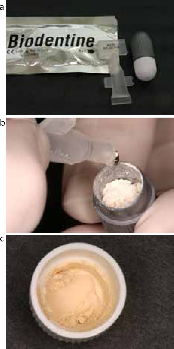
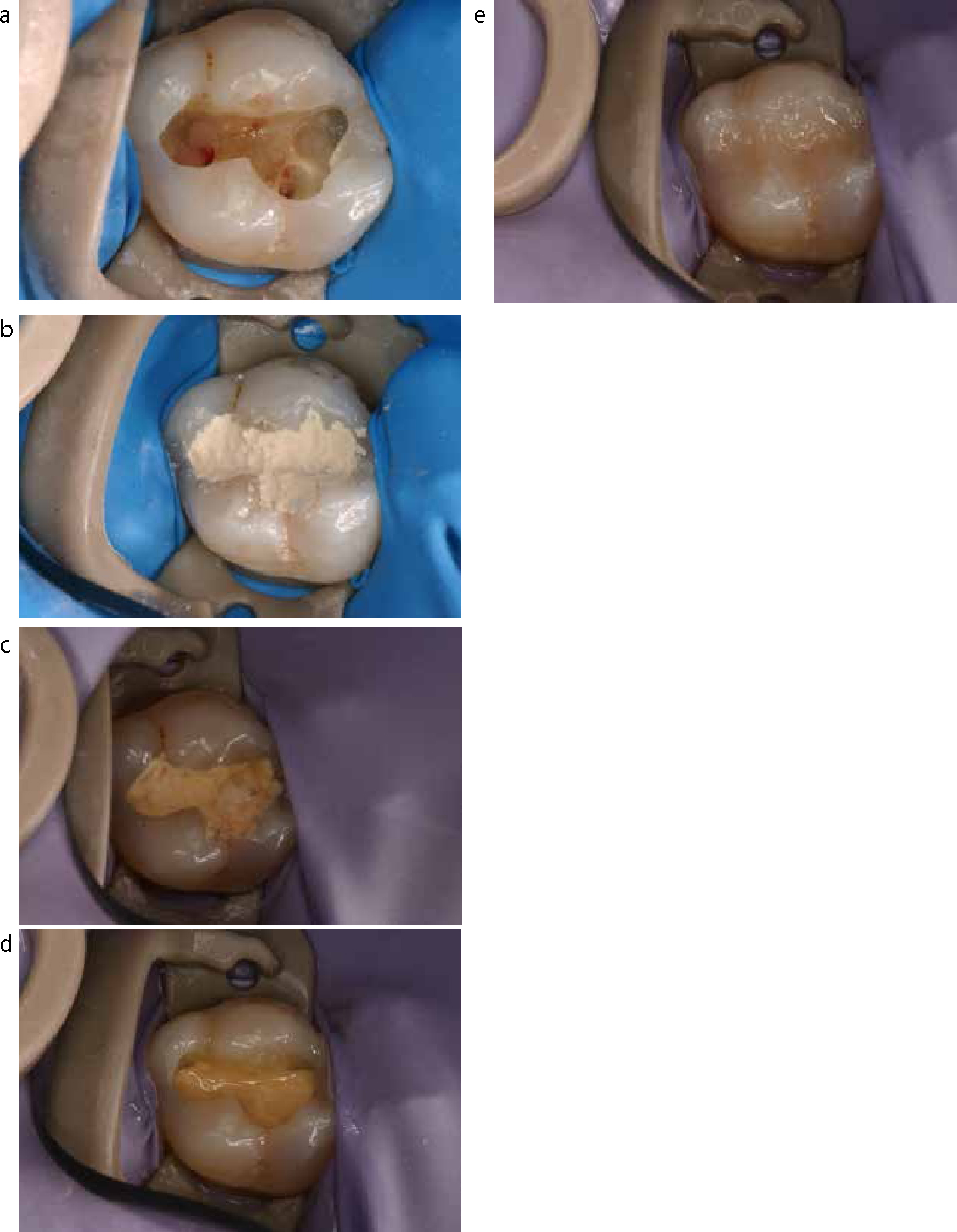
Figure 2 shows a pulp-capping case using Biodentine TM. After 6 weeks, the vitality of the pulp is checked and part of the Biodentine TM is partially removed and a ClI cavity is prepared using a pear-shaped diamond bur. The cavity is then restored conventionally using an adhesive system and resin-based composite.
A. When applied over an exposed pulp, the pulp-capping agent causes a series of inflammatory responses leading to the formation of a hard-calcified dentine bridge. Owing to a high pH (12.5), calcium hydroxide causes liquefaction necrosis of the superficial pulp removing up to 1.5 mm of the inflamed tissue. This is followed by the neutralization of toxicity in the deep layers with coagulative necrosis, which irritates the adjacent pulp and causes a minor inflammation leading to the formation of the dentine bridge. (This causes stimulation of the pleuripotential pulpal cells, which differentiate into odontoblasts, and these in turn lay down a calcified dentine bridge).
Investigators have reported that MTA induces pulpal cell proliferation, cytokine release, hard tissue formation and the synthesis of an interface with dentine similar to hydroxyapatite composition.19
A. It is important to avoid bacterial contamination of the pulp exposure in order to allow and promote healing. Caries should be removed at the periphery of the cavity first, moving circumferentially, removing the deepest remaining caries closest to the pulp last. In this way, an inadvertent exposure would be in a clean caries-free field. The exposure should be cleansed with 3% sodium hypochlorite. This will act as a bactericidal agent without causing an adverse pulpal reaction. Control of pulpal haemorrhage can be achieved with light pressure using a dry cotton pellet. Alternatively, the cotton pellet may be moistened with sodium hypochlorite, saline or local anaesthetic without epinephrine, ferric sulphate or slurry of calcium hydroxide.20,21
A. Rubber dam will reduce bacterial contamination of an exposed pulpal site and provide an appropriate environment for successful resin bonding, creating a good seal.
A. A patient's age has an influence on the success or failure rate of pulp capping. Bogen et al found, in an observational study, that pulpal repair and pulp-capping success appears to be more favourable in young patients.19 This success may be attributed to the presence of larger apical foramina and greater vascularization of the pulp. An increased blood flow allows for better immune cell surveillance and response.19 On the other hand, the success of pulp capping is not dependent on patient gender.22
A. The sealing potential of resin adhesive systems makes them an attractive alternative for direct pulp capping, preventing bacteria from entering the pulp. However, Stanley and Pameijer found that the application of acid to exposed pulps caused haemorrhage that was difficult to control.23 It is difficult to achieve an effective resin seal under such conditions, and consequently poor results were obtained. Modena et al also agree that adhesives result in lower pulp healing and induce chronic inflammation, even in the absence of bacteria. Inflammation is a poor environment for pulp healing; a pulp inflamed due to caries will have decreased healing capacity.24
According to Cui et al, self-etching adhesives are relatively toxic to pulp tissue. This may be due in part to poor marginal sealing at the tissue-material interface.25 Another issue when placing adhesives directly on the pulp is the heat generated by the light-curing devices. It has been shown that an intrapulpal temperature increase of more than 11°C can lead to irreversible pulp damage.26 It can be concluded that resin adhesives are therefore not suitable as a direct pulp-capping agent.
The literature is divided on when to measure the successful outcome of direct pulp capping. Some publications demonstrated that the survival rate of directly capped pulp tissue, using calcium hydroxide, decreases over time when compared to short-term evaluations and the most unfavourable treatment outcomes occur within the first year after the treatment. According to the European Society of Endodontology, direct pulp capping should be assessed no longer than six months post-operatively and then at five yearly intervals.29 Matsuo et al suggested that the necessary interval for an adequate post-operative follow-up examination is 21 months.30 It is obvious that long-term outcomes (10 years) are the most relevant for the patients but, for convenience, in most of the clinical studies, the evaluation period is shorter.
Success rates of direct pulp capping in a carious field have varied, depending on the technique and materials.8,22,27,28,29,30 In humans, success rates ranged between 13% and 98% in 1–10 year retrospective studies using calcium hydroxide (Table 2).
| Authors | Sample Size | Time Elapsed | Success Rate |
|---|---|---|---|
| Armstrong and Hoffman(1962)27 | 46 | 2–16 months | 97.8% |
| Heyduck and Wagner (1978)28 | 210 | 1-5 years | 61.4% |
| Haskell et al (1978)29 | 149 | 5 years | 87.2% |
| Matsuo et al (1996)30 | 44 | Group with follow-ups for 3–18 months | 80–83% |
| Group with follow-ups for 21 months | 91.7% | ||
| Group with follow-ups for 24 months | 100% | ||
| Barthel et al (2000)8 | 123 | 5 years | 37% |
| 10 years | 13% | ||
| Al-Hiyasat et al (2006)22 | 204 | 5 years | 59.3% |
A. Follow-up of pulp-capped teeth includes clinical and radiographical examinations. During recall appointments, patients' self-reports concerning pain and sensitivity are recorded and pulp-capped teeth are tested with a cold stimulus. Radiographs are taken to evaluate reparative dentine formation, pulpal calcification, continued normal root development and the absence of pathosis, such as internal root resorption and apical periodontitis.31 The marginal integrity of the final restorations should always be evaluated.19
Clinical signs indicative of a favourable outcome are a normal response to pulp sensitivity tests, absence of pain or tenderness to percussion and absence of swelling, sinus tract and pathological mobility. Also, in anterior teeth, absence of tooth discoloration is the most important factor to be considered.31,7
Partial pulpotomy is the removal of a small portion of the vital coronal pulp and preserving the vitality of the remaining coronal and radicular pulp tissue. This technique is preferred in order to encourage physiological development and formation of a root end.32
Other authors have reported a high level of success using partial pulpotomies in caries exposures.33,34
A. Novel techniques are being developed to regenerate rather than to restore lost hard dental tissue, including enamel, dentine and pulp tissue. The regeneration of dentine is considered feasible as it is in direct contact with a highly vascularized and innervated pulpal tissue. Odontoblastic cells in the pulp continue to produce secondary dentine, even after tooth eruption at low level, and they secrete tertiary dentine as a repair reaction with the help of progenitor cells that will be, at this point, triggered to differentiate into odontoblastic-like cells under the influence of specific growth factors. Research on pulpal tissue regeneration is based on growth factor delivery, stem cell delivery and gene-enhanced tissue regeneration.
With growth factor delivery, TGF-beta 1 (polypeptide responsible for the control of cell growth, cell proliferation, cell differentiation and apoptosis) is implanted intra-pulpally to induce odontoblastic-like cell differentiation and reparative dentine formation; nevertheless its action is limited. Other growth factors, such as insulin-like growth factor in mechanically exposed pulps, have been reported. This will reduce inflammation, preserve tooth vitality and promote pulp repair.35
Future trends on pulp capping also include the use of lasers, more specifically CO2 Laser, which was found to have anti-inflammatory, cell biostimulation and decontamination effects on pulpal cells and dentine.36 CO2 Laser used in direct pulp capping was found to increase the percentage of pulp vitality preservation and the thickness of the dentinal bridge newly formed after pulp exposure significantly.36
Preservation of the vitality of a tooth is the aim of the restorative clinician. However, the clinician is often faced with a direct pulp exposure as a result of the operative procedure or trauma. Criteria for success are outlined in this paper using an easy reference design.
Direct pulp capping, although a very technique and case selection sensitive procedure, is considered to be a basic conservative approach and, when it is found to be indicated, should be performed. The high success rate found after a long-term evaluation, and the variety of products that ensure such a rate, are plus factors when considering direct pulp capping.