Bernier JL. Report of the Committee on Classification and Nomenclature. J Periodontol. 1957; 28:56-58
Armitage GC. Development of a classification system for periodontal diseases and conditions. Ann Periodontol. 1999; 4:1-6
Sweeney EA, Alcoforado GAP, Nyman S, Slots J. Prevalence and microbiology of localized prepubertal periodontitis. Oral Microbiol Immunol. 1987; 2:65-70
Bimstein E, Sela MN, Shapira L. Clinical and microbial considerations for the treatment of an extended kindred with seven cases of prepubertal periodontitis: a 2-year follow-up. Pediatr Dent. 1997; 19:396-403
Marshall-Day CD, Shourie KL. A roentgenographic survey of periodontal disease in India. J Am Dent Assoc. 1949; 39:572-588
Ramfjord SP. The periodontal status of boys 11 to 17 years old in Bombay, India. J Periodontol. 1961; 32:237-248
Basu MK, Dutta AN. Report of ‘Prevalence of periodontal disease in the adult population in Calcutta’ by Ramfjord's technique. J All India Dent Assoc. 1963; 35:187-201
Hansen BF, Gjermo P, Bergwitz-Larsen KR. Periodontal bone loss in 15-year-old Norwegians. J Clin Periodontol. 1984; 11:125-131
Miyazaki H, Hanada N, Andoh MI Periodontal disease prevalence in different age groups in Japan as assessed according to the CPITN. Community Dent Oral Epidemiol. 1989; 17:71-74
Brown LJ, Albandar JM, Brunelle JA, Löe H. Early onset periodontitis: progression of attachment loss during 6 years. J Periodontol. 1996; 67:968-975
Perry DA, Newman MG. Occurrence of periodontitis in an urban adolescent population. J Periodontol. 1990; 61:185-188
Massler M, Cohen A, Schour I. Epidemiology of gingivitis in children. J Am Dent Assoc. 1952; 45:319-324
Massler M, Schour I. The P-M-A Index of gingivitis. J Dent Res. 1949; 28:(abstr)
Jenkins WMM, Papapanou PN. Epidemiology of periodontal disease in children and adolescents. Periodontology 2000. 2001; 26:16-32
Matsson L, Hjersing K, Sjödin B. Periodontal conditions in Vietnamese immigrant children in Sweden. Swed Dent J. 1995; 19:73-81
Matsson L, Sjödin B, Blomquist HK. Periodontal health in adopted children of Asian origin living in Sweden. Swed Dent J. 1997; 21:177-184
Melvin WL, Sandifer JB, Gray JL. The prevalence and sex ratio of juvenile periodontitis in a young racially mixed population. J Periodontol. 1991; 62:330-334
Löe H, Brown LJ. Early onset periodontitis in the United States of America. J Periodontol. 1991; 62:608-616
Position paper: Periodontal diseases of children and adolescents. J Periodontol. 1996; 67:57-62
Position paper: Periodontal diseases of children and adolescents. J Periodontol. 2003; 74:1696-1704
Parameter on plaque-induced gingivitis (Parameters of care supplement). J Periodontol. 2000; 71:851-852
Mariotti A. Dental plaque-induced gingival diseases. Ann Periodontol. 1999; 4:7-17
Mühlemann HR, Son S. Gingival sulcus bleeding – a leading symptom in initial gingivitis. Helv Odontol Acta. 1971; 15:107-113
Polson AM, Goodson JM. Periodontal diagnosis. Current status and future needs. J Periodontol. 1985; 56:25-34
Haffajee AD, Socransky SS, Goodson JM. Subgingival temperature (I) relation to baseline clinical parameters. J Clin Periodontol. 1992; 19:401-408
Rüdin HJ, Overdiek HF, Rateitschak K-H. Correlation between sulcus fluid rate and clinical and histological inflammation of the marginal gingiva. Helv Odontol Acta. 1970; 14:21-26
Moore WE, Holdeman LV, Smibert RM Bacteriology of experimental gingivitis in children. Infect Immunol. 1984; 46:1-6
Mariotti A. Sex steroid hormones and cell dynamics in the periodontium. Crit Rev Oral Biol Med. 1994; 5:27-53
Parfitt GJ. A five year longitudinal study of the gingival condition of a group of children in England. J Periodontol. 1957; 28:26-32
Sutcliffe P. A longitudinal study of gingivitis and puberty. J Periodont Res. 1972; 7:52-58
Hefti A, Engelberger T, Buttner M. Gingivitis in Basel school children. Helv Odontol Acta. 1981; 25:25-42
Stamm JW. Epidemiology of gingivitis. J Clin Periodontol. 1986; 13:360-366
Cianciola LJ, Park BH, Bruck E, Mosovich L, Genco RJ. Prevalence of periodontal disease in insulin-dependent diabetes mellitus (juvenile diabetes). J Am Dent Assoc. 1982; 104:653-660
Gusberti FA, Syed SA, Bacon G, Grossman N, Loesche WJ. Puberty gingivitis in insulin-dependent diabetic children. I. Cross-sectional observations. J Periodontol. 1983; 54:714-720
Albandar JM, Brown LJ, Genco RJ, Löe H. Clinical classification of periodontitis in adolescents and young adults. J Periodontol. 1997; 68:545-555
Kinane DF. Periodontal disease in children and adolescents: introduction and classification. Periodontology 2000. 2001; 26:7-15
Ranney RR. Classification of periodontal diseases. Periodontology 2000. 1993; 2:13-25
Tonetti MS, Mombelli A. Aggressive periodontitis. In: Lindhe J, Karring T, Lang N (eds). Oxford: Blackwell Munksgaard; 2003
Needleman H, Nelson L, Allred E, Seow K. Alveolar bone height of primary and first permanent molars in healthy 7 to 9-year-old children. ASDC J Dent Children. 1997; 64:188-196
Parameter on chronic periodontitis with slight to moderate loss of periodontal support (Parameters of Care supplement). J Periodontol. 2000; 71:853-855
Lindhe J, Ranney R, Lamster I Consensus Report: Chronic periodontitis. Ann Periodontol. 1999; 4
Clerehugh V, Lennon MA, Worthington HV. 5-year results of a longitudinal study of early periodontitis in 14- to 19-year-old adolescents. J Clin Periodontol. 1990; 17:702-708
Clerehugh V, Worthington HV, Lennon MA, Chandler R. Site progression of loss of attachment over 5 years in 14- to 19-year-old adolescents. J Clin Periodontol. 1995; 22:15-21
Clerehugh V. Periodontal problems in the young: myth or reality?. In: Clerehugh V, Tugnait A, Chapple ILC (eds). London: Quintessence Publishing Co Ltd; 2004
Clerehugh V. Periodontal diagnosis in young patients. In: Clerehugh V, Tugnait A, Chapple ILC (eds). London: Quintessence Publishing Co Ltd; 2004
Parameter on aggressive periodontitis (Parameters of Care supplement). J Periodontol. 2000; 71:867-869
Lang N, Bartold PM, Cullinan M Consensus Report: Aggressive periodontitis. Ann Periodontol. 1999; 4
Haffajee AD, Teles RP, Socransky SS. The effect of periodontal therapy on the composition of the subgingival microbiota. Periodontology 2000. 2006; 42:219-258
Ledder RG, Gilbert P, Huws SA Molecular analysis of the subgingival microbiota in health and disease. Appl Environ Microbiol. 2007; 73:516-523
Paster BJ, Boches SK, Galvin JL Bacterial diversity in human subgingival plaque. J Bacteriol. 2001; 183:3770-3783
Gemmell E, Yamazaki K, Seymour GJ. Destructive periodontitis lesions are determined by the nature of the lymphocyte response. Crit Rev Oral Biol Med. 2002; 13:17-34
Gemmell E, Seymour GJ. Immunoregulatory control of Th1/Th2 cytokine profiles in periodontal disease. Periodontology 2000. 2004; 35:21-41
Deas DE, Mealey BL. Response of chronic and aggressive periodontitis to treatment. Periodontology 2000. 2010; 53:154-166
Baer PN. The case for periodontosis as a clinical entity. J Periodontol. 1971; 42:516-520
Armitage GC. Periodontal diagnoses and classification of periodontal diseases. Periodontology 2000. 2004; 34:9-21
Armitage GC. Diagnosis and classification of periodontal diseases and conditions: current and future challenges. In: Bartold PM, Ishikawa I, Zhang J (eds). Adelaide, Australia: Asian Pacific Society of Periodontology; 2008
Armitage GC, Cullinan MP. Comparison of the clinical features of chronic and aggressive periodontitis. Periodontology 2000. 2010; 53:12-27
Periodontal diseases in children and adolescents: a clinician's perspective part 1 Sujata Surendra Masamatti Ashish Kumar Mandeep Singh Virdi Dental Update 2024 39:8, 707-709.
Authors
Sujata SurendraMasamatti
MDS
Reader, Department of Periodontics, ITS Centre for Dental Studies and Research, Murad Nagar, Ghaziabad, Uttar Pradesh, India
Contrasting forms of periodontal disease can affect children and adolescents with varying prevalence, severity and extent, leading to a diverse prognosis in these age groups. For an early diagnosis and treatment of periodontal conditions in young patients, it is essential for the dental practitioner to be able to identify and classify the disease correctly at the earliest opportunity, applying basic principles along with understanding of aetiology and risk factors. The first part of this article discusses the classification, plaque-induced and non-plaque-induced gingival diseases, localized and generalized forms of chronic, as well as aggressive, periodontitis.
Clinical Relevance: Knowledge of different forms of periodontal diseases affecting children and adolescents may help to distinguish between different forms of diseases and have value in screening and early diagnosis of the disease.
Article
Diseases affecting the periodontium can be limited to the gingival tissues or can be associated with destruction of the periodontal ligament and alveolar bone. Periodontal disease may affect children and adolescents. There have been various attempts to classify periodontal diseases. Various classifications have been developed over a period of time.1,2,3,4 Problems associated with the 1989 classification3 led to a 1999 international workshop on the classification of periodontal diseases.4 A new classification system was proposed in 1999 and is presently the most accepted classification system of periodontal diseases (Table 1). Periodontal diseases are classified as:
1989 Classification of Periodontal Diseases
1999 Classification of Periodontal Diseases
Gingival diseases (Plaque-induced and Non-plaque-induced)
Adult periodontitis
Chronic periodontitis (Localized and Generalized)
Early-onset periodontitis
Aggressive periodontitis (Localized and Generalized)
Periodontitis associated with systemic disease
Periodontitis as a manifestation of systemic disease
Necrotizing ulcerative periodontitis
Necrotizing periodontal disease
Refractory periodontitis
Abscesses of periodontium
Periodontitis associated with endodontic lesions
Developmental and acquired deformities and conditions
Gingival diseases (plaque-induced and non-plaque-induced);
Chronic periodontitis (localized and generalized);
Aggressive periodontitis (localized and generalized);
Periodontitis as a manifestation of systemic disease;
Necrotizing periodontal disease;
Abscesses of periodontium;
Periodontitis associated with endodontic lesions; and
Developmental and acquired deformities and conditions.
In the new classification,4 a category of gingival diseases (plaque-induced and non-plaque-induced) was introduced. Gingival diseases were not represented in the 1989 classification. The term ‘adult periodontitis’ was changed to ‘chronic periodontitis’ and ‘early-onset periodontitis’ to ‘aggressive periodontitis’. These changes were made to eliminate the age-dependent criteria. Chronic periodontitis was considered a less age-dependent description than adult periodontitis. Early-onset periodontitis in the 1989 classification was further classified into:
Pre-pubertal periodontitis (localized and generalized);
Juvenile periodontitis; and
Rapidly progressive periodontitis.
The term ‘early-onset periodontitis’ was discarded to eliminate age-dependent criteria as this form of disease can occur in children, adolescents and adults and the term was replaced by ‘aggressive periodontitis’. ‘Localized aggressive periodontitis’ replaced the older expression ‘localized juvenile periodontitis’ or ‘localized early-onset periodontitis’. ‘Generalized aggressive periodontitis’ replaced ‘generalized juvenile periodontitis’ or ‘generalized early-onset periodontitis’.
The categories of refractory periodontitis and rapidly progressive periodontitis were eliminated because of their heterogeneity. Prepubertal periodontitis was also eliminated, as it was not perceived to be a single entity. Many severe periodontitis cases in children are due to the presence of systemic disease(s).5,6
Various studies show that gingivitis is prevalent in children and adolescents.7,8,9,10,11 Studies have indicated that attachment loss and supporting bone loss is infrequent in the young, but that the incidence increases in adolescents aged 12 to 17 when compared to children aged 5 to 11.11,12,13 A study conducted on school children demonstrated that the prevalence and extent of gingivitis increased with age.14 Gingivitis starts in the deciduous dentition, reaching a peak at puberty. Gingivitis reduced during adolescence and was followed by a gradual rise throughout adult life.15 The increase in gingivitis levels may be ascribed to the increase in sites at risk, plaque accumulation and inflammatory changes related with tooth eruption and the influence of hormonal factors in puberty. The decline in gingivitis in adolescence may be due to improved social awareness and enhanced oral hygiene.16
Prevalence of periodontitis in the primary dentition is difficult to estimate because of scarcity of data. Exfoliation and eruption can lead to undependable information. A low prevalence of marginal bone loss in the primary dentition is found in children of European origin in comparison to Asian children.17,18 The prevalence of early onset periodontitis (aggressive periodontitis) in Blacks was 2.1%19–2.6%.20 The prevalence rate for Whites was 0.17%.20 In a survey in the United States, no significant difference was found in prevalence rates between males and females.20 Black males and white females were approximately three times more likely to have localized early-onset periodontitis (localized aggressive periodontitis) than black females and white males, respectively.20
Severe generalized periodontitis may be found in young children with rare systemic diseases, such as Papillon-Lefèvre syndrome, cyclic neutropenia, agranulocytosis, Down's syndrome, hypophosphatasia and leukocyte adhesion deficiency.21
According to the American Academy of Periodontology, periodontal diseases that can affect young individuals include:
Dental plaque-induced gingival diseases;
Chronic periodontitis;
Aggressive periodontitis;
Periodontitis as a manifestation of systemic diseases; and
However, a few other diseases, like primary herpetic gingivostomatitis, may also affect children.
Dental plaque-induced gingival diseases
Definition: Plaque-induced gingivitis is defined as inflammation of the gingiva in the absence of clinical attachment loss.23
Gingivitis associated with dental plaque only
Chronic marginal gingivitis is the most prevalent type of gingival change in childhood. Dental plaque causes inflammation within the gingival tissues, which manifests as clinical signs of gingivitis.
The universal features of gingival diseases24 associated with plaque, endogenous hormonal fluctuations, drugs, systemic diseases and malnutrition are listed in Table 2.
Clinical signs of inflammation
Signs and symptoms that are restricted to the gingiva
Reversibility of diseases by removing the aetiology
Presence of bacterial plaque to initiate and intensify the severity of the lesion
A potential role as a precursor to attachment loss
Histological changes including an inflammatory lesion;
Reversible with plaque removal.
Subgingival levels of Actinomyces sp, Capnocytophaga sp, Leptotrichia sp and Selenomonas sp have been found to be increased in experimental gingivitis in children when compared to gingivitis in adults.29
Gingival diseases modified by systemic factors associated with the endocrine system
Hormonal changes affect the periodontal diseases, although bacterial plaque is essential to initiate gingival disease.
Puberty-associated gingivitis
The rise in steroid hormone levels during puberty in both sexes has a transitory effect on gingivitis.30 There is an increase in gingival inflammation in circumpubertal age individuals of both sexes without a simultaneous increase in plaque levels.31,32,33
The predilection to develop candid signs of gingival inflammation in the presence of relatively small amounts of plaque during the circumpubertal period differentiates the disease. The incidence and severity of gingivitis in adolescents is also influenced by dental caries, mouth breathing, crowding of the teeth, and tooth eruption.34
Diabetes mellitus-associated gingivitis
Diabetes mellitus-associated gingivitis is found in children with poorly controlled Type 1 diabetes mellitus (insulin-dependent diabetes mellitus or juvenile onset).35,36 The features of gingivitis associated with diabetes mellitus are similar to plaque-induced gingivitis. The level of diabetic management along with plaque control is important in determining the severity of the gingival inflammation.35,36
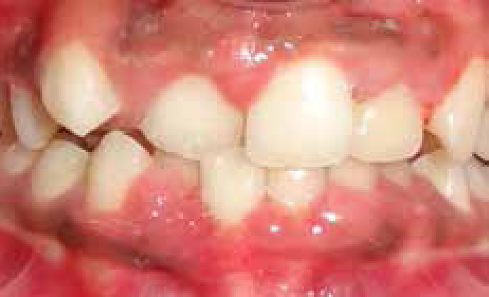
Gingivitis is frequently associated with tooth eruption. Tooth eruption by itself does not cause gingivitis. The inflammation results from plaque accumulation around erupting teeth. Partially exfoliated, loose deciduous teeth often cause gingivitis due to plaque accumulation. The incidence and severity of gingivitis is more around malpositioned teeth because of their increased tendency to accumulate plaque37 (Figure 1).
Figure 1. Clinical appearance of gingivitis in a 12-year-old child. Note the pronounced inflammation in the maxillary and mandibular anterior teeth.
Periodontics
Periodontitis, irrespective of the specific classification, shows irreversible loss of connective tissue attachment and apical migration of the junctional epithelium and true pocket formation. The correct diagnosis of the different types of periodontitis is important as the management of periodontitis depends on the correct diagnosis.
Incidental attachment loss – a precursor to periodontitis
The term ‘incidental attachment loss’38,39 has been used to describe loss of support in children and adolescents. Attachment loss in children that does not fit the diagnostic criteria of aggressive or chronic periodontitis is termed as incidental attachment loss. Incidental attachment loss may be isolated areas of attachment loss in otherwise healthy dentitions, including recessions associated with traumatic injuries and/or tooth position, attachment loss associated with impacted third molars, with endodontic infection, root fractures, subgingival caries and/or with restorations.40 These isolated areas of attachment loss may also represent initial clinical presentations of periodontitis. Children having such clinical presentation should be considered a high-risk group for chronic or aggressive periodontitis.41
Bite-wing radiographs have been used to determine the normal distance between the cemento-enamel junction and the alveolar crest of primary and permanent molars in 7 to 9-year-old children.42 Median distances recorded for primary molars were 0.8 to 1.4 mm. Most of the children present with distances significantly smaller than 2–3 mm. Greater distances have been identified at sites with caries, fillings or open contacts, indicating that these factors may contribute to bone loss in similar ways to adult patients. The presence of one of these local factors could be a reason for local cause of bone loss, other than periodontitis. A distance of 2mm between the cemento-enamel junction and the alveolar crest in the absence of the above-mentioned local factors could be an indication of periodontitis.41 The tentative diagnosis needs to be confirmed with complete periodontal examination.
According to The British Society of Periodontology, it is the responsibility of the dentist to screen patients for the presence of periodontal diseases, including the use of relevant radiographs to make a diagnosis and institute a treatment plan with defined therapeutic goals. Basic Periodontal Examination is a sensitive, easy, quick method of screening patients for periodontal diseases that condenses essential information. Basic periodontal examination will be explained in detail in the second part of this article. Appearance of loss of marginal bone on bite-wing radiographs, along with increased probing attachment loss, is a sign of periodontitis. Detection of bone loss during screening should necessitate comprehensive periodontal examination to establish periodontal diagnosis. After the diagnosis has been confirmed as periodontitis, a further differentiation between aggressive periodontitis and chronic periodontitis needs to be established, according to the criteria mentioned.41 The cases which do not conform to the criteria which define aggressive periodontitis should be diagnosed as chronic periodontitis.41
Chronic periodontitis
Definition: Chronic periodontitis is defined as inflammation of the gingiva extending into the adjacent attachment apparatus. The disease is characterized by loss of clinical attachment due to destruction of the periodontal ligament and loss of the adjacent supporting bone.43
Chronic periodontitis is the most common form of periodontal disease in adults but can be found in children and adolescents affecting both the primary and secondary dentitions.
The amount of periodontal destruction is proportionate to local factors.
The composition of microbial plaque is complex and varies to a great extent within and between patients and subgingival calculus is a frequent finding.
Chronic periodontitis can be classified on the basis of extent of disease as ‘localized’ when fewer than 30% of sites are affected, and ‘generalized’ when this level is exceeded.
Chronic periodontitis can also be classified on the basis of the severity of the periodontal destruction. Disease is mild (1–2 mm clinical attachment loss), moderate (3–4 mm clinical attachment loss), or severe (>5 mm clinical attachment loss).
Although chronic periodontitis is initiated by microbial plaque, factors such as systemic risk factors, including smoking, stress, diabetes, HIV and host factors, influence the pathogenesis and progression of the disease.
Progression can only be confirmed by repeated clinical examinations and is considered likely to occur in diseased sites that are left untreated. It usually has slow to moderate rates of progression, but may have periods of rapid progression.
Incipient chronic periodontitis
The change in nomenclature of adult periodontitis to chronic periodontitis implies that periodontitis is not just limited to adults, but can begin in adolescents and progress throughout the teens.45,46
The initial ‘incipient’ stage of periodontal destruction in a young patient will usually be characterized by loss of attachment of 1–2 mm, formation of true shallow periodontal pockets of 4–5 mm, and early crestal alveolar bone loss of at least 0.5 mm.47
It is realistic to identify initial stages of periodontal disease using a careful probing technique. A ‘one-stage’ loss of attachment measuring technique is used. The probe tip is located at the CEJ using tactile sensation and the tip advanced apically until the base of the pocket is reached, whilst at the same time directly observing the distance, in millimeters, advanced by the probe.47
The diagnostic features of incipient chronic periodontitis are:48
Age of onset can be in adolescence (13–14 years);
Interproximal clinical attachment loss of 1–2 mm (commonly seen on maxillary first molars, mandibular incisors), associated with presence of plaque, subgingival calculus;
Pockets of 4–5 mm.
Bone loss no more than 0.5 mm over an 18-month period (bite-wing radiographs usually show horizontal bone loss).
Aggressive periodontitis
Definition: Aggressive periodontitis encompasses distinct types of periodontitis that affect people who, in most cases, otherwise appear healthy. It tends to have a familial aggregation and there is a rapid rate of disease progression. Aggressive periodontitis occurs in localized and generalized forms.49
Aggressive periodontitis can be classified as localized aggressive periodontitis and generalized aggressive periodontitis.
Secondary features that are generally present but may not be present in all cases:
Amount of microbial deposits inconsistent with the severity of periodontal destruction;
Elevated proportions of Aggregatibacter actinomycetemcomitans (earlier named as Actinobacillus actinomycetemcomitans);
Phagocytic abnormalities;
Hyper-responsive macrophage phenotype, including elevated production of PGE2 and interleukin-1β in response to bacterial endotoxins;
Progression of attachment loss and bone loss may be self-arresting.
The diagnosis may be made on historical, radiographic and clinical data. In addition to primary and secondary features common to all aggressive periodontitis patients, the following features can be identified.
Localized aggressive periodontitis
Circumpubertal onset;
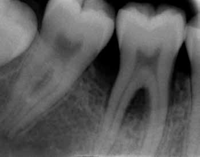
Localized first molar/incisor show interproximal attachment loss on at least two permanent teeth, one of which is a first molar (Figure 2), and involving no more than two teeth other than first molars and incisors;
Robust serum antibody response (IgG2 in particular).
Figure 2. Radiograph showing arc-shaped bone loss around first molar of a 13-year-old child.
Generalized aggressive periodontitis
Usually affects people under 30 years of age but patients may be older;
Generalized interproximal attachment loss affecting at least three permanent teeth, other than first molars and incisors;
Pronounced episodic nature of destruction of attachment and alveolar bone;
Poor serum antibody response.
It is not necessary for all the features to be present to assign a diagnosis or classify a disease. The diagnosis may be based on clinical, radiographic or historical data. Laboratory examination and testing is a useful tool to help in diagnosis but is not essential.50
Similarities in clinical features of chronic and aggressive periodontitis
Chronic and aggressive periodontitis have numerous common clinical features, but the common features are not necessarily alike in both forms of the disease. It is well recognized that both chronic and aggressive periodontitis are complex infections that occur in susceptible hosts and are caused by biofilms.51,52,53 In addition, host immune response to the biofilms is largely responsible for periodontal destruction.54,55 Successful management of both forms of periodontitis includes reduction of bacterial load.56 The untreated disease invariably leads to loss of tooth.
Individuals affected by chronic and aggressive periodontitis have no known medical or general health conditions that might contribute to development of their periodontitis. If an individual has a systemic disease that modifies the initiation and clinical course of periodontal infections, the resulting periodontitis should be classified as periodontitis as a manifestation of systemic disease.4
Differences in clinical features of chronic and aggressive periodontitis
The clinical differences are the primary basis for classifying individuals into one of the categories of periodontitis. Chronic and aggressive forms of periodontitis have a number of significant clinical differences.
Age of onset
Despite the fact that criteria of age has been removed, age still remains an important distinguishing factor in differentiating between chronic and aggressive forms of periodontitis. Individuals suffering from aggressive periodontitis are considerably younger than individuals with chronic periodontitis for similar extents of periodontal damage. This factor can help in basically deciding whether a patient has chronic or aggressive periodontitis.
Amount of plaque and calculus
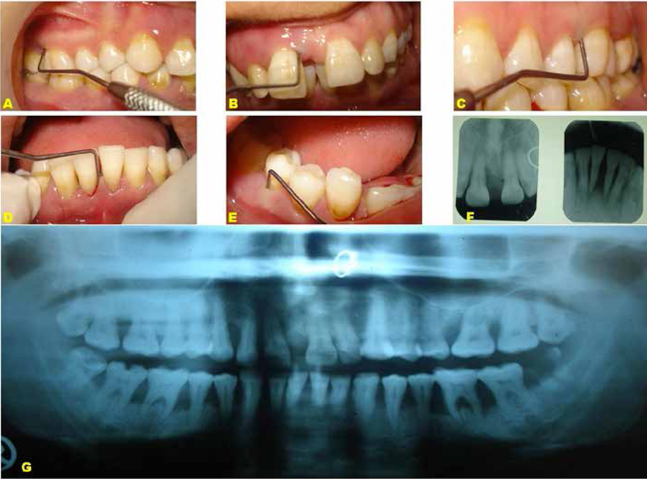
In aggressive periodontitis, there are generally thin deposits of dental plaque, with little or no calculus (Figures 3 A–E). Chronic periodontitis sufferers usually have thick deposits of plaque and calculus on affected root surfaces.
Figure 3. Clinical photographs of an untreated generalized aggressive periodontitis in a 21-year-old female. (A–E) Gingival tissues show slight clinical signs of inflammation. There is slight bleeding on probing of the mesial and distal interproximal surfaces. Probing shows presence of deep periodontal pockets at various sites. (F) Radiographic appearance of the maxillary and mandibular central incisors. Note the severe bone loss in incisors. (G) OPG shows generalized bone loss.
Clinical signs of inflammation
Gingival inflammation may or may not be evident because of thin biofilms in cases of localized and generalized aggressive periodontitis (Figures 3 A–E). Chronic periodontitis usually presents with moderately severe gingival inflammation.
Patterns of destruction
There is no consistent pattern to the number and types of teeth involved and there is no definite pattern in most cases of chronic periodontitis. In cases of generalized aggressive periodontitis, most permanent teeth are usually affected (Figures 3 F, G). Localized aggressive periodontitis cases generally have first molar/incisor involvement (Figures 2 and 4).
Rates of progression
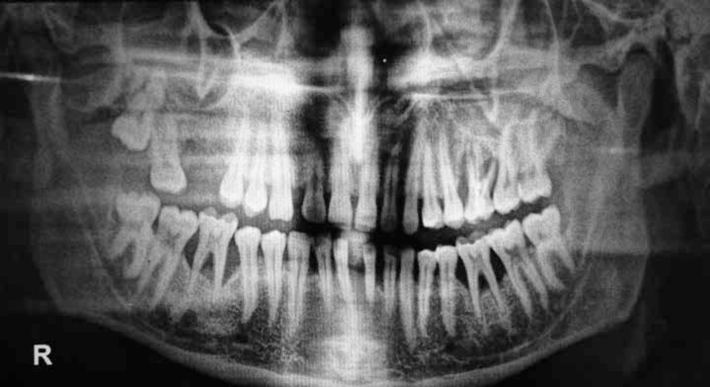
Chronic periodontitis is usually a slowly progressing disease, whereas aggressive periodontitis progresses at a rapid rate. Loss of attachment in aggressive periodontitis patients has been estimated to progress three or four times faster than chronic periodontitis.57 A self-limiting feature of localized aggressive periodontitis is called ‘Burn out’ phenomenon57 (Figure 4). Figure 4 shows an orthopantomogram (OPG) of a 24-year-old Indian male in whom periodontal destruction was localized to all first molars and mandibular incisors, as observed in cases of localized aggressive periodontitis. His dental history revealed exfoliation of maxillary right first molar at the age of 17 years. His first ever visit to a dentist was at 24 years of age for mobility in maxillary left first molar, for which an intra-oral periapical radiograph was taken and root canal treatment was advised. The patient was referred to a periodontist after the root canal therapy, for treatment of deep pockets in relation to molars and mandibular incisors. He had no history of any periodontal therapy and his periodontal tissues had minimal to no signs of clinical inflammation. An OPG was advised which revealed classic arc-shaped bone loss in molars and angular defects on mandibular incisors. But it should not be expected that all cases of localized aggressive periodontitis would be self-limiting.
Figure 4. OPG of self-limiting untreated localized aggressive periodontitis in a 24-year-old Indian male. Radiograph shows bone loss localized to first molars (arc-shaped) and mandibular anterior teeth. Maxillary right first molar exfoliated at the age of 17.
Localized aggressive periodontitis vs localized incipient chronic periodontitis
Localized aggressive periodontitis can present around puberty and specifically affects first molars and incisors, whereas incipient chronic periodontitis may start around adolescence or even earlier and can affect these and other sites as well.
Plaque and calculus levels are inconsistent with the severity of periodontal destruction in localized aggressive periodontitis and subgingival calculus may or may not be a significant factor, whereas periodontal destruction is proportionate to local factors in incipient chronic periodontitis when subgingival calculus is present.
In localized aggressive periodontitis, localized first molar/incisor involvement is seen with interproximal attachment loss of >3 mm,48 whereas in incipient chronic periodontitis interproximal clinical attachment loss of 1–2 mm is generally seen. Bone loss is no more than 0.5 mm over an 18-month period.48 The rate of periodontal destruction is rapid in aggressive periodontitis in comparison to chronic periodontitis.
Despite all these differences, the clinical distinction between chronic and aggressive periodontitis may be difficult, especially in initial stages of disease in children. A diagnosis is a summary statement of the clinician's best estimate regarding the disease or condition detected in a given patient. It is derived from a thorough analysis of all information collected during a review of relevant data from medical/dental histories, the results of diagnostic tests, and findings from a careful clinical examination.58,59 A diagnosis gives an idea of disease present in a specific patient. It provides a foundation about appropriate treatment approaches. The diagnosis may not precisely be according to the classification system. The exact definition of a case is not a main issue in the management of specific patients in clinical practice, as the diagnosis is tailor-made for the individual.60 The detection of loss of attachment in children and adolescents should warrant further periodontal examination and such clinical presentation should be considered a high-risk group for chronic or aggressive periodontitis. Proper anti-infective therapies need to be instituted for successful resolution of both forms of the disease.60
Summary
Children and adolescents experience several periodontal diseases, with plaque being the key contributing agent. Local and systemic risk factors can modify the response of periodontal tissues. Although severe periodontal diseases are less common, children can still develop severe forms of periodontitis.20 Dental practitioners need to keep key features of the different periodontal conditions in mind to help in diagnosing the disease in order to stop it from progressing to adolescence where it might follow a more severe course.