Zebenholzer K, Wober C, Vigl M, Wessely P, Wober-Bingol C. Facial pain and the second edition of the International Classification of Headache Disorders. Headache. 2006; 46:(2)259-263
Renton T. Pain Part 1: Introduction to pain. Dent Update. 2015; 42:109-124
Ravaghi V, Farrahi-Avval N, Locker D, Underwood M. Validation of the Persian short version of the Oral Health Impact Profile (OHIP-14). Oral Hlth Prevent Dent. 2010; 8:(3)229-235
Renton T, Yilmaz Z. Profiling of patients presenting with posttraumatic neuropathy of the trigeminal nerve. J Orofac Pain. 2011; 25:(4)333-344
Woda A, Tubert-Jeannin S, Bouhassira D, Attal N, Fleiter B, Goulet JP Towards a new taxonomy of idiopathic orofacial pain. Pain. 2005; 116:396-406
Affolter B, Thalhammer C, Aschwanden M, Glatz K, Tyndall A, Daikeler T. Difficult diagnosis and assessment of disease activity in giant cell arteritis: a report on two patients. Scand J Rheumatol. 2009; 38:(5)393-394
Pareja JA, Kruszewski P, Sjaastad O. SUNCT syndrome. Diagnosis morbi. Shortlasting Unilateral Neuralgiform headache attacks, with Conjunctival injection, Tearing and rhinorrhoea. Neurologia. 1997; 12:66-72
Balasubramaniam R, Klasser GD, Delcanho R. Trigeminal autonomic cephalalgias: a review and implications for dentistry. J Am Dent Assoc. 2008; 139:(12)1616-1624
Teixeira MJ, de Siqueira SR, Bor-Seng-Shu E. Glossopharyngeal neuralgia: neurosurgical treatment and differential diagnosis. Acta Neurochirurgica. 2008; 150:(5)471-475
Closmann JJ, Fielding CG, Pogrel MA. Prevention and management of trigeminal herpes zoster and postherpetic neuralgia. Gen Dent. 2008; 56:(6)563-566
Baron R, Mayoral V, Leijon G, Binder A, Steigerwald I, Serpell M. Efficacy and safety of 5% lidocaine (lignocaine) medicated plaster in comparison with pregabalin in patients with postherpetic neuralgia and diabetic polyneuropathy: interim analysis from an open-label, two-stage adaptive, randomized, controlled trial. Clin Drug Invest. 2009; 29:(4)231-241
London: National Institute for Health and Clinical Excellence; 2010
Renton T, Adey-Viscuso D, Meechan JG, Yilmaz Z. Trigeminal nerve injuries in relation to the local anaesthesia in mandibular injections. Br Dent J. 2010; 209:(9)
Renton T, Yilmaz Z. Managing iatrogenic trigeminal nerve injury: a case series and review of the literature. Int J Oral Maxillofac Surg. 2012; 41:(5)629-637
Birch R, Bonney G, Dowell J, Hollingdale J. Iatrogenic injuries of peripheral nerves. J Bone Joint Surg. 1991; 73:(2)280-282
Nixdorf DR, Moana-Filho EJ, Law AS, McGuire LA, Hodges JS, John MT. Frequency of nonodontogenic pain after endodontic therapy: a systematic review and meta-analysis. J Endod. 2010; 36:(9)1494-1498
MacDermid JC. Measurement of health outcomes following tendon and nerve repair. J Hand Therapy. 2005; 18:(2)297-312
Cruccu G, Anand P, Attal N, Garcia-Larrea L, Haanpaa M, Jorum E EFNS Guidelines on Neuropathic Pain Assessment. Eur J Neurol. 2004; 11:(3)153-162
Haanpaa M, Attal N, Backonja M, Baron R, Bennett M, Bouhassira D NeuPSIG Guidelines on Neuropathic Pain Assessment. Pain. 2011; 152:(1)14-27
Mason DA. Lingual nerve damage following lower third molar surgery. Int J Oral Maxillofac Surg. 1988; 17:(5)290-294
Ngeow WC, Nair R. Injection of botulinum toxin type A (BOTOX) into trigger zone of trigeminal neuralgia as a means to control pain. Oral Surg Oral Med Oral Pathol Oral Radiol Endod. 2010; 109:(3)e47-e50
Argoff CE, Galer BS, Jensen MP, Oleka N, Gammaitoni AR. Effectiveness of the lidocaine patch 5% on pain qualities in three chronic pain states: assessment with the Neuropathic Pain Scale. Curr Med Res Opin. 2004; 20:S21-S28
Mongini F, Rota E, Evangelista A, Ciccone G, Milani C, Ugolini A Personality profiles and subjective perception of pain in head pain patients. Pain. 2009; 144:(1–2)125-129
Aggarwal VR, Lovell K, Peters S, Javidi H, Joughin A, Goldthorpe J. Psychosocial interventions for the management of chronic orofacial pain. Cochrane Database Syst Rev. 2011; (11)
Dworkin RH, O'Connor AB, Audette J, Baron R, Gourlay GK, Haanpaa ML Recommendations for the pharmacological management of neuropathic pain: an overview and literature update. Mayo Clinic Proceedings. 2010; 85:S3-14
Tan T, Barry P, Reken S, Baker M. Pharmacological management of neuropathic pain in non-specialist settings: summary of NICE guidance. Br Med J. 2010; 340
Baad-Hansen L. Atypical odontalgia – pathophysiology and clinical management. J Oral Rehabil. 2008; 35:(1)1-11
List T, Leijon G, Svensson P. Somatosensory abnormalities in atypical odontalgia: a case-control study. Pain. 2008; 139:(2)333-341
Morley S, Williams A, Hussain S. Estimating the clinical effectiveness of cognitive behavioural therapy in the clinic: evaluation of a CBT informed pain management programme. Pain. 2008; 137:(3)670-680
Arch JJ, Eifert GH, Davies C, Plumb Vilardaga JC, Rose RD, Craske MG. Randomized clinical trial of cognitive behavioral therapy (CBT) versus acceptance and commitment therapy (ACT) for mixed anxiety disorders. J Consult Clin Psychol. 2012; 80:(5)750-765
Aggarwal VR, Tickle M, Javidi H, Peters S. Reviewing the evidence: can cognitive behavioral therapy improve outcomes for patients with chronic orofacial pain?. J Orofac Pain. 2010; 24:(2)163-171
Fledderus M, Bohlmeijer ET, Pieterse ME, Schreurs KM. Acceptance and commitment therapy as guided self-help for psychological distress and positive mental health: a randomized controlled trial. Psychol Med. 2012; 42:(3)485-495
Jurgens TP, Muller P, Seedorf H, Regelsberger J, May A. Occipital nerve block is effective in craniofacial neuralgias but not in idiopathic persistent facial pain. J Headache Pain. 2012; 13:(3)199-213
Yang HW, Huang YF. Treatment of persistent idiopathic facial pain (PIFP) with a low-level energy diode laser. Photomed Laser Surg. 2011; 29:(10)707-710
Professor of Oral Surgery, King's College London; Honorary Consultant in Oral Surgery, King's College Hospital NHS Foundation Trust and Guy's and St Thomas' NHS Foundation Trust, London
Neuropathic pain is a significant social and economic burden. Back pain, joint pain and headaches affect over 30% of the population. Chronic orofacial pain is a common condition and is difficult to diagnose and manage. This two-part paper aims to provide an overview of novel understanding of neuropathic pain, and furnish clinical teams with an update on the less common and less well-recognized chronic orofacial conditions. Headaches and temporomandibular disorders are the most common conditions and are covered in separate papers (6 and 10). Trigeminal neuralgia, burning mouth, and trigeminal autonomic cephalgias are also covered in separate papers (7, 8 and 9). The remaining conditions: post-traumatic neuropathy (nerve injury); and persistent idiopathic facial pain and atypical odontalgia are discussed in this and the following paper.
Clinical Relevance: Neuropathic pain, though rare, is a consequence of dental treatment. Nerve injury in relation to M3M surgery, dental implants, endodontics and local anaesthesia result in 70% of affected patients experiencing chronic neuropathic pain.
Article
Neuropathic pain is defined as ‘pain initiated or caused by a primary lesion or dysfunction in the nervous system’1 and is characterized by intense, chronic pain in the absence of any overt tissue damage or dysfunction. While chronic pain is an umbrella term referring to any type of neuropathic or dysfunctional (Table 1) pain affecting the body, there is evidence to suggest that neuropathic pain affecting the trigeminal nerve, which innervates most of the head and face tissues, is distinct from painful conditions seen elsewhere in the body.
Pain is normally caused by tissue damage of intense noxious stimuli, which activate high threshold receptors that may be pain specific (nociceptors) or polymodal receptors.2 Peripheral nervous system (PNS) neuropathic pain manifests in many ways, however, key signs include:
Spontaneous pain either constant or intermittent;
Elicited pain caused by non noxious stimuli (allodynia) resulting in neuralgic type pain (sharp, shooting, penetrating);
Pain that is dysaesthetic (burning, throbbing) in nature.
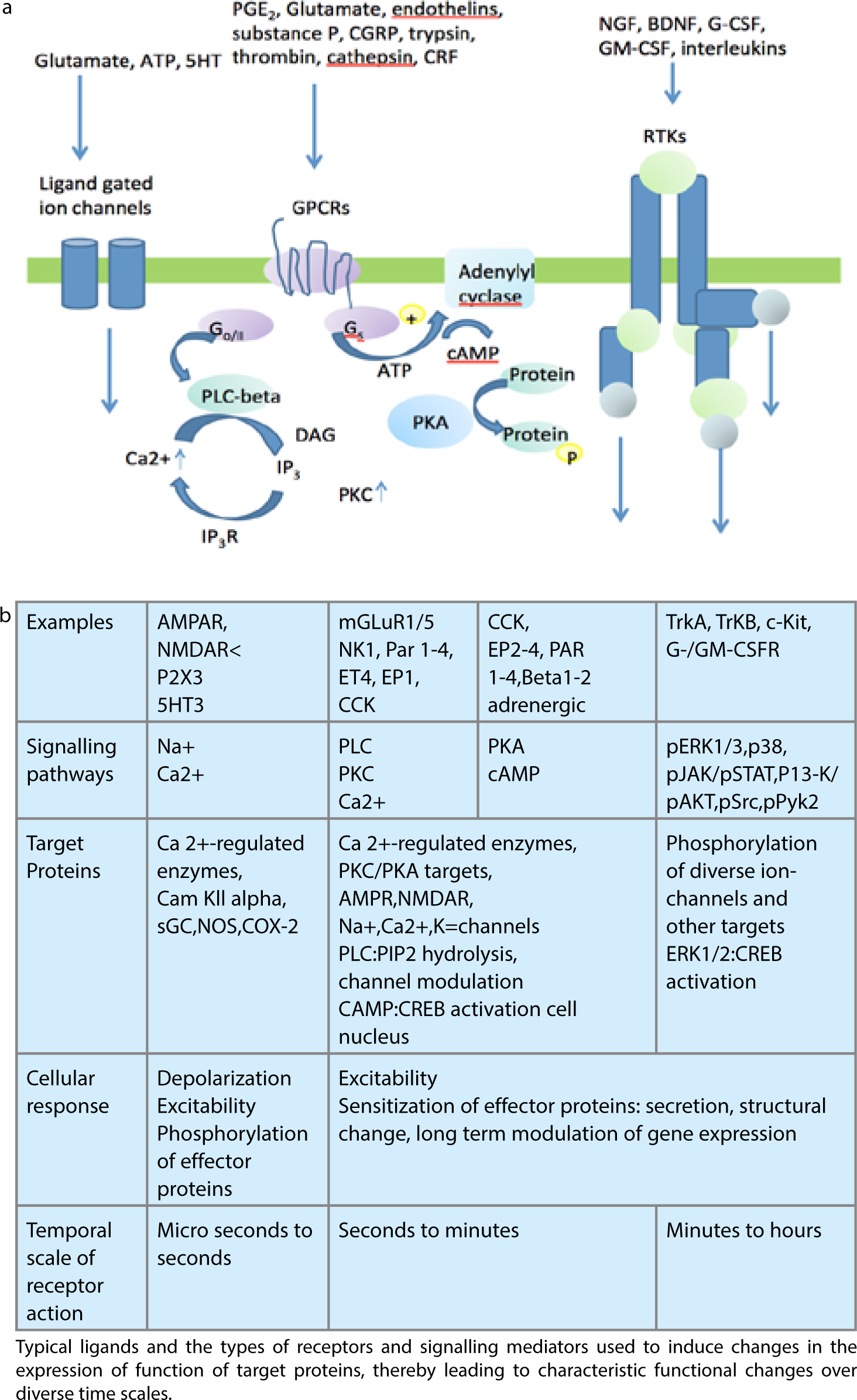
Mechanisms for the development and perpetuation of chronic pain are discussed earlier in this series.2 See figures illustrating overall central and peripheral mechanisms related to symptoms of neuropathic pain (Figure 1). Peripheral mechanisms related to persistent pain (Figure 2) and central mechanisms of neuropathic pain (Figure 3).
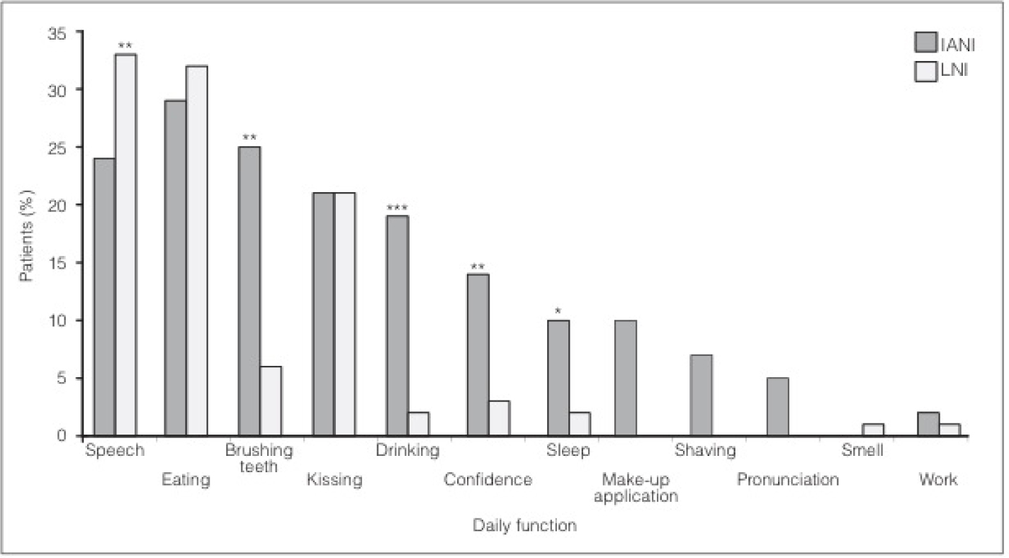
Sleep interruption is rare and pathoneumonic of neuropathic pain (NePain), instead it may be elicited by touch, thermal changes, taste changes (mechanical and cold allodynia) during normal daily function (Figure 4), with escalation with stress, anxiety or tiredness. There is also a lack of response to anti-inflammatory drugs, antibiotics and opiates.3
Figure 4. Interference of symptoms with functionality of the IANI and LNI patients. The majority of IANI and LNI patients had problems with speech and eating, where speech significantly affected more LNI patients than IANI patients (**P<0.001). Significantly more IANI patients had difficulties with brushing their teeth (**P<0.001), drinking (***P<0.0001), confidence (**P<0.001) and sleep (*P<0.05).4
The unique nature of trigeminal neuropathic pain has been supported by evidence, including:
The facial area is made up of a variety of unique tissues, such as the cornea and oral mucosa;
Tooth pulp requires specialized sensory innervations, including input by other cranial nerves and cervical nerves;
The bony structure of the facial area provides the opportunity for nerves to become entrapped;
The sensory cerebral cortex area dedicated to the processing of orofacial sensation is represented by over 50%, and therefore the impact of nerve injury may be significantly larger than in other areas of the body.
The orofacial area routinely undergoes regular tissue trauma due to: daily consumption of spicy food and crusty bread, the removal of teeth, root canal treatment, and exfoliation of the deciduous teeth, all which could produce nerve damage leading to a neuropathic pain condition.
It remains a mystery as to why trigeminal neuropathic pain is not more prevalent, likely due to the regular use of local anaesthesia which may prevent central sensitization (Table 2).
Estimated chronic severe (disabling) pain (<5 out of 10 score)
Amputation
30–50%
5–10%
159 (lower limb only)
Breast surgery (lumpectomy and mastectomy)
20–30%
5–10%
479
Thoracotomy
30–40%
10%
Unknown
Inguinal hernia repair
10%
2–4%
609
Coronary artery bypass surgery
30–50%
5–10%
598
Caesarean section
10%
4%
220
*National Centre for Health Statistics, Ambulatory and Inpatients procedures, USA 1996.
Chronic orofacial pain syndromes represent a diagnostic challenge for any practitioner.3 Patients are frequently misdiagnosed or attribute their pain to a prior event, such as a dental procedure, ENT problem or facial trauma.3 Psychological or psychiatric co-morbidities of depression and anxiety are prevalent in this population and compound the diagnostic conundrum.4 Treatment is less effective than in other pain syndromes, thus it often requires a multidisciplinary approach to address the many facets of this pain syndrome.
Aetiology of orofacial pain
Facial pain can be associated with pathological conditions or disorders related to somatic and neurological structures.1 There are a wide range of causes of chronic orofacial pain and these have been divided into three broad categories:1
Neurovascular (Musculoligamentous) – the most common cause of chronic orofacial pain is temporomandibular disorders, principally myofascial in nature;
As our understanding of pain develops, more accurate classifications, which are mechanism-based, may come to be used.1,4 Recent developments include identifying burning mouth syndrome as a neuropathic condition rather than a psychological condition,6 understanding the significant psychological burden both causative and produced by these conditions, and reduction in grey matter density and volume in several areas, including the ipsilateral anterior cingulate gyrus and insular cortex, both areas related to antinociception and anticipation of pain.7 Changes in the somatosensory cortex in relation to chronic orofacial pain are also reported, similar to other chronic pain conditions.8 As with psychological features identified in patients with chronic orofacial pain, it is not entirely clear whether these changes are a result of the condition or predispose to it.7,8 What is accepted is that both peripheral and central mechanisms play significant roles in chronic neuropathic pain due to various mechanisms, including central sensitization, peripheral hypersensitivity (Figure 2) and structural neuroplastic changes (Figure 3).9
Pain in the oral and facial region (orofacial pain) has a significant biopsychosocial impact.9,10,11 A recent US Surgeon General's report states that ‘… oral health means much more than healthy teeth, it means being free of chronic orofacial pain conditions’.10 Epidemiologists report a significant burden of orofacial pain affecting the community: estimated at 39 million (22%) of Americans (18 years +) suffering from orofacial pain.10,11 The estimated prevalence of chronic orofacial pain in the UK is also large at 7%.3
Risk factors for chronic orofacial pain include chronic widespread pain, older age (>50 years), gender and psychological factors.11 Most population-based studies have shown that women report more facial pain than men, with rates approximately twice as high among women compared to men.12 In contrast, other condition-specific studies have found no gender difference in the prevalence of orofacial pain.13 This may be due to the extensive variety of orofacial pain conditions, which may have differing gender ‘predilections’.
Diagnosis
The International Headache Society (IHS)14 has published diagnostic criteria for primary and secondary headaches as well as facial pain. Criteria have also been published by the International Association for the Study of Pain (IASP)1 and by the American Academy of Orofacial Pain (AAOP),15 the EACDS16 and the Research Diagnostic Criteria for Temporomandibular Disorders17 covered elsewhere in this series. Debate about the appropriate classification of orofacial pain continues.18,19 The impact of trigeminal pain must not be underestimated. Consequences include interruption of daily social functions' such as eating, drinking, speaking, kissing, applying make-up, shaving and sleeping20,21 (Figure 4). A recent validated tool, OHIP-14, has been developed for the assessment of disability related to oral function.20
Symptoms of neuropathic pain
Many patients with neuropathic pain present with paroxysmal background pain that is independent of a stimulus but exacerbated with a stimulus. The characteristics of the pain may be neuralgic in nature – shooting, lancinating, electric shock-like or burning sensations. Frequently focal neuropathies, regardless of aetiology, will present with a mixture of numbness (anaesthesia), altered sensation (paraesthesia, dysaesthesia) and pain. Common symptoms include:
The background burning pain which is thought to be related to ongoing spontaneous activity of the c fibres and sensitization of the dorsal horn neurons (for spinal sensory nerves) or mesencephalic nucleus neurons (for the trigeminal nerve) (Figures 1–3);
Spontaneous activity in large myelinated A fibres (which normally signal innocuous sensations [general sensory pressure, touch, proprioception]) which is related to stimulus independent paraesthesia and felt after central sensitization dysaesthesia and pain (allodynia).
Stimulus evoked pain is a common presentation of peripheral nerve injury or damage and has two key features:
– Mechanical hyperalgesia (increased pain with painful stimulation) possibly due to abnormal processing of nociceptive inputs (peripherally and centrally);
– Mechanical allodynia (pain elicited by non-noxious stimuli) which can be caused either by:
– Lowered threshold of low threshold A beta fibres on an altered CNS; or – Lowered threshold of nociceptor terminals in PNS.
Thermal allodynia may be related to hyperexcitability of A delta fibres (pain and cold).
Psychological assessment
Many recommendations have been made for the psychological assessment of orofacial pain patients.4,9,18 The authors' preferred pain questionnaires include:
PSEQ – Personal Self-Efficacy Questionnaire;
PCS – Pain Catastrophizing Scale;
HADs – Hospital Anxiety Depression score;
EuroQOL – Quality of Health;
OHIP 14 – Oral Health Impact Questionnaire.
(Further discussion is provided in paper 5b of this series.)
Classification of orofacial pain
The aim of this section is to address the causes of chronic orofacial pain (lasting >3 months) that are not covered in other sections of this series.
Currently, there are four major pain classification systems of relevance to orofacial pain:
The International Association for the Study of Pain (IASP);1
International Classification of Headache Disorders (ICHD-II);14
The American Academy of Orofacial Pain (AAOP);15 and
The Research Diagnostic Criteria for Temporomandibular Disorders (RDC/TMD).1,17
Of the four the RDC/TMD is the most biopsychosocial system with the remaining three focusing more on the biomedical aspect. Unsurprisingly, clinical scientists and clinicians have both reported perceived deficiencies in the published systems and have proposed further modified classifications and nomenclature for orofacial pain.18,19
Establishing a standardized biopsychosocial classification of orofacial pain is essential for ensuring continuity of patient care as it creates a standard language with which to communicate healthcare information.9 Thus it enables improved and more specific (epidemiological) research and patient care. Despite multiple attempts, an accepted overarching classification of orofacial pain is still a work in progress.19
The authors' favoured pragmatic classification, based on a cluster analysis of patients presenting with chronic orofacial pain (OFP), is shown in Table 1.5
The various suggested classifications of chronic orofacial pain do conflict with each other. Several classifications of chronic orofacial pain have been presented but this article will use the fourth classification (Groups 1–3) as it presents a pragmatic and clinically useful alternative.5
Group 1: Neurovascular pain (predominantly in the 1st division of trigeminal (V) nerve)
The 1st division innervates the globe of the eye and the skin in the area above the eye and forehead. Parasympathetic fibres delivered to the trigeminal system by the occulomotor, facial and glossopharyngeal nerves may activate neurovascular episodes with pain. Migraine, tension-type headaches, medication overuse headaches and chronic daily headaches, giant cell temporal arteritis,22 trigeminal autonomic cephalgias (cluster headaches) will be covered separately in this review series.11,23
Group 2: Neuralgia
This group includes primary neuropathies (trigeminal neuralgia (classical or symptomatic) and glossopharyngeal neuralgia), and secondary neuropathies including post-herpetic neuralgia and post-traumatic V neuralgia, and other peripheral neuropathies affecting the trigeminal system. Nutritional neuropathy, diabetes mellitus, human immunodeficiency virus (HIV), chemotherapy, and multiple sclerosis (MS) are not covered in this review but can present as orofacial pain.
Primary neuropathies
Trigeminal neuralgia
Trigeminal neuralgia (typical or atypical) is covered later in this series (Part 7).
Glossopharyngeal neuralgia
Glossopharyngeal neuralgia is characterized by pain attacks similar to those in trigeminal neuralgia, but is located unilaterally in the distribution of the glossopharyngeal nerve. Pain is most common in the posterior pharynx, soft palate, and base of tongue, ear, mastoid or side of the head. Swallowing, yawning, coughing or phonation may trigger the pain. Management is similar to that for trigeminal neuralgia.24
Secondary neuropathies
Many conditions can cause peripheral sensory neuropathies25 that may present with pain, these include:
Diabetes;
Post-herpetic neuralgia;
Human Immunodeficiency Virus;
Chemotherapy;
Multiple Sclerosis;
Post-surgical traumatic neuropathy;
Parkinson's disease;
Malignancy;
Drugs, eg Growth hormone injections;
Nutritional neuropathy.
The most common causes of trigeminal neuropathy include post-herpetic neuralgia, post-traumatic neuropathy and idiopathic persistent post-surgical pain.26
Post-herpetic neuralgia (PHN)
In patients over 50 years of age there is a 60% incidence of developing post-herpetic pain. Herpetic skin eruption is caused by the reactivation of latent varicella zoster virus from the sensory nerve ganglia. The reactivated virus is carried via the axons distally to the skin where it produces a painful rash with crusting vesicles in a dermatomal distribution. The trigeminal nerve is the second most commonly affected after nerves in the thoracic region. Ramsay Hunt syndrome occurs when herpes zoster infection of the geniculate ganglion causes earache and facial palsy. Pain that persists for two or more months after the acute eruption is known as post-herpetic neuralgia (PHN). The pain is neuropathic in nature, severe, and it is associated with allodynia and hyperalgesia, most commonly affecting the 1st (ophthalmic) distribution of the trigeminal nerve. High doses of antivirals, steroids, and amitriptyline are often used for the acute eruption in otherwise healthy individuals.24 At diagnosis, GMPs are routinely prescribing high dose antivirals, steroids, NSAIDs and either amitriptyline or pregabalin in order to minimize neuroinflammation and attempt to prevent post-herpetic pain. The numbers of cases presenting in secondary care have significantly decreased with the introduction of this prophylactic regimen. Management of PHN is very difficult and is underpinned by systemic medication with membrane stabilizing drugs, including oxcarbazepine, gabapentin and pregabalin. More recently, there is evidence that topical 5% lidocaine plasters (Versatis, (Grunenthal®)) worn alternatively every 12 hours are very effective.26 NICE have provided prescribing guidance for neuropathic pain.27
Post-traumatic trigeminal neuropathy
Post-surgical traumatic painful neuropathy is becoming recognized as a significant problem (Table 2). The most problematic outcome of dental surgical procedures with major medico-legal implications is injury to the trigeminal nerve.21 The prevalence of temporarily impaired lingual and inferior alveolar nerve function is thought to range between 0.15–0.54%, whereas permanent injury caused by injection of local analgesics is much less frequent at 0.0001–0.01%.28 Traumatic injuries to the lingual and inferior alveolar nerves may induce a pain syndrome owing to the development of a neuroma. The most commonly injured trigeminal nerve branches, the inferior alveolar nerve (IAN) and lingual nerves are different entities, whereby the lingual nerve sits loosely in soft tissue compared with the IAN that resides in a bony canal. Injury to the third division of the trigeminal may occur due to a variety of different treatment modalities, such as major maxillofacial and minor oral surgery.21 Peripheral sensory nerve injuries are more likely to be painful and persistent when the injury is severe, if the patient is older, if the time elapsed between the cause of the injury and the review of the patient is of longer duration, and when the injury is more proximal to the cell body.29,30
Post-traumatic painful neuropathy and its significance is a phenomenon which has been coming to light over the last 5–10 years.30 The prevalence is high in many general surgical procedures and is related to the age of the patient, co-morbidity including other ongoing chronic pain conditions, smoking and pre-surgical infection and surgical intervention rates. Surgical factors may include duration of surgical procedure and the degree of retraction. Many patients presenting at general pain clinics are now diagnosed with post-surgical neuropathic pain (Table 2).30 Increased education of professionals is required to screen out patients with neuropathic pain (NePain) rather than inflammatory pain, thereby helping to prevent unwarranted surgery. In addition, further research is required to assess how effective local anaesthesia and/or pre-surgical membrane stabilizing drugs (gabapentin and pregabalin) may be in reducing post-surgical NePain.27
For general surgical procedures, the overall 30% of patients are diagnosed with post-surgical NePain and 10% are severely affected31(Table 2). However, 70% of patients presenting with post-traumatic trigeminal nerve injuries present with pain.21 Considering the frequency of dental surgical interventions, the incidence of trigeminal post-surgical NePain is surprisingly low, but it is estimated that approximately 0.1% of patients are affected with persistent post dental surgical NePain.32
Subsequent to iatrogenic trigeminal nerve injury, the patient often complains about a reduced quality of life, psychological discomfort, social disabilities and handicap.21,31 Patients often find it hard to cope with such negative outcomes of dental surgery, since the patient usually expects significant improvements not only regarding jaw function, but also in relation to dental, facial, and even overall body image, after oral rehabilitation.33 Altered sensation and pain in the orofacial region may interfere with many daily functions as illustrated in Figure 4.21
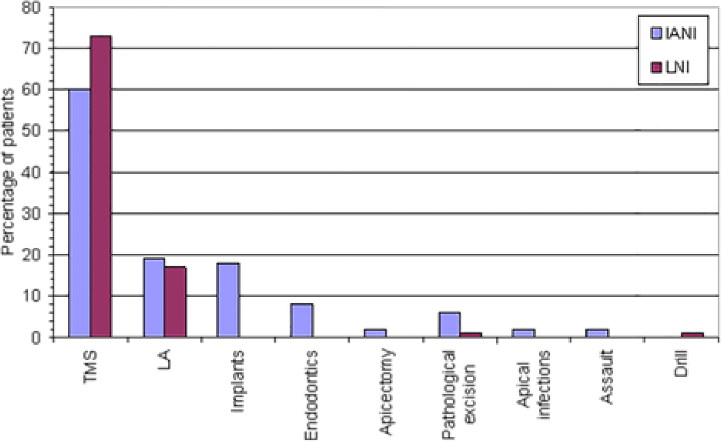
In a prospective assessment of 252 patients with iatrogenic trigeminal nerve injuries,34 most were caused by third molar surgery (TMS), but implants and local anaesthesia were significant contributors (Figure 5).21
Figure 5. Factors associated with iatrogenic trigeminal nerve injury.4
The diagnosis of post-traumatic neuralgia/neuropathy is based upon a history of surgery or trauma temporally correlated with the development of the characteristic neuropathic pain. Age, poor wound closure, infections, foreign material in the wound, haematoma, skull fracture, diabetes mellitus or peripheral neuropathy elsewhere in the body predispose to neuroma development. The pains commonly persist after the injury and can be permanent. Medical therapy is similar to that used in neuropathic pain conditions, depending on the patients' symptoms.27
In a recent survey of 252 iatrogenic trigeminal nerve injuries related to dental treatment, 70% of patients presented with pain. This highlights the problems related to post-surgical neuropathy aggravated by the fact that many patients may not have been warned at all about nerve injury or told that they would risk numbness.21
Many patients experienced significant daily disability, predominantly caused by elicited mechanical or cold allodynia, resulting in pain on eating, drinking, kissing, sleeping and other essential functions (Figure 4).
Current assessment of these nerve injuries is inadequate.21 The focus remains on surgical correction or laser therapy, with little or no attention to medical or counselling intervention and the patients' psychological, functional or pain related complaints. The fault partly rests with how we assess these patients. Assessment tends to show little regard for the functional or pain evaluation, with the main focus on basic mechanosensory evaluation, which is not necessarily reflective of the patients' difficulties. Oral Surgery specialists assessing these injuries should, therefore, follow guidelines from the World Health Organization, which suggest that nerve injury outcomes should be assessed in terms of impairment, activity limitations and participation restrictions.35 Guidelines set out by the International Association for the Study of Pain and European Federation of Neurological Societies should also be followed.36 Without exception, these recommendations are holistic when compared with reports evaluating the management of trigeminal nerve injuries.
Traumatic injuries to peripheral nerves pose complex challenges and treatment of nerve injuries must consider all aspects of the inherent disability. Pain control is of paramount importance and rehabilitation needs to be instituted as first-line treatment. Several guidelines for the assessment of chronic NePain have been published recently.33,34,37
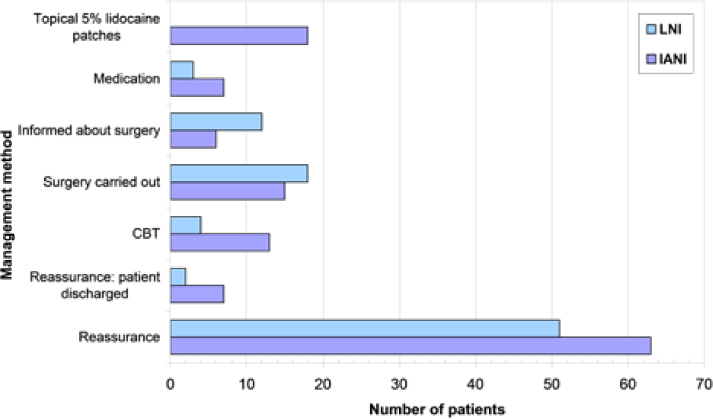
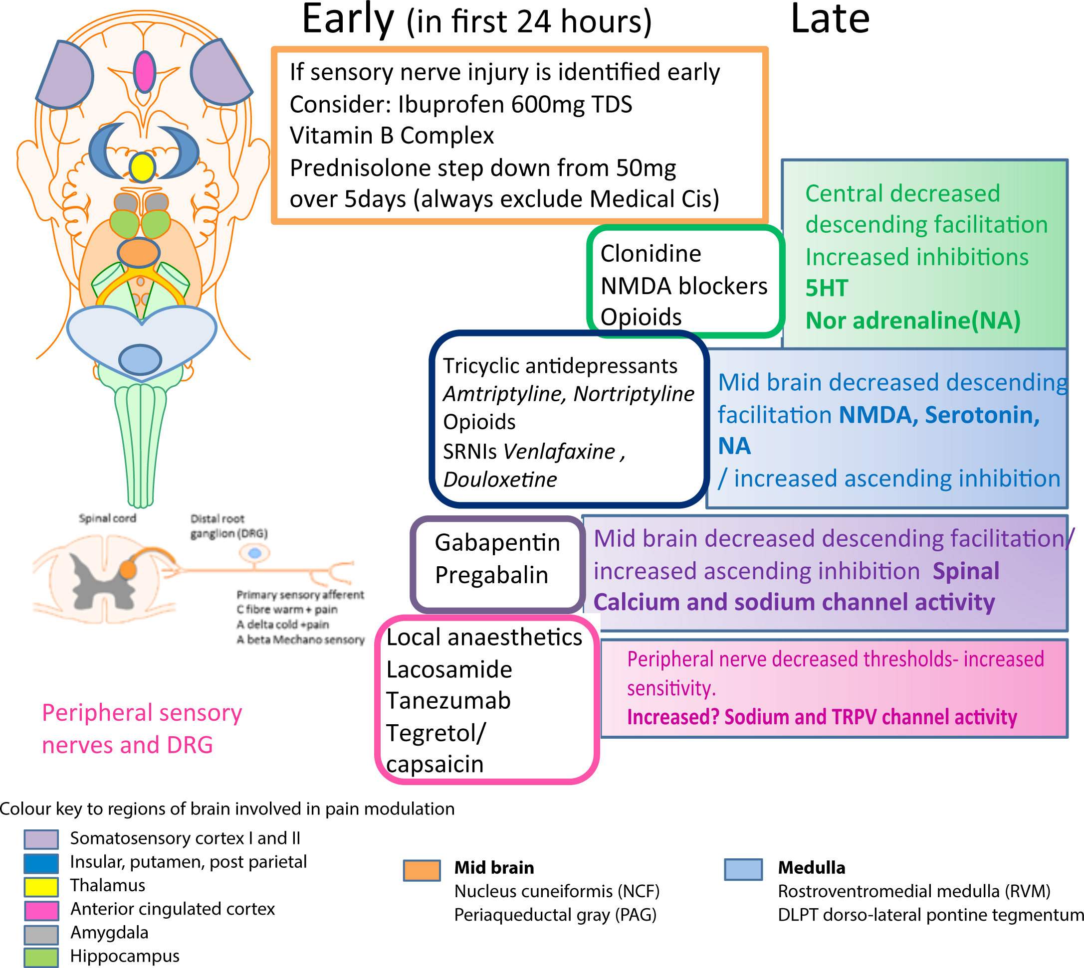
Figure 6. Multiple strategies used for management of post traumatic neuropathy and chronic pain.4Figure 7. Medical management for chronic pain related to mechanisms of known action of the recommended drugs. (Neuropathic pain – pharmacological management: The pharmacological management of neuropathic pain in adults in non-specialist settings NICE guidelines [CG173]. Published date: November 2013 https://www.nice.org.uk/guidance/cg173).
Management for post-traumatic neuropathy may be early or late (Table 3). Prevention of the nerve injuries is recommended, but when they do occur, prevention of persistent pain is paramount in these patients. The intervention will depend upon duration and mechanisms of injury. Intervention include surgery (indicated urgently post injury as ineffective later), medical (Figure 7) and psychological interventions.
Mechanism
Duration
Treatment
Known/suspected nerve section
Immediate exploration
TMS IANI – retained root
<30 hours
Immediate exploration
Implant
<30 hours
Remove implant
Endodontic
<30 hours
Remove tooth/overfill
Implant/Endodontic
>30 hours
Treat therapeutically
TMS IANI – large neuropathic area, pain and disability
<3 months
Consider exploration
TMS LNI – large neuropathic area, pain and disability
<3 months
Consider exploration
TMS IANI
>6 months
Treat therapeutically
TMS LNI
>6 months
Treat therapeutically
LA, fracture, orthognathic, other surgery
Treat therapeutically
For all peripheral sensory nerves, excluding the trigeminal nerve, early surgical exploration and intervention is important for optimal physiologic and functional recovery.35 Reparative surgery for the trigeminal nerve is urgent for any injury, with the exception of lingual nerve injury in relation to lingual access third molar surgery.28 Recommendations for delay in intervention for these injuries are based upon the likelihood of the temporariness of the injury, in that 90% recover post-operatively at 3 months.38 There remains a significant deficiency in evidence base to support this practice. The patients presenting complaints may include functional problems due to the reduced sensation, intolerable changed sensation or pain. Patients who present with pain are usually intransigent to surgery.29,30 Patients rarely present worried about psychological issues relating to the iatrogenesis of the injury and chronic pain. Generally, for lesions of the peripheral sensory nerves in man, the gold standard is to repair the nerve as soon as possible after injury.35 However, the relatively few case series of trigeminal nerve repair on human subjects have carried out surgery immediately after injury.
It is accepted that multiple strategies must be used when managing patients with post-traumatic trigeminal neuropathy (Figure 6), which are mostly non-surgical. This reflects a more holistic approach to patient management (pain control, functionality and psychological factors) rather than fixating on the mechanics of the nerve repair.
Medical management may be early or late (Figure 7). There is limited evidence base for early medical intervention but it never the less has become routine practice to prescribe steroids, NSAIDs and Vitamin B complex when surgical nerve injury is recognized. Nice guidelines for medical management of neuropathic pain (2013) are illustrated in Figure 7.
Novel strategies, including Botox injections for peripheral post-traumatic painful neuropathy36 and Versatis (Grunenthal®) (topical 5% Lidocaine patches),26 may prove of benefit in localized, less systemically toxic pain management control.39,40
Conclusion
It is evident from the literature review that there needs to be a cultural change in the choice of intervention, timing and outcome criteria for trigeminal nerve injuries. To date, there have been a very limited number of prospective randomized studies to evaluate the effect of treatment delay on the surgical, medical or counselling outcomes for trigeminal nerve injuries in humans. A recommended protocol for the management of iatrogenic nerve injuries is presented in Table 3.