Saha A, Seth J, Gorai S, Bindal A. Dermatitis artefacta: a review of five cases: a diagnostic and therapeutic challenge. Indian J Dermatol. 2015; 60:613-615 https://doi.org/10.4103/0019-5154.169139
Mohandas P, Bewley A, Taylor R. Dermatitis artefacta and artefactual skin disease: the need for a psychodermatology multidisciplinary team to treat a difficult condition. Br J Dermatol. 2013; 169:600-606 https://doi.org/10.1111/bjd.12416
Chatterjee SS, Mitra S. Dermatitis artefacta mimicking borderline personality disorder: sometimes, skin could be misleading. Clin Psychopharmacol Neurosci. 2016; 14:311-313 https://doi.org/10.9758/cpn.2016.14.3.311
Alcántara Luna S, García Bravo B, Rodríguez Pichardo A, Camacho Martínez FM. Dermatitis Artefacta in childhood: a retrospective analysis of 44 patients, 1976-2006. Pediatr Dermatol. 2015; 32:604-608 https://doi.org/10.1111/pde.12625
Thiele J, Kaatz M, Schmidt S Recurrent erythematous dermatitis artefacta in the face induced by benzyl nicotinate. Exogenous Dermatology. 2002; 1:242-245 https://doi.org/10.1159/000068794
Shivakumar S, Jafferany M, Kumar SV, Sood S. A brief review of dermatitis artefacta and management strategies for physicians. Prim Care Companion CNS Disord. 2021; 23 https://doi.org/10.4088/PCC.20nr02858
Tittelbach J, Peckruhn M, Elsner P. Histopathological patterns in dermatitis artefacta. J Dtsch Dermatol Ges. 2018; 16:559-564 https://doi.org/10.1111/ddg.13504
Maio P, Santos R, Cardoso J. Letter: factitial dermatitis: an unusual presentation in an old woman. Dermatol Online J. 2012; 18
Woolf RT, Bewley AP, Taylor RE A difficult case of dermatitis artefacta requiring surgical intervention. Br J Dermatol. 2013; 168:889-891 https://doi.org/10.1111/bjd.12086
Zarei M, Kamali M, Bidaki R. Bullous dermatitis artefacta in a 17 year-old girl induced by a native herb. Iran Red Crescent Med J. 2013; 15:862-864 https://doi.org/10.5812/ircmj.8886
Bhalla M, Thami GP. Photoletter to the editor: bullous dermatitis artefacta induced with a hot spoon. J Dermatol Case Rep. 2014; 8:81-83 https://doi.org/10.3315/jdcr.2014.1181
Sarin A, Ummar SA, Ambooken B, Gawai SR. Dermatitis artefacta presenting with localized alopecia of right eyebrow and scalp. Int J Trichology. 2016; 8:26-28 https://doi.org/10.4103/0974-7753.179395
Dermatitis artefacta in the orofacial region: a case report with literature review Nutan Patel Shadaab Mumtaz Florence Deroide Ali Amini Dental Update 2024 50:4, 707-709.
Authors
NutanPatel
BDS
Dental Core Trainee Year 2, Department of Oral and Maxillofacial Surgery, Alder Hey Children's Hospital, Liverpool
In spite of wide prevalence, deliberate self-injury in the oro-facial region is rarely reported in literature. It is also associated with misinterpretation related to ‘attention seeking’ or ‘mental health crises’ leading to deficient understanding of this phenomenon. A literature review was performed using online search databases looking at dermatitis artefacta in the head and neck region. A case of a patient who was seen in our unit is also presented to give important insights into this condition. In total, 54 cases from 15 publications were included in this observational study. Female gender predilection was notable (4:1) with an average presenting age of 30 years. The face itself was more frequently injured, along with the neck and scalp. Only one-third (34%) of the patients were known to have psychiatric conditions, such as depressive and personality disorders. Dermatitis artefacta is a well-known skin condition caused by deliberate self-injury. It is a complex entity that is frequently unrecognized and underdiagnosed.
CPD/Clinical Relevance: Understanding dermatitis artefacta will facilitate correct diagnosis and improve patient care.
Article
Skin abnormalities in the head and neck region occur frequently and can range from benign inflammatory conditions to malignant neoplasms. Psychiatric disorders manifesting as cutaneous and/or oral abnormalities are often unrecognized. In the head and neck region, the face is frequently affected due to ease of access. Dermatitis artefacta (DA) is a factitious disorder seen more frequently in females. However, there is evidence to suggest that the face is a site that is affected relatively frequently in males.1 Other areas that are commonly affected are the scalp, neck, abdomen and forearms.
DA has a close association with psychiatric illnesses including borderline personality disorder and Münchausen syndrome in cases whereby deliberate injuries are made to mimic forms of skin disease. In these cases, the skin changes may be mistaken for primary dermatological conditions.2 The condition may present intermittently and is triggered by stressful episodes which appears to be a factor in the majority of cases.3
Literature review
A literature review of DA affecting the head and neck region was undertaken using multiple search databases, including Ovid Medline, PubMed, EMBASE and Google. All cases/series looking at DA in the head and neck region published in the English Literature between 1987 and 2020 were included. The search terms used included dermatitis artefacta of ‘head’, ‘neck’, ‘face’, ‘oral’, ‘nose’, ‘mouth’, ‘ear’, ‘scalp’ in both title and abstracts.
A total of 14 publications were included in this literature review in addition to the case presented here.1,6,8,9,14,15,16,17,18,19,20,21,22,23 The publications reported 106 cases of DA, although only 54 of these (51%) involved the head and neck region. The face was the most frequent site to be affected (75%), with predominance of forehead, malar, cheek and peri-ocular regions. Neck and scalp were the other areas affected by this condition. There was a predominance of females in the study sample (82%) with an overall mean age of 30 years. There was only one case of intra-oral DA noted, that being of the patient reported here.
In the majority of cases, the exact mechanism of method of injury was unknown, mainly due to non-divulgence of information by the patients. In fact, denial of such acts is common and the main reason for under-reporting of this condition in the literature. Thermal, chemical and physical injury with sharp instruments/weapons was noted in the few cases that reported these findings. The clinical features of the injuries included ulceration, excoriation, blistering and hypopigmentation.
Approximately 35% of patients were reported to have concomitant mental health disorders, which may have been a contributory factor. Among these patients, anxiety and depressive disorders predominated, followed by mood and personality disorders. Four cases were reported to have associated substance and/or physical abuse resulting in self-injurious behaviour. Although patients were referred for psychological evaluation and counselling, many declined or were lost to follow-up.
Case report
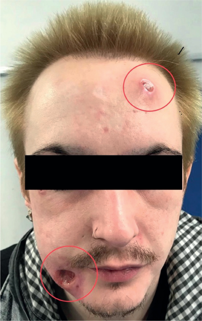
A 33-year-old male presented at the emergency department with ulcers of 6 months' duration on his face (Figure 1). He gave a history of self-injecting collagen, hydrogen peroxide and some ‘fillers’ on different areas of his face. He reported that he bought these ‘injectables’ from a popular online ‘web-shop’. The patient's general practitioner had treated him with multiple courses of antibiotics and topical antiseptics, but with no observable relief.
Figure 1. Skin abnormalities in the right cheek and left forehead
The patient's past medical history consisted of paranoid schizophrenia, which was controlled with procyclizine and olanzapine. He also reported recreational drug use and smoked 5–10 cigarettes per day.
Examination revealed a punched-out ulcer of the right cheek measuring 3.5 cm in diameter with a depth of 0.5–0.8 cm, exposing the underlying musculature (Figure 1). The surrounding area was indurated. No obvious discharge was noted. Additionally, there was a smaller punched-out ulcer of the left forehead of 1.5 cm diameter with a healing base. Both ulcers were non-tender on palpation. Owing to the persistent and suspicious nature of the ulcers, a diagnostic biopsy was organized for both. Unfortunately, the patient failed to attend his appointments for another 3 months and therefore, no biopsy was undertaken.
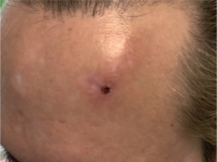
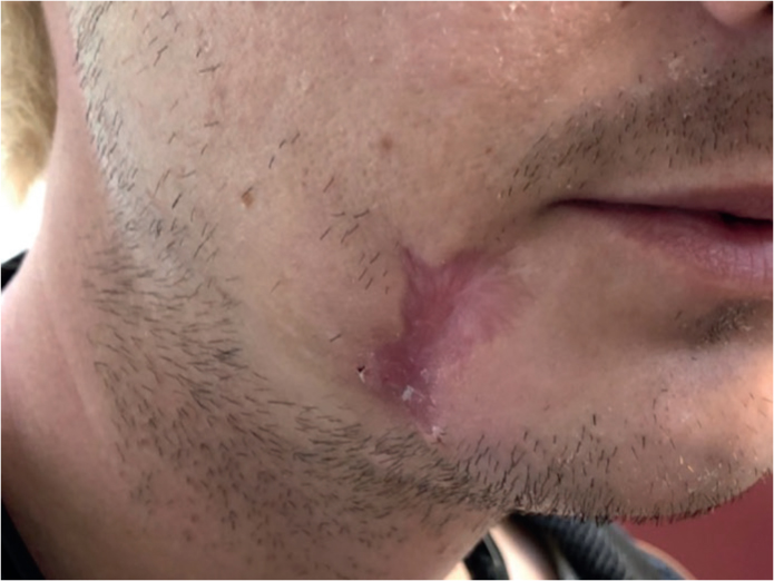
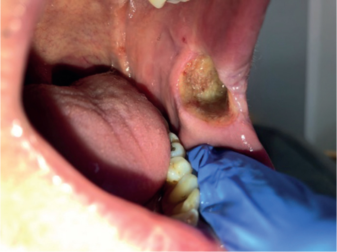
When the patient next attended, both areas of ulceration had healed with scarring (Figures 2 and 3). However, the patient reported the sudden occurrence of a new deep ulcer in the left buccal mucosa measuring 2.5 cm in diameter involving the underlying musculature (Figure 4). A diagnostic biopsy was carried out that revealed a thick crusted acanthotic epidermis with no dysplasia or vasculitis. Mixed inflammation and fibrotic changes reactive to filler were noted, consistent with the diagnosis of dermatitis artefacta. Unfortunately, the patient failed to attend multiple review appointments after the biopsy and remained non-contactable.
Figure 2. Healing left forehead.Figure 3. Healing right cheek.Figure 4. Deep oral ulcer in the left buccal mucosa.
Discussion
Dermatitis artefacta (DA) represents skin abnormalities caused by deliberately self-inflicted injury. The patient does not usually admit to self-injurious behaviour and the tissue changes may resemble known skin conditions, causing difficulty in diagnosis. This also means that the true incidence of DA is unknown, although some papers describe an incidence of 0.05–0.4% in the population.13 A patient with DA will often present with psychological issues that are alleviated by self-harm. Exploration of associated psychiatric illness is an important aspect of treatment to prevent further self-injury. Unfortunately, many of these patients are either non-compliant with treatment or lost to follow-up.
The tissue changes seen in DA are widely variable and often strange in appearance. They can be caused by self-injecting foreign material, which can lead to full-thickness skin loss, requiring surgery.2 The areas involved tend to be those that are easily accessible by the hands, and clear history is frequently challenging to elicit from patients, making it difficult to reach a diagnosis. It has been found that patients experience high levels of dissociation during self-infliction, thus accounting for their poor recollection.2
Diagnosis of DA is reached by a process of elimination, and differentials include contact dermatitis, pyoderma gangrenosum, infections including carbuncle and ecthyma, immunobullous disease and malignancy.4 Histopathological examination is not diagnostic, but necessary to eliminate other sinister differential diagnoses. DA characteristically affects the epidermis with features including epidermal necrosis and subepidermal blisters. In cases where foreign material is self-injected, epidermal acanthosis and granuloma may also be found.5
Multidisciplinary team input is essential for adequate management of DA.6 This condition requires treatment of the underlying mental health condition. Psychotherapy remains the mainstay management approach in these cases and includes psychodynamic therapy and cognitive–behavioural therapy. Family therapy is useful if there is available support from a patient's family and relatives.3,7 Supportive medical management with antidepressants and/or anti-psychotics is frequently necessary. Selective serotonin reuptake inhibitors help in countering impulsive and aggressive behaviours in these patients.12
The tissue damage involved in DA is usually chronic and may need topical regimens such as antibiotics and emollients. Systemic antibiotics may also be prescribed in cases with evidence of severe infection.4,7 In cases of extensive tissue loss, loco-regional skin grafts may be necessary to reconstruct these defects.
Conclusion
Regarding DA, this review and case report highlights the need for general dental practitioners to recognize unusual clinical features, both intra- and extra-orally at an early stage so that an appropriate specialist referral can be made. Establishing a good rapport with the patient will help to achieve relevant insight and appropriate holistic management.