
Article
Masters of imitation: necrotizing sialometaplasia management quandaries
We report the case of a 69-year-old women with necrotizing sialometaplasia, a condition caused by ischaemia and well recognized for mimicking squamous cell and/or muco-epidermoid carcinoma. An equivalent cutaneous example in its malignant resemblance is kerato-acanthoma. Both are self-limiting and monitored for resolution, whereas if cancerous, the most important prognostic factor is early intervention.1,2 Therein lies the consequence of diagnostic accuracy. This case discussion summarizes the learning necessary to support confident navigation by the general dental practitioner of this newly classified non-neoplastic lesion.3
The patient attended via the urgent suspected cancer route through her general medical practitioner. She complained of an 8-week history of sudden onset palatal discomfort that was consistent in nature. Soluble prednisolone, dispersible doxycycline, artificial saliva gel and benzydamine hydrochloride had all been used to treat the condition, but to no avail. She had an unremarkable medical history of long-standing stable comorbidities and had not smoked for 15 years (~25 pack-years).
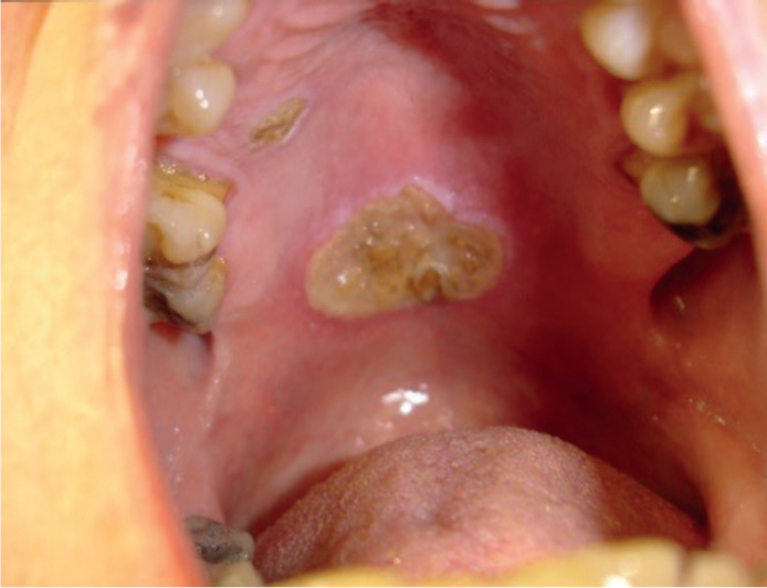
Examination found no lymphadenopathy, but intra-orally, there was an ulcerated mass on the hard palate measuring approximately 2 x 2 cm with raised keratinized margins (Figure 1). Anterior to this was also a smaller lesion displaying identical features. At this stage, the differential diagnoses were squamous cell and muco-epidermoid carcinoma, trauma, necrotizing sialometaplasia, adenocarcinoma, tuberculosis and histoplasmosis. Further probing questions covered nasal and respiratory symptoms and history of trauma or local anaesthetic by palatal infiltration. Negative responses helped eliminate sinus neoplasm with oral extension and lung infection. To narrow down the remainder, a biopsy was performed. This was suggestive of early necrotizing sialometaplasia and clinical review recommended with repeat investigation conditional on persistence, recurrence or continued concern. After 2 weeks, there were promising signs of healing; however, at 4 months this was incomplete. A second specimen was sent that confirmed the original diagnosis, and within 2 months the affected mucosa had returned to normal.
Necrotizing sialometaplasia constitutes under 1% of oral lesions biopsied.4 While this case is not correlated with an obvious aetiological event, there are other classical aspects of the clinical presentation, specifically site and age.1,2 The utility of this information is heightened when interpreted collectively with the pathological findings from well-orientated tissue sections. Although histological appearances vary, attributed to five stages by Anneroth and Hansen, preservation of lobular architecture and ductal squamous metaplasia are the mainstay features.5 The alarming atypia of reactive squamous epithelium with single-cell necrosis has prompted the use of immunohistochemistry that may be helpful as an adjunct only. Treatment is symptomatic. Resolution will occur within 6–10 weeks and recurrence is unusual.4
To summarize, the most important differentials include squamous cell and muco-epidermoid carcinoma. No one characteristic is pathognomonic, and proper diagnosis avoids overtreatment. Where suspected in primary care, a two-week wait referral is necessary and should contain sufficient detail to support histopathological confirmation. Interim analgesia and topical steroids can also be prescribed as needed.