Eltahlah D, Lynch CD, Chadwick B, Blum IR, Wilson NHF. An update on the reasons for placement and replacement of direct restorations. J Dent. 2018; 72:1-7
Blum IR. The management of failing direct composite restorations: replace or repair?. In: Lynch CD, Brunton PA, Wilson NHF (eds). London: Quintessence Publishing Company; 2008
Blum IR, Lynch CD, Wilson NHF. Factors influencing repair of dental restorations with resin composite. Clin Cosmet Investig Dent. 2014; 17:81-88
Blum IR, Schriever A, Heidemann D, Mjör IA, Wilson NHF. The repair of direct composite restorations: an international survey of the teaching of operative techniques and materials. Eur J Dent Educ. 2003; 7:41-48
Gordan VV, Mjör IA, Blum IR, Wilson NHF. Teaching students the repair of resin based composite restorations: a survey of North American dental schools. J Am Dent Assoc. 2003; 134:317-323
Blum IR, Lynch CD. Repair versus replacement of defective direct dental restoration in posterior teeth of adults. Prim Dent J. 2014; 3:62-67
Blum IR. Restoration repair as a contemporary approach to tooth preservation. Prim Dent J. 2019; 8:38-42
Blum IR, Özcan M. Reparative dentistry: possibilities and limitations. Curr Oral Health Rep. 2018; 5:264-269
Green D, MacKenzie L, Banerjee A. Minimally invasive long-term management of direct restorations: the ‘5Rs’. Dent Update. 2015; 42:413-426
Combe EC, Burke FJT, Douglas WH.USA: Kluwer Academic Publishers; 1999
Burke FJT, Lucarotti PSK. The ultimate guide to restoration longevity in England and Wales. Part 10: key findings from a ten million restoration dataset. Br Dent J. 2018; 225:1011-1018
Deliperi S, Bardwell DN. Clinical evaluation of direct cusp coverage posterior composite resin restorations. J Esthet Restor Dent. 2006; 18:256-267
Burke FJT, Crisp RJ, James A, MacKenzie L, Thompson O, Pal A, Sands P, Palin WM. Five-year clinical evaluation of restorations placed in a low shrinkage stress composite in UK general dental practices. Eur J Prosthodont Rest Dent. 2017; 25:108-114
Burke FJT. Repair of metal-ceramic restorations using an abrasive silica-impregnating technique: two case reports. Dent Update. 2002; 29:398-402
Burke FJT. Repair of fractured metal-ceramic restorations using tribochemical impregnation. Dent Update. 2016; 43
Stewardson DA, Creanor S, Thornley P, Biggs T, Burke FJT The survival of Class V restorations in general dental practice. Part 3: five-year survival. Br Dent J. 2012; 212
Heintze S, Ruffieux C, Rousson V. Clinical performance of cervical restorations: a meta analysis. Dent Mater. 2010; 26:993-1000
Gwinnett AJ, Kanca J. Interfacial morphology of resin composite and shiny erosion lesions. Am J Dent. 1992; 5:315-317
Zimmerli B, De Munck J, Lussi A, Lambrechts P, van Meerbeck B. Long-term bonding to eroded dentin requires superficial bur preparation. Clin Oral Invest. 2012; 16:1451-1461
Nordbo H, Leirskar J, von der Fehr FR. Saucer-shaped cavity preparations for posterior approximal resin composite restorations: observations up to 10 years. Quintessence Int. 1998; 29:5-11
Peumans M, DeMunck J, Van Landuyt KL, Poitevin A, Lambrechts P, Van Meerbeck B. Eight year clinical evaluation of a 2-step self etch adhesive with and without selective enamel etching. Dent Mater. 2010; 26:1176-1184
Burke FJT, Crisp RJ, Cowan AJ, Raybould L, Redfearn P, Sands P, Thompson O, Ravaghi V. A randomised controlled trial of a universal bonding agent at three years: self etch vs total etch. Eur J Prosthodont Rest Dent. 2017; 25:220-227
Burke FJT, Mackenzie L, Shortall ACC. Survival rates of resin composite restorations in loadbearing situations in posterior teeth. Dent Update. 2019; 46:523-535
Burke FJT, Lucarotti PSK. The ultimate guide to restoration longevity in England and Wales. Part 9: Incisor teeth: restoration time to next intervention and to extraction of the restored tooth. Br Dent J. 2018; 225:964-975
Lucarotti PSK, Burke FJT. Patient history as a predictor of future treatment need? Considerations from a dataset containing over nine million courses of treatment. Br Dent J. 2019; 228:345-350
Suggestions for Non-Aerosol or Reduced-Aerosol Restorative Dentistry (for as Long as is Necessary) FJ Trevor Burke Louis Mackenzie Peter Sands Dental Update 2024 47:6, 707-709.
The advent of coronavirus and the associated disease COVID-19 has led to the closure of dental practices in the UK and, indeed, in many parts of the world. In order to get dental practices operating again, it is suggested that it is necessary to adopt a new way of working. Principal among concerns has been the potential carriage of droplets (from an infected patient) into the aerosols resulting from the use of the turbine handpiece and from ultrasonic and sonic scalers, and other instruments used in restorative dentistry (current terminology being Aerosol Generating Procedures [AGPs]). It is therefore the aim of this paper to review restorative techniques and suggest those which are appropriate to aerosol-free, or reduced-aerosol restorative dentistry.
CPD/Clinical Relevance: With anxieties regarding aerosol generating procedures abounding, it may be helpful to review procedures which either reduce or avoid these AGPs.
Article
FJ Trevor Burke
The advent of coronavirus and the associated disease COVID-19 has led to the closure of dental practices in the UK and, indeed, in many parts of the world. At the time of writing, dental practices in many countries have re-opened and, in some countries, practices did not close. The “green light” to re-open dental practices in the UK has therefore come later than in many places. In order to get dental practices operating again, the authors suggest that it is necessary to adopt a new way of working. Principal among concerns has been the potential carriage of infected droplets (from an infected patient) into the aerosols resulting from the use of the turbine handpiece and from ultrasonic scalers, and other instruments used in restorative dentistry (current terminology being Aerosol Generating Procedures [AGPs]). It may be of interest to note that the World Health Organization has produced a list of AGPs in healthcare and dentistry is not mentioned.
It is therefore the aim of this paper to review restorative techniques and suggest those which are appropriate to aerosol-free, or reduced-aerosol restorative dentistry.
The solution to ultrasonic instrumentation in periodontal treatment is simple – a return to hand scaling and an increased focus on prevention. The solution to the aerosol-generating procedures in restorative dentistry is not quite so straightforward, but the authors suggest that there are a variety of techniques which can be used without the need, or with a significant reduction in the need, for a turbine handpiece.
The new armamentarium
The authors suggest that the new armamentarium without an aerosol or with a reduced aerosol will include the following:
A speed increasing handpiece attached to an electric motor to be used when absolutely necessary: these offer a considerable reduction in aerosol emission compared to a turbine, and that the aerosol may be proportional to the revolutions per minute of the rotary instrument (AS Brietenbach (NSK UK), personal communication, May 2020). However, at present, there is ongoing research with a view to elucidating this more fully;
High volume suction;
Rubber dam isolation;
Hypochlorite solution, as used for disinfecting root canals, to disinfect the tooth surfaces exposed through the rubber dam (not as a pre-operative mouthrinse);
Hand scalers;
A reliable dentine bonding agent and resin composite material;
Glass ionomer restorative materials (resin-modified and conventional);
Stainless steel crowns;
Stainless steel bands with associated placement and removal devices;
The authors propose to examine the various cavity types and tooth preparations which are common in restorative dentistry in light of aerosol-free or reduced-aerosol dentistry. However, it may first be appropriate to reinforce the concept of reparative dentistry, rather than the wholesale removal/replacement of defective restorations.
Reparative dentistry
Igor Blum and Nairn Wilson have been the vanguard in promotion of the repair concept, providing a large number of publications on the subject.1,2,3,4,5,6 In that regard, in a recent paper, Blum7 considered that ‘replacement of failing restorations, particularly those with localized defects, may be considered as excessively interventional as most of the restorations may be clinically and radiologically sound’, citing the fact that a replacement restoration often leads to unnecessary sacrifice of tooth structure and an acceleration of the tooth on the restoration cycle. Notwithstanding the fact that every intervention on a tooth carries with it the risk of pulpal damage and, as Blum states ‘misuse of the patient's time and financial resources.’ Furthermore, in a review of the literature, Blum and Özcan8 stated unequivocally that ‘restoration replacement should be considered as the last resort when there are no other viable alternatives’. Their review also assessed the literature on survival of repaired restorations, concluding that ‘numerous longitudinal clinical studies have shown that restoration repairs in permanent teeth are able to significantly increase the lifetime of restorations and the restored tooth unit’.
Readers are also directed to the paper by Green and colleagues9 in which they describe, in detail, the long-term clinical management of deteriorating/failing restorations, utilizing the 5Rs principles, namely, reviewing, resealing, refurbishment, repair and, where necessary, replacement, thereby ‘encouraging the preservation of tooth structure and extending the clinical life of the tooth-restoration complex’.
Given the minimally invasive nature of repairs, it is considered that the majority of these may be carried out with a minimum of operative intervention and that, indeed, only debridement of the defect may be needed, ie may be carried out by way of aerosol-free dentistry.
Which materials are appropriate for repairs? It goes without saying that these should be materials which are adhesive to tooth substance, namely, resin composite (in conjunction with a dentine bonding agent), and glass ionomer (GI) and its derivatives.
Composite repair
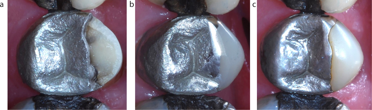
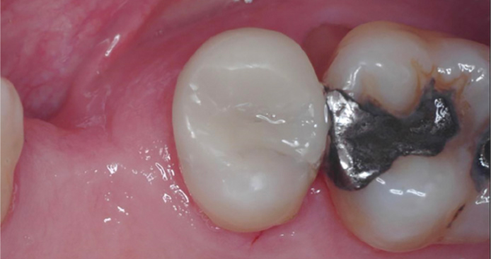
If the defect to be repaired is adjacent to an amalgam restoration, then either composite or amalgam may be considered appropriate, but if the defect is adjacent to an existing composite restoration, then resin composite may be considered as the appropriate material, having first roughened/cleaned the adjacent surface (for example using a diamond bur without water, run slower than normal) before applying a dentine bonding agent. Minor mechanical retention may be considered necessary if the adjacent composite restoration has not been recently placed, as there will be minimal or no unpolymerized resin to bond to. Figure 1 presents the repair/restoration of a fractured buccal premolar cusp.
Figure 1.
(a) Fractured buccal cusp. (b) MI composite repair. (c) Restoration at 5 years.
Glass ionomer (GI)
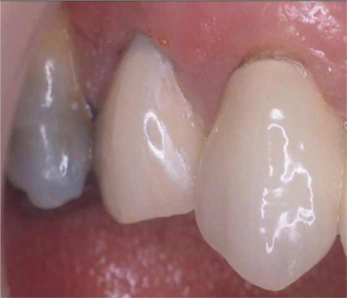
Early versions of this material had poor wear resistance and low mechanical properties,10 therefore reinforced GIs are considered appropriate, although they still have less than ideal fracture and wear resistance and the potential for dissolution in weak organic acids (such as are found in plaque), which afflicts GI materials in general. However, the potential of GIs to adhere to tooth substance may be considered to outweigh these disadvantages (Figures 2 and 3).
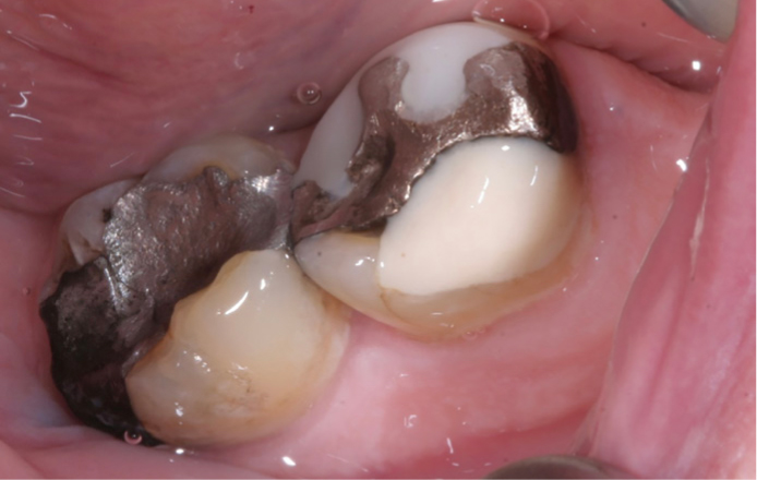
Figure 2. A ‘patchwork molar’ – one cusp repaired with resin composite, another with GI.Figure 3.
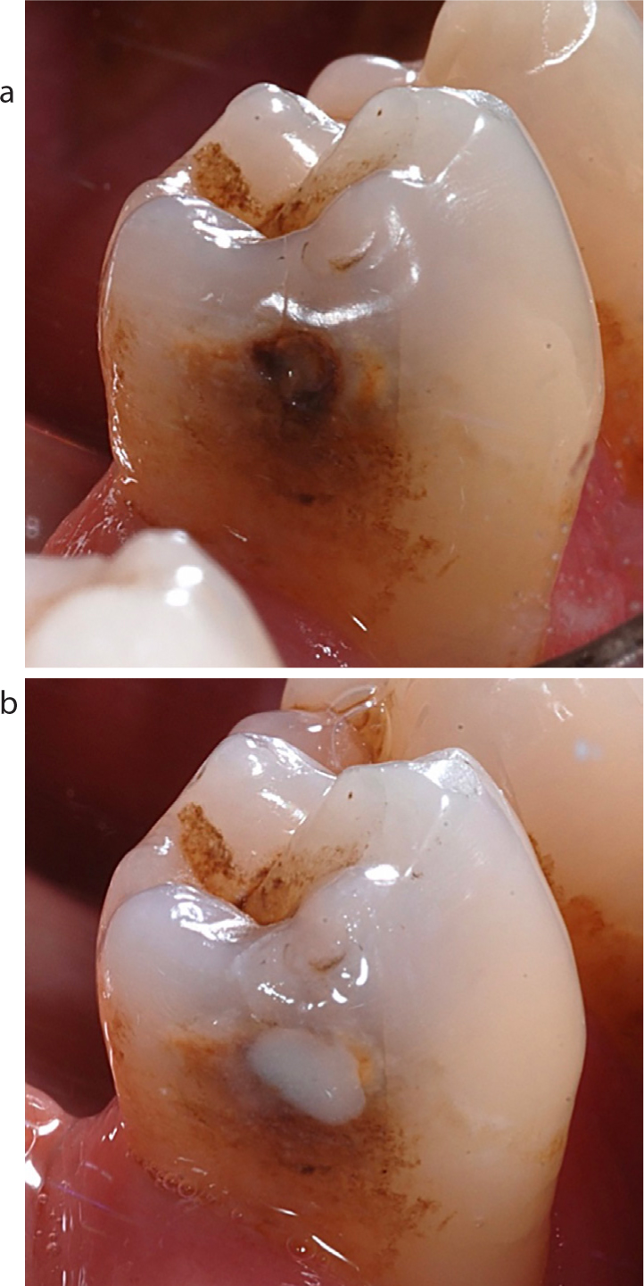
(a, b) Stabilizing GI restoration without the use of high-speed instrumentation.
Resin Modified Glass Ionomers (RMGIs) are also appropriate, especially given that these materials have largely overcome the problem of dissolution. The more recently introduced successor to Fuji IX, Equia Forte (GC), holds promise in the authors' opinion, given the improvements in its physical properties (such as 20% improved flexural strength, 21% improvement in acid resistance, 40% better wear resistance) claimed by the manufacturers, although not yet verified by independent testing. The application of 20% polyacrylic acid solution (such as GC Cavity Conditioner) prior to placing the restoration will provide improved adhesion of the GI.
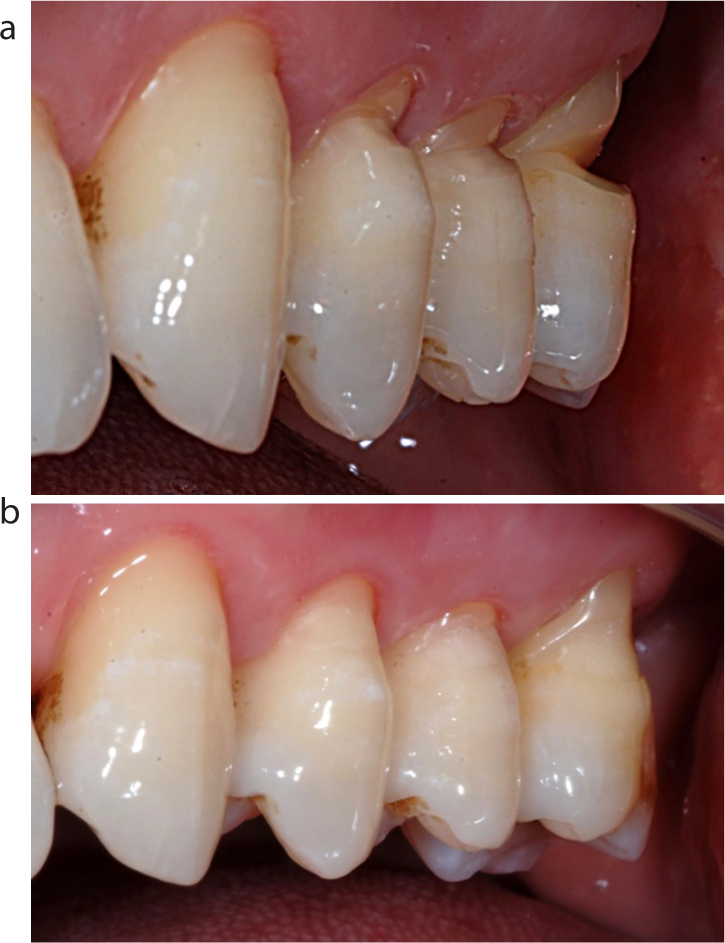
A conventional GI, such as that repairing the fractured cusp of the upper premolar for a 70-year-old patient (Figure 4) will adhere to enamel and dentine (and also potentially micromechanically to the amalgam ‘cliff face’) but might not be considered a definitive long-term restoration because of the material's poor wear resistance and less-than-ideal aesthetics: however, it may provide a solution until such time as the landscape changes re COVID-19, and when the remaining amalgam is removed and the tooth restored with a cusp-replacement composite (Figure 5). A crown is not indicated here because research has indicated poor performance in posterior teeth,11 and the fact that the only remaining tooth substance (the palatal cusp) would have to be reduced during crown preparation: the composite restoration is therefore much more biologically and financially friendly. It is the authors' view that this treatment could be carried out without AGPs, given that the non-retentive amalgam should be readily removed. There is some evidence indicating the satisfactory performance of such large cusp-replacement resin composite restorations.12,13
Figure 4. Fractured premolar cusp temporarily repaired with GI.Figure 5. Tooth in Figure 4: amalgam and GI removed and tooth restored with cusp replacement composite.
While resin composite may be the material of choice for large cusp replacement restorations, other materials which are adhesive to tooth substance, such as RMGI, may also be appropriate (Figure 6).
Figure 6.
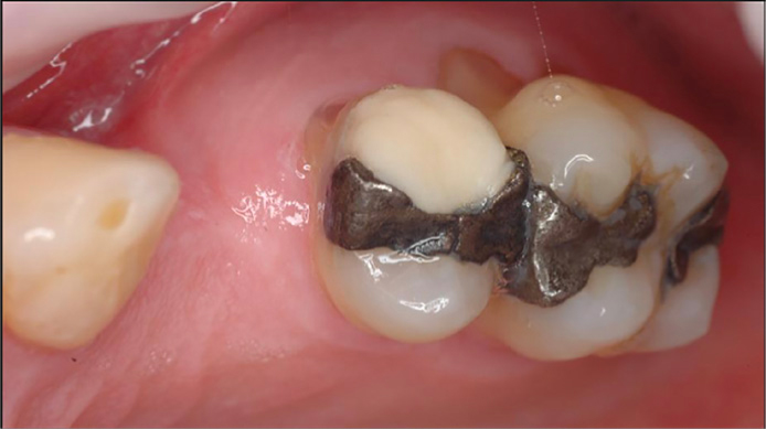
(a–c) Glass ionomer restoration of an MOL cavity in a lower right second permanent molar.
A technique whereby fractured or chipped metal ceramic restorations may be repaired using the Cojet (3M ESPE) system has previously been described in Dental Update.14,15 This will generally only apply to anterior crowns or bridges where there is an aesthetic compromise, as opposed to chipped posterior metal-ceramic crowns, which are often, in the experience of the authors, of little or no concern unless there is a sharp edge. Briefly, this involves the use of silica-coated alumina particles being directed at the fractured surface (be it metal or ceramic) using an intra-oral sandblaster under rubber dam, the application of a silane solution, an opaquer, followed by resin composite to effect the repair. Although this technique necessitates the use of an intra-oral sandblaster, this is not an aerosol (Table 1), so may be considered appropriate during the current situation. Figures 7, 8 and 9 present the repair of a crown which was being removed: the patient decided that she could not tolerate any further treatment and left the surgery. Subsequent examination did not reveal any defects at the crown margin (other than at the gingival margin), so the crown defect was repaired.
Instrument/device
Effect
High speed handpieces (air turbines & speed increasing electric motors)
AEROSOL
Ultrasonic and sonic scalers
AEROSOL
Slow speed handpieces (burs and prophylaxis)
Splatter
Air and water syringe if used together
AEROSOL
Water syringe (used alone)
Splatter
Particle air abrasion
Splatter
Electrosurgery units
Splatter
Lasers
Largely splatter
Figure 7. Crown at UR4 with obvious evidence of operative intervention.Figure 8. Metal substructure being sandblasted with Cojet Sand (3M ESPE).Figure 9. Defect repaired with the components of Cojet (3M ESPE) and resin composite.
General suggestions for restorative dentistry
Use rubber dam (RD) routinely: rather than placing only for restoration placement, as in pre-pandemic times. It should be applied prior to cavity preparation as, if it is necessary to use a turbine or speed-increasing handpiece, it could be anticipated that the aerosol will pose less of a threat with RD in place. Research is ongoing on this subject;
Swab the tooth pre-operatively with hypochlorite (as used in root canal disinfection);
Use the most minimal cavity preparation;
In all cavity configurations removal of the biofilm from the tooth surface is important in achieving a good bond to resin-based or GI materials. Tooth preparation will mostly achieve this but, if parts of the tooth to which the restoration will be bonded are not prepared in any way, then there is likely to be biofilm on the surface. It is suggested that a brief prophylaxis using pumice and water in a slow handpiece should remove the biofilm.
Class V cavities
A proportion of Class V cavities are non-carious, and will require treatment due to sensitivity, food stagnation, compromised aesthetics, or for protection against further abrasive wear. Such cavities are the simplest to treat using aerosol-free adhesive dentistry, given that these will be readily accessible to a slow handpiece or hand instruments. Results of a survey of 1,000 Class V cavities placed in UK dental practices16 indicated that RMGI provided the best results at 5 years (75% survival), with composite providing 68% success. It may therefore be considered that, if aesthetics is the overriding concern, composite should be used but, if not, then RMGI will provide the optimum results. Conventional GIs produced the worst results (51% survival), so these may not be recommended. Large restorations performed less well. Furthermore, it is worth noting that, if the dentine surface was roughened (for example by a steel bur in a slow handpiece), the results improved, this finding being appropriate for GI and resin composite restorations. This finding is confirmed by a meta-analysis by Heintze and colleagues17, and the value of roughening a sclerotic Class V dentine surface is confirmed by Gwinnett and Kanca18 and by Zimmerli and colleagues.19Figures 10 a and b present the restoration, using RMGI, of abrasion cavities with no cavity preparation.
Figure 10.
(a, b) Restoration of Class V cavities using RMGI with no cavity preparation.
Class I and II cavities
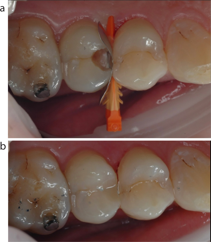
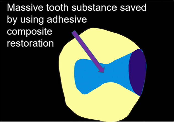
Ease of access to these is more problematic than for Class V but, if the cavity preparation is kept minimal (as in the cavity in Figure 11), the authors suggest that access through the enamel to the caries lesion can be kept to a minimum without the use of a turbine, and the remaining caries removal carried out using hand instruments and a slow handpiece. The use of a typical Black Class II cavity design would be difficult without using a turbine, notwithstanding the fact that it is unnecessarily destructive of tooth substance (Figure 12), therefore this is contra-indicated.
Figure 11.
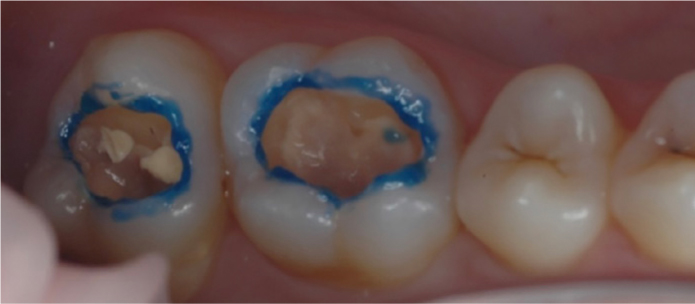
(a) A mini saucer-shaped cavity design which is appropriate to adhesive restorative technology. (b) Cavity in (a) restored using Filtek Bulk Fill Restorative (3M).Figure 12. A comparison of the tooth substance prepared by a typical Black's design Class II cavity and a contemporary saucer-shaped adhesive design cavity (dark blue)
There is a paucity of research on the success of such minimal restorations, but the work of Nordbo et al,20 on minimal Class II restorations followed for 7 years, albeit using materials which may be considered outmoded at the present time, provide satisfactory data on success and a conclusion that the saucer-shaped resin composite restoration represents a viable treatment modality for small cavities', adding ‘that the time may have come to include it in dental curricula as a routine operative treatment for small Class II lesions’.
During the placement of the resin composite restoration, selective etching of the enamel (Figure 13) may be considered necessary, given that this has been shown to provide superior margins at 8 years when using a so-called self-etch adhesive (Clearfil SE, Kuraray).21 Rather than using a 3-in-1 (air/water) syringe to wash off the etchant (with an associated aerosol) the etchant may be removed with a damp cotton roll or pledget. Alternatively, the etchant may be washed off (water only) and the tooth dried using air alone. Better still, the use of a Universal bonding agent in self etch mode will obviate the need for etchant, with results of one paper22 indicating no difference in cavity margins when Scotchbond Universal (3M) was used in total etch mode (ie enamel margins etched) or in self-etch mode (ie not etched). While the instructions for use with dentine bonding agents frequently advise gentle air thinning to evaporate solvent, it is suggested that this will evaporate if the layer of bonding agent is left undisturbed for a short time.
Figure 13. The majority of etchant should be removed with a damp cotton roll rather than a 3-in-1 syringe.
It should be added that success with ‘posterior composites’ is dependent also upon the operator's knowledge of, and familiarity with, the various technique sensitivities which have been described by Mackenzie and colleagues.23 In this regard, success rates of posterior composite restorations have been evaluated in a recent review,24 with the results indicating, both from cohort studies and meta-analyses which fulfilled the inclusion criteria (among these being that the studies were based in primary care), that resin composite restorations have acceptable survival rates when placed in loadbearing situations in posterior teeth, with AFRs generally within the range 2% to 3%, which the authors consider to be compatible with successful clinical practice. Risk factors for premature failure include patients at high risk of caries and the presence of a liner or base beneath the resin composite restoration.
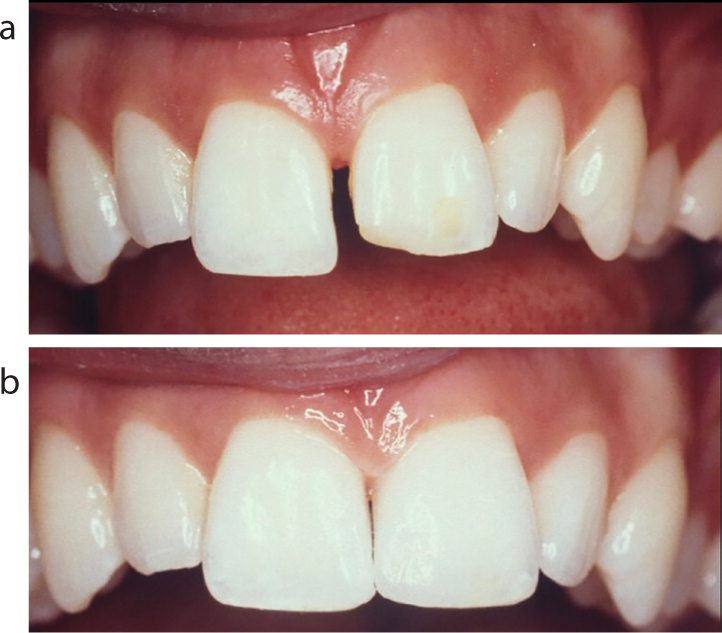
Class III and IV cavities
Access to these cavity types presents less of a problem, therefore these are readily amenable to aerosol-free dentistry. While Class III cavities require preparation, it should not be necessary to use a turbine. Class IV cavities generally require little preparation (Figures 14 a and b), given that these arise as a result of trauma or the fracture of an incisal corner in a tooth already restored with a Class III restoration. For these, often the only preparation which is needed is the placement of a short bevel along the buccal incisal edge of the cavity: again, this may be carried out without the need for a turbine. Resin composite is the material of choice, and, if it is necessary to etch the cavity margins, in order to ensure a long-term defect-free margin, or to provide retention for a Class IV restoration then, as mentioned above, the etchant may be removed using a damp cotton roll or pledget. At one time there were fears that using such a procedure, rather than using a 3-in-1 spray, could damage the etch pattern, but such fears have not materialized.
Figure 14.
(a, b) Often no, or minimal, preparation is needed for a Class IV restoration or a composite build-up.
Crown preparations
Given that preparation of a tooth for a crown would be a challenging procedure without using a turbine drill (with its associated aerosol), the authors therefore suggest that crown preparations are not appropriate to aerosol-free or reduced-aerosol dentistry. There are other reasons! Results of research carried out on a 10 million restoration dataset have indicated that, when the actual survival of the restoration is examined, crowns perform optimally, but when the longevity of the crowned tooth is assessed, crowns perform poorly, while direct restorations do not reduce the tooth's survival.25 The explanation for this is that, when a crown fails, it may do so catastrophically, whereas direct-placement restorations may be repaired or replaced.
Total crown failure
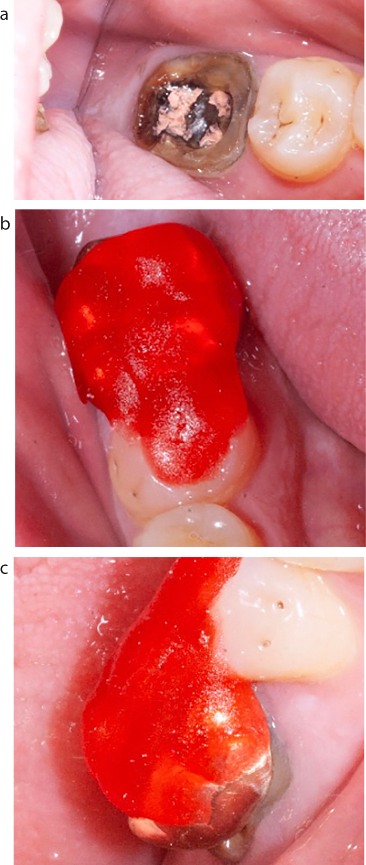
Given the difficulties in preparation or repreparation of a tooth for crown, it would appear desirable, in the current climate, to ‘salvage’ a crown, if possible. In that regard, if a crown does ‘fail catastrophically’, but the patient retains the crown and it can be reseated, albeit maybe not perfectly then, if the crown is re-seated and a sectional impression can be taken, upon the removal of the old crown and any hand removal of soft caries, it may be possible to make a provisional crown from a Bis-Acryl material such as ProTemp (3M). This can be adjusted easily with a slow handpiece and abrasive discs and cemented with an active resin cement. In the experience of one of the authors (PS), these have been shown to last many years in some instances.
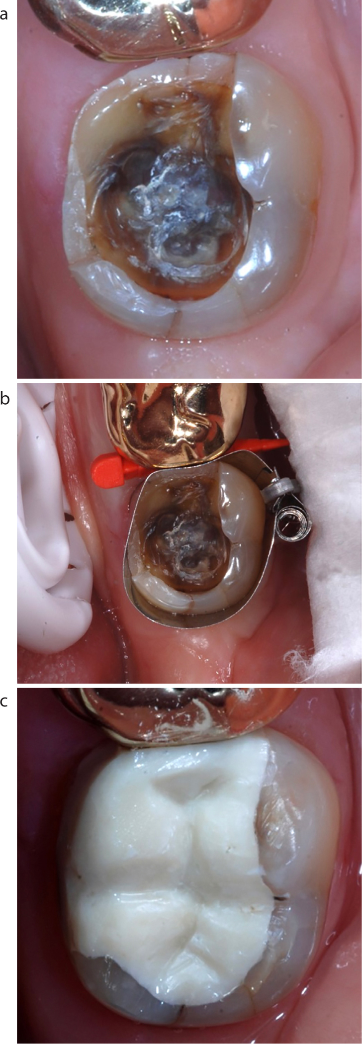
Alternatively, if the crown can be recemented, following the removal of any soft caries, then recement the crown. The problem here is that, following the removal of the caries, the crown may no longer ‘seat’ perfectly on the remaining tooth structure and cementation in the correct place, so as to conform with the existing occlusion, can be very easily compromised. This problem can be managed if, prior to the removal of the crown and caries, a ‘jig’ can be made which will allow the crown (onlay) to be cemented in exactly the right place. The technique is illustrated in Figures 15 a, b and c.
Figure 15.
(a) Lost full coverage crown and core. (b) Construction of Duralay ‘jig’. (c) Crown relocated on tooth.
In this case, a gold crown and the entire core has come off. Some caries removal was necessary along with some of the old gutta percha. Prior to this, the crown was relocated and, in this case, a Duralay ‘jig’ was constructed. This author (PS) prefers a rigid jig as this allows accurate reseating and ‘grips’ the restoration better than a non-rigid material. A Duralay jig was made prior to removal of any tooth tissue (Figure 15b). Upon removal of caries with hand excavation or a slow handpiece, the crown can be relocated in exactly the same special relationship to its adjacent tooth and antagonist and recemented.
It is the authors' view that crowns generally fail, initially, at their margins and that such marginal failures can be repaired readily without a turbine, given that access should not be a problem with a slow handpiece. A more catastrophic failure of a crowned tooth presents a more difficult restorative problem, as described above, which may only be solved by placement of a long-term provisional crown to tide the patient over until such times as re-preparation may be undertaken.
Stainless Steel (SS) bands
It's not just orthodontists who need a SS band!
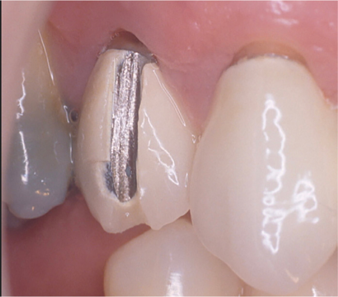
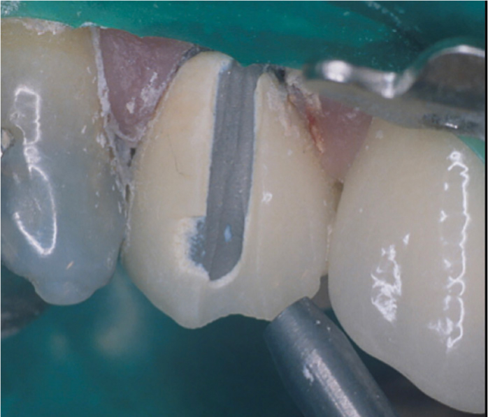
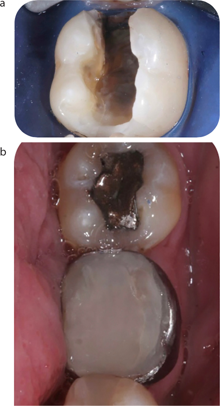
If a patient attends with pain and the diagnosis is made of a cracked tooth, conventional treatment may have involved the provision of an indirect restoration such as a crown or onlay. However, an alternative solution to this which will minimize any aerosol would be to place a SS band around the tooth and cement with either a resin or glass ionomer cement. In this example (Figure 16), undertaken prior to ‘lockdown’ it was felt necessary to remove the existing restoration, obviously creating an aerosol, but this technique can quite easily be used without removing the existing restoration provided it is felt that there is no pressing reason to do so.
Figure 16.
(a) Diagnosis made of cracked tooth. (b) Stainless steel band cemented with GI cement.
Having suspected a cracked tooth, with the affected tooth testing vital, upon the removal of the existing restoration, a significant crack along the internal line angle of the lingual wall was detected. Using wedges to separate the mesial contact point slightly, a stainless steel band was fitted and cemented with a GI luting cement. The tooth was then provisionally restored with a composite resin material.
This actual restoration dates back to 2016 and presently remains in place. The patient remains able to eat comfortably, without pain and, while a crown might have been thought to be a preferred option, the patient can't see the need for any further intervention.
Stainless steel crowns
Finally, the Hall crown is an obvious addition to the list of treatments which can be carried out without an aerosol and, as such, may become an increasingly utilized non-preparation treatment for carious cavities of any size in primary teeth: with minimal preparation, the technique may also be adapted for permanent teeth.
Discussion
It has been beyond the scope of this paper to deal with the organizational issues which will need to be put in place in order to allow dental practices to open safely in the current climate, but it is hoped that some suggestions made in this paper, while hopefully readily achievable, will have advantages in terms of the time required for tooth preparation. The turbine drill facilitates fast tooth preparation and, with slower handpieces, preparations will take longer. However, it could be hoped that this will be mitigated by the use of minimal cavity preparation designs, or repairs. Patients may require education in expecting to pay as much (or more!) for minimally invasive techniques which cause less damage to their teeth.
As has been stated above, it will be necessary to try and avoid AGPs for as long as is necessary, until clear guidance from evidence emerges. This is going to require dentists to be more ‘imaginative’ than in the past and this may require us to employ techniques that may not have the full support of ‘conventional wisdom’ or ‘Ivory Towers’ dentistry. In that regard, suggestions made by the authors in this paper may be considered to fly in the face of conventional dental wisdom and teaching. However, the time during which this paper is being written is unusual, difficult and frightening, and therefore demands a new approach to restorative dental treatments if dental practices are to re-open for treatment of patients, some of whom will have suffered as a result of the closures. At the time of writing, no-one knows the duration of this new norm for restorative dentistry: perhaps we will ascertain, in due course, as has been suggested by Dominic O'Hooley, writing in the current issue, that aerosol generating procedures can be handled satisfactorily without a major alteration to practice procedures. If this proves to be the case, then this article will fast be confined to history, which is what the authors, unusually, hope for!
It is worth adding that a significant proportion of a dentist's time in his/her practice does not involve the restoration of teeth, given the results from a recent paper which indicated that only half of the patients seen by NHS dentists required ‘active’ treatment – defined in the paper as restorative or periodontal treatment.26 There is therefore no reason why non-invasive treatments, such as examination/diagnosis, cannot commence at the earliest opportunity once arrangements for social distancing and enhanced PPE are in place. Preventive advice may be given both in practices and remotely. With that as a starting point, it is hoped that the aerosol-free or reduced-aerosol dentistry suggested here will not be far behind.
Conclusions
The authors have suggested that a wide variety of restorative dental treatments are amenable to aerosol-free or reduced-aerosol dentistry, with indirect preparations ruled out by being the only treatment requiring high speed rotary instruments. A reduction in the number of teeth prepared for traditional crowns may be considered an unexpected benefit of this national and professional crisis.