Physical signs for the general dental practitioner

From Volume 40, Issue 6, July 2013 | Page 505

Authors

Steve Bain

Professor of Medicine (Diabetes), University of Wales, Swansea

Articles by Steve Bain

Melanie Jones

Foundation Year 1 Doctor, Singleton Hospital, Swansea

Articles by Melanie Jones

Article

Steve Bain
Melanie Jones

‘Physical Signs for the General Dental Practitioner’ aims:

  • To increase awareness of the value of identifying general clinical signs.
  • To enable the interpretation of selected clinical signs that are visible in the clothed patient.
  • To indicate the potential relevance of these clinical signs to the dental management of the patient.
  • The series will contribute to non-verifiable CPD requirements.

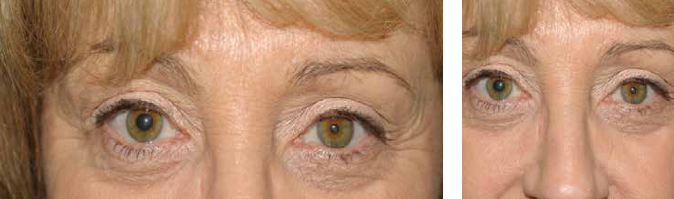
    A friend has pointed out that this person has one pupil bigger than the other. This has not been noticed before.

  • What clinical signs are seen?
  • What is the diagnosis?
  • What are the causes of this appearance?
  • How is it investigated?
  • Answers: Case 105

  • This lady has partial ptosis (drooping of upper eye-lid) and miosis (decreased pupil size). The other features of this syndrome are anhidrosis (reduced sweating on the affected side of the face) and enophthamos (small eye – the opposite of exophthalmos).
  • These clinical signs are typical of Horner's syndrome. This is caused by an interruption to the sympathetic nerve supply causing the above signs.
  • Lesions to the central neuron could be caused by a cerebrovascular accident, syringomyelia or tumours of the pituitary. Lesions to the peripheral nerve could result from an apical lung tumour, eg Pancoast's tumour, carotid artery dissection or lower brachial plexus trauma.
  • In the acute onset of Horner's syndrome, where carotid artery dissection is suspected, angiography or carotid ultrasound should be carried out. A chest X-ray is suggested to look for an apical tumour. CT head would be recommended if a cerebrovascular accident is suspected. Children with new onset Horner's syndrome should be investigated with imaging for neuroblastoma. Often (as in this case) no cause is found.