Perez CR. Alternative technique for Class V resin composite restorations with minimum finishing/polishing procedures. Oper Dent. 2010; 35:375-379
Hazen SP, Chilton NW, Mumma RD The problem of root caries. I. Literature review and clinical description. J Am Dent Assoc. 1973; 86:137-144
Chilton NW, Hazen SP, Mumma RD The problem of root surface caries. 1. Clinical lesions. J Dent Res. 1972; 50
Sumney DL, Jordan HV, Englander HR. The prevalence of root surface caries in selected populations. J Periodontol. 1973; 44:500-504
Banting DW, Courtright PN. Distribution and natural history of carious lesions on the roots of teeth. J Can Dent Assoc. 1975; 41:45-49
Banting DW, Ellen RP, Filley ED. Prevalence of root surface caries among institutionalised older persons. Community Dent Oral Epidemiol. 1980; 8:84-88
Warren JL, Levy SM, Wefel JS. Explorer probing of root surface caries lesions: an in vitro study. Spec Care Dentist. 2003; 23:18-21
Rosen B, Birkhed D, Nilsson K Reproducibility of clinical caries diagnosis on coronal and root surfaces. Caries Res. 1996; 30:1-7
Katz RV. Assessing root caries in populations: the evolution of the root caries index. J Public Health Dent. 1980; 40:7-16
Nyvad B, Machiulskiene V, Bælum V. Reliability of a new caries diagnostic system differentiating between active and inactive caries lesions. Caries Res. 1999; 33:252-260
Baelum V, Machiulskiene V, Nyvad B, Richards A, Væth M. Application of survival analysis to carious lesion transitions in intervention trials. Community Dent Oral Epidemiol. 2003; 31:252-260
Yip K, Smales R. Oral diagnosis and treatment planning: part 5. Preventive and treatment planning for dental caries. Br Dent J. 2012; 213:211-220
Marinova-Takorova M. Incidence of secondary root caries lesions in patients referred for treatment in the faculty of dental medicine – Sofia. Journal of IMAB Annual Proceeding (Scientific Papers). 2014; 20:537-541
Mount GJ. Clinical considerations in the prevention and restoration of root surface caries. Am J Dent. 1988; 1:163-168
Titus HW. Root caries: Some facts and treatment methods. Am J Dent. 1991; 4:61-68
Mjör IA, Toffenetti F. Secondary caries: a literature review with case reports. Quintessence Int. 2000; 31:165-179
Fusayama T. Two layers of carious dentin: diagnosis and treatment. Oper Dent. 1979; 4:63-70
Anderson MH, Charbeneau GT. A comparison of digital and optical criteria for detecting carious dentin. J Prosthet Dent. 1985; 83:643-646
Van de Rijke JW. Use of dyes in cariology. Int Dent J. 1991; 41::111-116
Kidd EA, Joyston-Bechal S, Smith MM, Allan R, Howe L, Smith SR. The use of caries detector dye in cavity preparation. Br Dent J. 1989; 167::132-134
Kidd E, Fejerskov O, Nyvad B. Infected dentine revisited. Dent Update. 2015; 42:802-809
Fusayama T. Clinical guide for removing caries using a caries detecting solution. Oper Dent. 1988; 19:397-401
Banerjee A, Kidd EAM, Watson TF. In vitro validation of carious dentine using different excavation criteria. Am J Dent. 2003; 16:228-230
Hosoya Y, Taguchi T, Arita S, Tay FR. Clinical evaluation of polypropylene glycol-based caries detecting dyes for primary and permanent carious dentine. J Dent. 2008; 36:1041-1047
Wefel JS, Clarkson BH, Heilman JR. Natural root caries: a histologic and microradiographic evaluation. J Oral Pathol. 1985; 14:615-623
Burke FJ, Wilson NH, Cheung SW, Mjör IA. Influence of patient factors on age of restorations at failure and reasons for their placement and replacement. J Dent. 2001; 29:317-324
Stewardson DA, Thornley P, Bigg T, Bromage C, Browne A, Cottam D The survival of Class V restorations in general dental practice. Part 2, early failure. Br Dent J. 2011; 210::1-6
Stewardson D, Creanor S, Thornley P, Bigg T, Bromage C, Browne A The survival of Class V restorations in general dental practice: part 3, five year survival. Br Dent J. 2012; 212:1-8
Frencken JE, Makoni F, Sithole WD. ART restorations and glass ionomer sealants in Zimbabwe: survival after 3 years. Community Dent Oral Epidemiol. 1998; 26:372-381
Lopez N, Simpser-Rafalin S, Berthold P. Atraumatic restorative treatment for prevention and treatment of caries in an underserved community. Am J Public Health. 2005; 95:1338-1339
Franca C, Colares V, van Amerongen E. The operator as a factor of success in ART restorations. Braz J Oral Sci. 2011; 10:60-64
Hayes M, Brady P, Burke FM, Allen PF. Failure rates of class V restorations in the management of root caries in adults – a systematic review. Gerodontology. 2016; 33:299-307
Burrow MF, Stacey MA. Management of cavitated root caries lesions minimum intervention and alternatives. Monogr Oral Sci. 2017; 26:106-114
Billings RJ, Brown LR, Kaster AG. Contemporary treatment strategies for root surface dental caries. Gerodontics. 1985; 1:20-27
Duke ES, Trevino DF. A resin-modified glass ionomer restorative: three-year clinical results. J Indiana Dent Assoc. 1998; 13:13-18
Levy SM, Jensen ME. A clinical evaluation of the restoration of root surface caries. Spec Care Dentist. 1990; 10:156-160
Carrigan PJ, Morse DR, Furst ML, Sinai H. A scanning electron microscopic evaluation of human dentinal tubules according to age and location. J Endod. 1984; 10:359-363
Tagami J, Hosoda H, Burrow MF, Nakajima M. Effect of aging and caries on dentin permeability. Proc Finn Dent Soc. 1992; 88:149-154
Toto PD, Kastelic EF, Duyvejjonck KJ, Rapp GW. Effect of age on water content in human teeth. J Dent Res. 1971; 50:1284-1285
Wilson AD, Kent BE. A new translucent cement for dentistry. Br Dent J. 1972; 132:133-135
McLean JW, Prosser HJ, Wilson AD. The use of glass ionomer cements in bonding composite resins to dentine. Br Dent J. 1985; 15:410-414
Kidd EA. Cavity sealing ability of composite and glass ionomer cement restorations. An assessment in vitro. Br Dent J. 1978; 144:139-142
Frencken JE, Peters MC, Manton DJ, Leal SC, Gordan VV, Eden E. Minimal intervention dentistry for managing dental caries – a review: report of a FDI task group. Int Dent J. 2012; 62:223-243
Lohbauer U. Dental glass ionomer cements as permanent filling materials? – properties, limitations and future trends. Materials. 2009; 3:76-96
Nicholson J. Adhesion of glass-ionomer cements to teeth: a review. Int J Adhes Adhes. 2016; 69::33-38
Walsh LJ. Minimal intervention management of the older patient. Br Dent J. 2017; 223:151-161
Ngo H, Opsahl-Vital S. Minimal intervention dentistry II: part 7. Minimal intervention in cariology: the role of glass-ionomer cements in the preservation of tooth structures against caries. Br Dent J. 2014; 216
Nicholson JW. Glass ionomers in medicine and dentistry. Proc Inst Mech Eng H. 1998; 212:121-126
Van Meerbeek B, Yoshida Y, Inoue S, De Munck J, van Landuyt K, Lambrechts P. Glass-ionomer adhesion: the mechanisms at the interface. J Dent. 2006; 34:615-617
Nicholson JW, Czarnecka B. Biocompatibility of resin-modified glass-ionomer dental cements. Dent Mater. 2008; 24:1702-1708
Yap AU. Effectiveness of polymerization in composite restoratives claiming bulk placement: impact of cavity depth and exposure time. Oper Dent. 2000; 25:113-120
St Georges AJ, Wilder AD, Perdigão J. Microleakage of Class V composites using different placement and curing techniques: an in vitro study. Am J Dent. 2002; 15:244-247
Minakuchi S, Munoz CA, Jessop N. Effect of flexural load cycling on microleakage of extended root caries restorations. Oper Dent. 2005; 30:234-238
Takehara J, Takano T, Akhter R, Morita M. Correlations of non-carious cervical lesions and occlusal factors determined by using pressure-detecting sheet. J Dent. 2008; 36:774-779
Wood ID, Kassir ASA, Brunton PA. Effect of lateral excursive movements on the progression of abfraction lesions. Oper Dent. 2009; 34:273-279
Attar N, Tam LE, McComb D. Flow, strength, stiffness and radiopacity of flowable resin composites. J Can Dent Assoc. 2003; 69:516-521
Sensi LG, Marson FC, Monteiro S, Baratieri LN, de Andrada MAC. Flowable composites as “filled adhesives”: a microleakage study. J Contemp Dent Pract. 2004; 5::32-41
Ozgunaltay G, Yazici Ar, Gorucu J. Effects of finishing and polishing procedures on the surface roughness of new tooth coloured restoratives. J Oral Rehab. 2003; 30:218-224
Hondrum SO, Fernandez R. Contouring, finishing and polishing Class V restorative materials. Oper Dent. 1997; 22:30-36
Yap AUJ, Sal C, Lye KW. Effects of finishing/polishing time on surface characteristics of tooth-coloured restoratives. J Oral Rehab. 1998; 25::456-461
Mitchell CA, Pintado MR, Douglas WH. Iatrogenic tooth abrasion comparisons among composite materials and finishing techniques. J Prosthet Dent. 2002; 88::320-328
Magni E, Zhang L, Hickel R, Bossu M, Polimeni A, Ferrari M. SEM and microleakage evaluation of the marginal integrity of two types of Class V restorations with or without the use of a light-curable coating material and of polishing. J Dent. 2008; 36::885-891
Hayes M. Evaluation of biodentine in the restoration of root caries. J Dent Res. 2016; 1:51-58
Grech L, Mallia B, Camilleri J. Investigation of the physical properties of tricalcium silicate cement based root end filling materials. Dent Mater. 2013; 29:c20-c28
Banerjee A, Watson TF, Kidd EAM. Dental caries: take it or leave it?. Dent Update. 2000; 27:272-276
Banerjee A, Watson TF., 9th edn. New York: Oxford University Press; 2011
Hara AT, Magalhães CS, Serra MC, Rodrigues Jr Cariostatic effect of fluoride-containing restorative systems associated with dentifrices on root dentin. J Dent. 2002; 30:205-212
Fure S, Lingstorm P, Birkhed D. Evaluation of Carisolv for the chemo-mechanical removal of primary root caries in vivo. Caries Res. 2000; 34:275-280
Navarro MFL. Introduction to the symposium. Two decades of ART: success through research. J Appl Oral Sci. 2009; 17:(Spec No)76-77
Ratledge DK, Kidd EA, Treasure ET. The tunnel restoration. Br Dent J. 2002; 193:92-99
Hunt PR. A modified Class II cavity preparation for glass ionomer restorative materials. Quintessence Int. 1984; 15:1011-1018
Knight GM. The use of adhesive materials in the conservative restoration of selected posterior teeth. Aust Dent J. 1984; 29:324-331
Kinomoto Y, Inoue Y, Ebisu S. A two-year comparison of resin-based composite tunnel and Class II restorations in a randomized controlled trial. Am J Dent. 2004; 17:253-256
Pyk N, Mejara I. Tunnel restorations in general practice. Influence of some clinical variables on success rate. Acta Odontol Scand. 1999; 57:195-200
With the increasing prevalence of root caries of varying complexity, clinicians will face challenges in deciding how best to manage such lesions. Non-operative caries control should be used whenever possible. In this paper factors that can affect success when restoring root caries are discussed.
CPD/Clinical Relevance: The restorative management of root caries can be challenging. Careful attention to detail when restoring root carious lesions is essential to optimize treatment outcome.
Article
Restoration of root caries can present a number of problems even for the most experienced clinician. It is well known that the failure rate of Class V root carious lesions (RCLs) can be high.1 In this article, difficulties in the restoration of root caries are discussed, factors affecting success are reviewed and treatment approaches with the aim of improving treatment success are considered.
Root surface caries has been defined as a soft progressive lesion that is found anywhere on the root surface that has lost connective tissue attachment and is exposed to the oral environment.2,3,4
Banting and Courtright have classified RCLs as round, elliptical or band-like.5 The band-like lesion has been described as the most invasive, usually involving more than one root surface. The majority of lesions are between 0.5 and 1 mm deep, but may range from slight surface etch to a 3 mm cavity, and discoloration is a prominent feature. Historically, a root surface has been diagnosed as carious when the following conditions are met:6
There is a discrete, well-defined and discoloured soft area;
The lesion is located either at the cement-enamel junction (CEJ) or wholly on the root surface;
When explored, a probe enters the lesion easily and there is resistance to displacement.
However, the use of a probe in this way is controversial (as discussed in Part 1)7,8 and the current evidence indicates that gentle probing with a blunt probe should be used and forceful probing with a sharp probe should be avoided when diagnosing root caries. The Root Caries Index (RCI) was proposed by Katz in 1980 as an objective classification to enable a more complete epidemiologic description of root caries and could be used for monitoring the regression of lesions following non-operative management.9 (Again, based on current evidence,7,8 suggested recommendations to this Index are italicized):
Grade I: Incipient (a) surface texture − smooth/rough − should not be probed (b) no surface defect (c) pigmentation variable: light tan to brown.
Grade II: Shallow (a) surface texture − smooth/rough/irregular − should not be probed (b) surface defect less than 0.5 mm deep (c) pigmentation variable: tan to dark brown.
Grade III: Cavitation (a) surface texture − smooth/rough/irregular − soft with judicious gentle blunt probing (b) penetrating lesion, cavitation present >0.5 mm no pulpal involvement (c) pigmentation variable: light brown to dark brown
Grade IV: Pulpal (a) deeply penetrating with pulp or root canal involvement (b) pigmentation variable: brown or dark brown
However, current scoring systems for dental caries do not differentiate between cavitated and non-cavitated, active and inactive carious lesions and do not consider the dynamic nature of the disease. More recently, there has been an interest in developing clinical diagnostic criteria for assessing the activity state of carious lesions.10 This is reflected in the surface features of the lesion with the knowledge that non-operative management of ‘active’ lesions, such as effective oral hygiene or through the use of fluoride toothpastes, can tip the balance from demineralization to remineralization.11
Restoration of root caries lesions
It is necessary to have well-defined criteria for deciding when to attempt to arrest and when to restore a RCL.12 There are a number of indications for restoring RCLs as listed in Table 1.
Patient has symptoms from RCLs
Previous attempts to arrest the RCL have failed
Cavitated lesions with active caries
Recurrent caries
There is a risk of pulp involvement
When effective plaque control is difficult or impossible
Aesthetic concerns
Root surface lesions are difficult to restore and recurrent lesions are frequently observed.13 Both access and cavity preparation can become complex problems for the clinician. Unfortunately, clinicians have been provided with very little useful information for diagnosing, managing and restoring root surface caries. ‘Contemporary principles for cavity preparation and restoration with guidelines for the choice of restorative materials are incompletely developed. Both are fertile areas for pioneering efforts and are urgently needed’.14 Over 30 years later there is still scope for further development in materials and techniques for managing RCLs.
Difficulties
Problems associated with the management of root caries include:15
Decision on non-operative treatment regimen to arrest active RCLs;
Limited assistance of radiography (as outlined in Part 1);
Access − direct vision may be difficult especially in restoration of subgingival and proximal root caries;
Decision-making on extent of caries removal − how to identify infected dentine from affected demineralized dentine and how best to remove it;
Moisture and tissue control;
Bonding to mixed cavity margins;
Dilemma on best material to use for restorations.
Further considerations include:
The potential close proximity of the pulp to the surface of the carious lesion;
The high risk of porosities or overhangs created during placement of restorations;
The high level of care and expertise needed with aproximal and subgingival root caries;
The need to inspect restorations regularly for recurrent caries, voids or poorly sealed margins.
Use of caries detector dyes (CDDs)
In addition to primary caries, secondary caries is frequently found on root surfaces, recurring more frequently at cervical margins than on occlusal surfaces.16 Could this in fact be residual caries left at the time of preparation?16 A carious dentinal lesion consists of two layers with different characteristics.
The outer layer is infected with bacteria and should be removed as it cannot be remineralized. The inner layer is partially demineralized without bacterial contamination: it can be remineralized and should therefore not be removed.17 As previously discussed, clinicians may experience difficulty in detecting cariously affected dentine by tactile discrimination or visual cues based on natural discoloration.18 Unsurprisingly, a significant number of failed restorations reported as caused by recurrent caries may result from residual caries: ie caries not removed at the initial excavation. Whilst dyes have been investigated to enhance visual recognition of carious dentine, their efficacy is controversial.
It is pertinent to consider the background research in the use of dyes to emphasize how little progress has been made in their use over the last 30 years. During the preparation of a carious tooth for restoration, removal of all carious dentine from the amelo-dentinal junction (ADJ) and cemento-enamel junction (CEJ) is considered imperative. In order to provide a more objective guide, attempts have been made to develop caries detector dyes (CDDs) to enhance the visual recognition of carious dentine. In a study by Anderson and Charbeneau, fuchsin-stained dentine was most commonly observed in the ADJ. Both colour and hardnesss/texture are inconsistent guides for caries removal, as indicated by the finding that, in 72% of the prepared teeth, fuchsin-stained dentine was present at the completion of caries removal.18 However, a later study on propylene-based indicator dyes developed to act as a CDD found the evidence on their efficacy to be conflicting19
Kidd et al published a study which compared the conventional visual and tactile method of detecting carious dentine during cavity preparation with a visual method enhanced using a dye. The results indicated that dye stain could be seen in the ADJ in 57 cavities out of 100 (57%) prepared by students, in cavities which had been assessed as caries free by visual and tactile means and passed as clinically satisfactory by their teachers. Subsequent laboratory work on extracted carious teeth confirmed histological evidence that dye stains demineralized dentine.20As it is well recognized that all caries should be removed from the ADJ, Kidd et al concluded that it might be prudent to use a CDD as an aid to diagnosis when restoring carious lesions.20 In a 2015 paper, Kidd et al stated that ‘caries disclosing dyes are notoriously contra-indicated. Originally designed to stain infected dentine, they encourage overcutting of tissue that should not be removed…and there is no need to remove all infected dentine prior to placing the restoration’.21 The original rationale in using CDD for identifying caries-infected dentine was based on preferential staining of demineralized dentine.22 However, the increased porosity of the dentine substrates following caries removal can result in staining, which may mislead the clinician and lead to subsequent removal of demineralized yet caries-free dentine.23 CDDs with a higher molecular weight than earlier conventional CDDs have been developed which, in the long term, may be more effective in correctly identifying infected dentine by minimizing over-staining of uninfected porous dentine.24 Excessive removal of sound dentine can therefore be reduced or avoided when removing caries, which can be more objective with less chance of false positive results. The problem for the clinician is to distinguish between dentine which should be removed and dentine that is safe to leave, none more so than when restoring RCLs. At best, colour and hardness are only guides for caries removal, as judgements on these criteria are subjective. It is therefore clear that detection and removal of all cariously affected dentine during operative procedures is uncertain. Nevertheless, based on current evidence, the use of CDDs is not recommended.21
Factors affecting success of restorations
Many restorations end on dentine or cementum where access is difficult and adequate isolation, determination of appropriate caries removal and satisfactory restoration are problematic. Restoration of RCLs can also present problems as the cavities may be broad and saucer-shaped with ill-defined margins which can be positioned in enamel as well as dentine.25 Dentists should aim to improve longevity of restorations in order to protect the pulp, promote patient satisfaction, limit the enlargement of cavities which often accompanies replacement and reduce the overall cost of dental care.26 There is a need for a good understanding of biological failure to control caries through the patient's efforts in diet control, good oral hygiene and effective use of toothpastes in order to reduce restorative interventions and improve the longevity of restorations. Awareness of the mechanical and technical factors associated with restoration failure may guide clinicians to use more reliable materials and techniques and identify situations which demand greater skill or specific management options to improve success. Factors associated with failure of RCLs are listed in Table 2.
Position of cavity;
Preparation method;
Operator – dependent variables;
Restorative material used.
Age of patient
There have been several studies on the failure of Class V restorations in which many variables were considered in an attempt to understand how to improve success and longevity of dental restorations.27,28 However, whilst the studies include cervical carious and non-carious lesions, the findings can, to some extent, be extrapolated to improve restoration success of RCLs, as there are no clear guidelines on the restorative management of root caries.
Stewardson et al 2011 compared the survival rate of Class V restorations placed by UK GDPs and found that 32/272 (10.5%) Class V carious restorations failed.27 Within this group of dentists, significant associations were identified between failure of Class V restorations within two years and included the dentist who placed the restoration, the cavity preparation method, the restoration material used and the age of the patient. Amalgam and resin-modified glass ionomer recorded the smallest proportion of early failure, while glass ionomer restorations (GICs) showed the greatest failure.27 In a later study, the findings relating to the 5-year survival of Class V restorations could also be applied to reduce failure rate of these restorations; after five years 275/989 restorations had failed (27.8%).28 However, for carious cavities, there was insufficient evidence of a significant difference in the time to failure of restorations being prepared with or without rotary instruments, although only 16 carious cavities were not prepared with a bur.
Amongst the variables that can affect treatment success when restoring Class V cavities are:28
Cavity position: restorations involving dentine were associated with a shorter time to failure than those involving enamel and dentine.28
Preparation method: use of a rotary instrument was associated with an increase in time to restoration failure compared to no preparation.28
The clinician placing the restorations: operator-related factors may contribute to the failure of the restorations, and include incorrect clinical indications to perform the procedure, insufficient removal of caries, inadequate management of moisture and poor manipulation of the restorative material.29,30,31
Choice of restorative material: the five-year survival of the different materials varied – resin-modified glass ionomer 78.6%, amalgam 75%, compomer 71.2%, flowable composite 69%, composite 68.3% and glass ionomer 50.6%.28
Age of the patient: increasing cavity size was associated with a shorter time to failure and there is a positive correlation between increasing patient age and increasing cavity size.28
Some or all of the above variables could impact on longevity of restorations like the one shown in Figure 1. Recommendations for improving success when restoring RCLs are listed in Table 3.
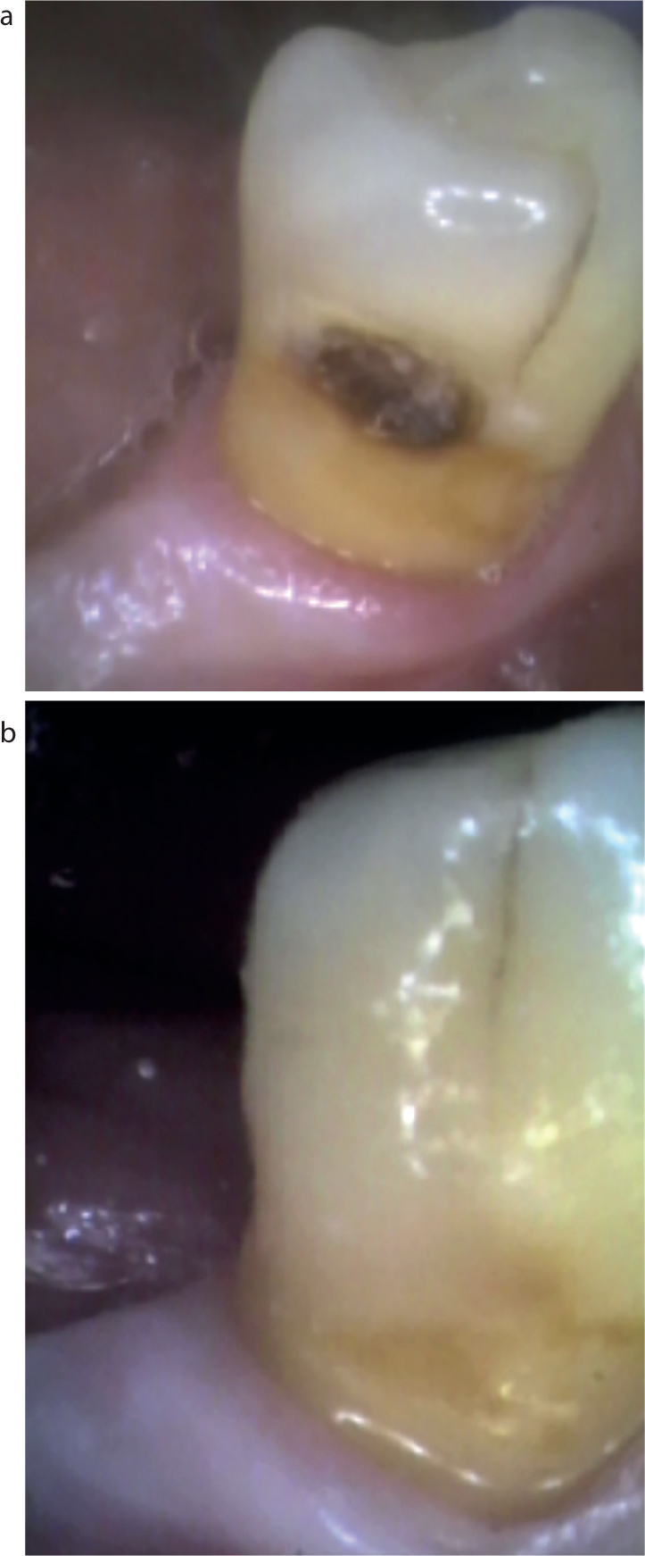
Figure 1.
(a, b) LL5 Class V supra-gingival RCL involving enamel and dentine. Rotary instruments and excavators used; dentine conditioned with 20% polyacrylic acid; restored with glass ionomer and layered composite.
Use rotary instruments and excavators during caries removal;
Condition dentine with polyacrylic acid;
Control moisture;
Use RMGI rather than GIC when possible.
Restorative material
In a systematic review of the literature, different restorative treatments of RCLs were compared. Failure rates were found to be high across all restorative materials and, at present, it was found that there is no gold standard restorative material.32 However, only five clinical trials were identified and these were confined to post-radiation, xerostomic patients, and may be atypical of the older adult dental patient.32 Nevertheless, it was concluded that there is insufficient evidence to recommend any specific material for routine use in the restoration of RCLs and highlighted the need for further research to guide dentists in the evaluation of restorative materials.32 This has implications on treatment outcome and efficacy, especially in the light of high failure rate of Class V restorations, which appears to be a common occurrence in practice.1 Restoration of RCLs can be challenging, especially as they may have dentine and enamel cavity margins, and because of the lack of restorative materials which bond equally well to both tissues.25 Deciding when and how to treat may improve operator skill in developing effective treatment protocols for the management of Class V root caries. There is a poor evidence base for the choice of restorative material for the treatment of RCLs1 restored with amalgam, glass ionomer cement(GIC), resin-modified glass ionomer cement (RMGIC), modified polyacid resins (‘compomers’) or composite resins, all of which have been considered in previous studies.33,34,35
Age-related changes in the structure of teeth may contribute to high restoration failure. Over time, dentine tubules decrease in diameter and number,36 with decrease in permeability,37 water content38 and altered mechanical properties.39 Consequently, clinicians need to modify their approach when restoring the teeth of older patients to provide longer lasting restorations.28
Glass ionomers
Glass ionomer cements (GICs) were first described in the dental literature by Wilson and Kent 1972.40 Since then the physical properties and potential uses of GICs have been extensively investigated and reported. McLean et al, in 1985, described GICs as a ‘replacement dentine’.41 They are self-adhesive to contaminated tooth structure and release fluoride into the restored tooth, thereby remineralizing and slowing caries progression.42,43 Chemical adhesion of GIC to the tooth tissues is the most important advantage of GICs.44 These well-established attributes should make GIC the material of choice for restoring RCLs. Unfortunately, early problems, such as high solubility, surface crazing and porosity, have not been overcome and restoration longevity is poor.28 Glass ionomer therefore requires careful handling to improve success. Even with contemporary GICs, there is a limited choice and shade matching can sometimes be a problem, resulting in poor aesthetics (Figure 2). RMGIs have been shown to give much better results.28
Figure 2.
(a, b) A 67-year-old patient with LL5 Class V root carious lesion involving enamel and dentine. Risk factors: gingival recession, drug-induced xerostomia, high sugar intake and use of removable partial dentures (RPDs). Active and non-active RCLs restored with GIC. Caries control: saliva substitutes, Duraphat toothpaste (1.1%), diet advice and oral hygiene instruction (OHI). Preparation: rotary instruments and excavators, 20% polyacrylic acid. Restoration: Fuji IX. Note internalized stain and extension onto sound root surface as a preventive measure.
The new systems are encapsulated and, when triturated, produce a homogeneous and consistent mix. They can be overlaid with composite, are water resistant, set quickly and can be finished in a wet field after initial set.45 Applying GIC in thin layers onto exposed root surfaces, as shown in Figure 2, is an effective preventive measure when the patient is at risk of developing root caries at these sites due to deteriorating health and dexterity.46 In addition to its use to protect sound roots, GIC can also be applied in thin layers over incipient lesions of root surface caries which have an intact leathery surface. Such lesions can remineralize over time, obviating the need for more involved restorative procedures.47 The inherent adhesion of these materials enables them to be used with relatively minimal cavity preparation.44 However, bonding to dentine is known to be a particular challenge because this tissue contains more water than enamel and less mineral phase for bonding.44 Whilst GICs appear to be relatively simple materials to use, they exhibit low fracture strength and low tensile strength, which results in a much lower effective bond strength than other adhesive materials.48 GIC demands careful handling and the high failure rate of GIC restorations reported in a recent study indicates that this is not fully appreciated by some clinicians.27
Therefore, to optimize bonding, the smear layer blocking the dentinal tubules on the cut dentine surface should be removed. This allows the GIC material to penetrate the surface with development of a micromechanical bond.49
Removal of the smear layer before placing RMGI and GIC is usually carried out through surface pre-treatment, or conditioning with 10% or 20% polyacrylic acid solutions applied for 20 seconds prior to rinsing.50 Conditioning partially demineralizes the dentine substrate and increases the surface area and exposure of micro-porosities, which allows micro-mechanical attachment.49
Resin composite
Restoring Class V root caries can be challenging, because many lesions are subgingival and usually located in dentine or cementum, factors that can affect bond strength and marginal adaptation. It may be possible with OHI to improve gingival health and convert subgingival lesions to more accessible supragingival lesions. Whilst resin composites may be considered to be the material of choice for the restoration of these cavities, when aesthetics is most important, inherent polymerization shrinkage is a major disadvantage that can result in microleakage at the tooth-restoration interface.51,52 The consequent restoration failure can be minimized by good isolation, which is frequently difficult, and careful placement technique restoring in incremental multiple layers to reduce the risk of polymerization shrinkage. Figures 3 and 4 show approaches to restoring subgingival root caries.
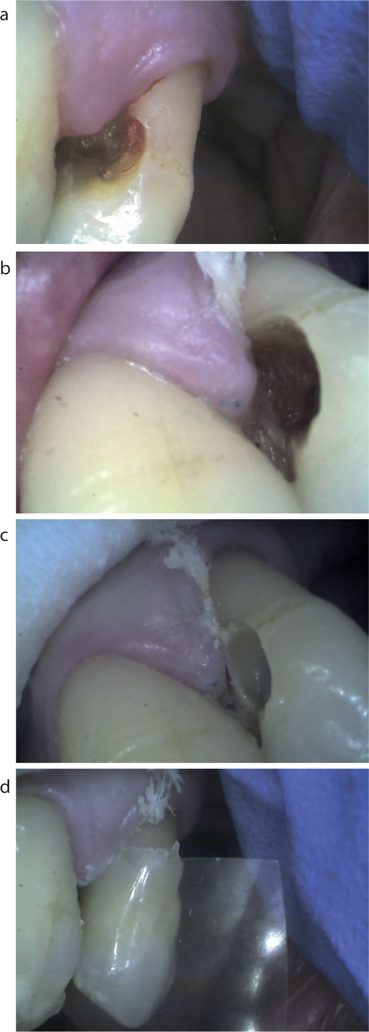
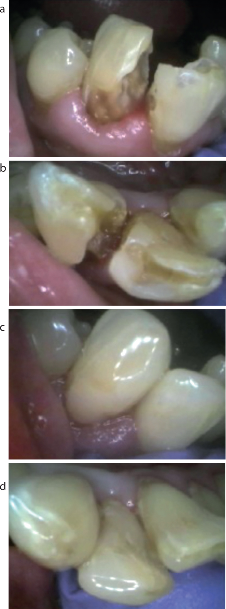
Figure 3.
(a) Direct access to the root caries in UL4 (b) during caries removal with retraction cord in place. (c) Good tissue retraction and moisture control; clear subgingival margins; flowable composite placed over GIC base. (d) Bulk composite placed with celluloid matrix strip in place; minimal finishing needed.Figure 4.
(a, b) A 72-year-old patient with Class V RCL in UR2. Sub- and supra-gingival lesion enamel and dentine. Risk factors: drug-induced xerostomia, high sugar intake, smoking, poor interdental cleaning. Restoration: GIC and composite. Note good moisture control and no signs of tissue trauma. Caries control: OHI, diet advice, Duraphat toothpaste (1.1%) and saliva substitutes.
Flowable composite resins (FCRs) can be used for the restoration of a Class V RCL, involving enamel and dentine, to facilitate adaptation due to their low viscosity.1 Moreover, FCRs have often been advocated for Class V restorations of RCLs because of their lower modulus of elasticity than hybrid resin composites.53 The results of an in vitro study indicated that FCR and dentine adhesive may be suitable for restoring advanced root caries when the proximal carious lesion is large, is wrapped around the root surface and is exposed to flexural load.53 An improved flow is likely to facilitate adaptation, and the reduced elastic modulus may provide the material with stress absorbing ability, demonstrated mostly in non-carious lesions but, nevertheless, applicable to restoration of root caries.54,55,56,57 Restorations in close proximity to gingival tissues require surface smoothness for optimal gingival health. As with all other restorative materials, special attention is needed when using FCR flowable composites to create smooth surfaces in order to avoid plaque retention, surface discoloration and gingival inflammation.58,59 Finishing and polishing of cervical restorations are important procedures that enhance aesthetics, gingival health, periodontal integrity and longevity of restorations and restored teeth. However, achieving an optimal finish can be problematic, especially when restorations extend subgingivally. The smoothest surfaces are achieved when resin material is cured against a cellulose strip or cervical former. However, their use is not always possible and, therefore, properly contoured restorations are seldom achieved without the need to remove excess material.60,61,62 Consequently, finishing and polishing Class V restorations carries a high risk of iatrogenic damage to the soft and hard tissues. In some cases, it may be prudent to recall the patient to ensure finishing is optimal and reinforce oral hygiene measures.
Biodentine
In a 2016 review, Hayes evaluated biodentine, a quick-setting, calcium silicate-based cement which was developed in 2011 as a dentine replacement material.63 As RCLs are often confined to dentine and biodentine produces mineral tags in dentinal tubules, it has the potential to offer high microleakage resistance. As many of the RCLs were typically found to be broad and shallow, conservative preparation resulted in restorations with a thin cross-section, which may have contributed to the high failure rate. The risk of failure was found to be higher when placed in a cavity that was in close proximity to the gingival margin within 1 mm or that extended subgingivally. The subsequent poor moisture control may be detrimental to biodentine restorations, which demonstrate a high level of washout.64 Furthermore, the caries-affected dentine substrate was thought to affect the bond strength and restoration success. The poor aesthetics of biodentine can be another disadvantage. It was therefore concluded, based on this limited evidence, that biodentine cannot be recommended for restoration of RCLs and Fuji products continue to be the best options for operative treatment of root caries.63
Minimally invasive treatment
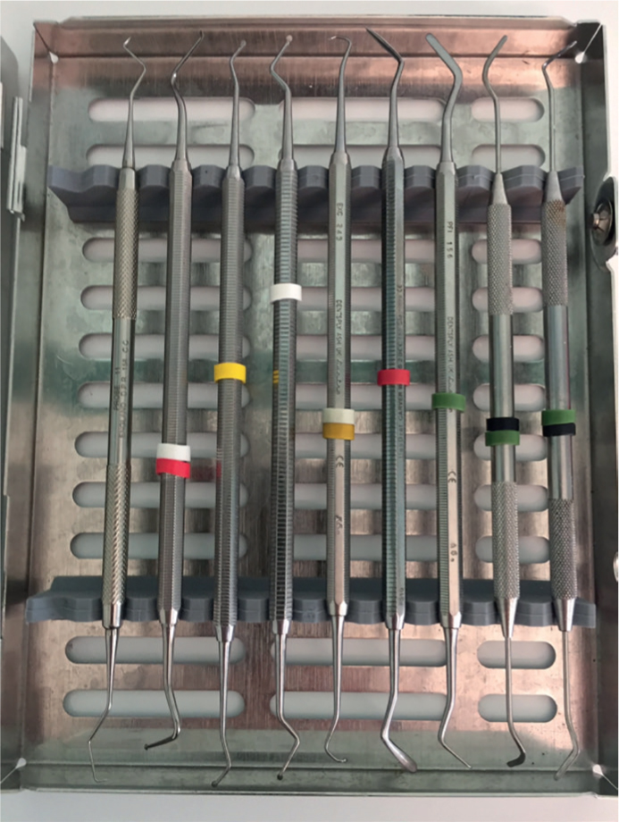
Minimally invasive dentistry is a philosophy of healthcare provision and can be defined as ‘the maintenance and monitoring of oral health through continuous care, comprising comprehensive preventive management, a longitudinal approach to risk assessment and diagnosis of chronic disease with a minimally interventive approach to any necessary operative intervention’.43 Minimally invasive treatment should be at the forefront of management of all patients, including the elderly, as decisions to drill and fill RCLs may not always be the best options when less invasive, non-operative management aimed at improving oral hygiene and remineralization of incipient lesions may be more appropriate, less challenging and more cost-effective. However, even when the decision to restore RCLs has been made, the clinician can often be faced with a dilemma as the clinical guidelines used by dentists in caries removal are based on subjective criteria that can vary from one operator to the next and, if inadequate, can lead to early restoration failures.65 The use of rotary instruments to remove carious tissue has often resulted in considerable removal of tooth structure and extensive tissue loss, which is now considered to be overly destructive.65 Ideally, only caries infected dentine should be removed as a principle of modern minimally invasive caries removal.66 Deciding on how best to remove carious dentine and, in fact what constitutes infected dentine, has to be determined to ensure effective treatment protocols. Undoubtedly, tactile appreciation with various patterns of hand excavators (Figure 5) should optimize treatment aims and outcomes to include removal of infected dentine and create a sound substrate to which restorations can be bonded.
Figure 5. Hand instruments with a variety of excavators and flat plastics used when restoring root carious lesions.
Atraumatic restorative treatment
Alternative approaches in the management of root caries can range from conventional removal with rotary instruments and restoration to the use of fluoride toothpastes aimed at remineralizing RCLs. Atraumatic restorative treatment (ART) can be used for root carious management when it may not be feasible to use rotary instruments in some older individuals who may be cognitively or physically impaired and unable to visit a dental practice. ART involves removal of softened tooth tissue with hand instruments followed by sealing of the partially demineralized dentine defect with GIC.67 Fure et al conducted a study to evaluate the clinical efficacy, treatment time and patient perception of Carisolv, a chemo-mechanical method of primary caries removal that could be used in ART, that was compared to conventional use of rotary instruments.68 The Carisolv group of patients were treated without anaesthesia and none experienced any pain during caries removal. There were no complications or adverse effects found in follow-up a year later. In conclusion, whilst Carisolv was found to be an effective method of root caries removal, it entailed a longer duration of treatment.68 This technique is advantageous insofar as only the softened tissue is removed, the cavity is prepared according to the shape of the lesion, anaesthesia is normally unnecessary, expensive equipment is not required, it has a low cost and reduced patient anxiety.69 However, the failure rate was found to be higher in restorations in close proximity to the gingival margin, within 1 mm or those that extended sub-gingivally. Many of the RCLs are shallow and it has been confirmed that caries-affected dentine as the bonding substrate could affect bond strength.65 So two questions remain unanswered:
Is the bond strength of restoration affected in such circumstances?;
When restorations are deemed to fail through secondary caries is this failure in fact due to residual caries that was left?
Proximal RCLs
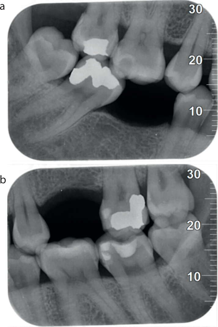
Banting and Courtright have pointed out that proximal lesions start as small circular lesions at the cemento-enamel junction and then spread laterally onto facial and lingual surfaces.5 When proximal root caries develops in this way, extensive sound tooth tissue often has to be removed to gain access following a conventional Class II cavity. Figures 6 and 7 show cases in which tunnel restorations (TRs) were contra-indicated and conventional access cavities were prepared.
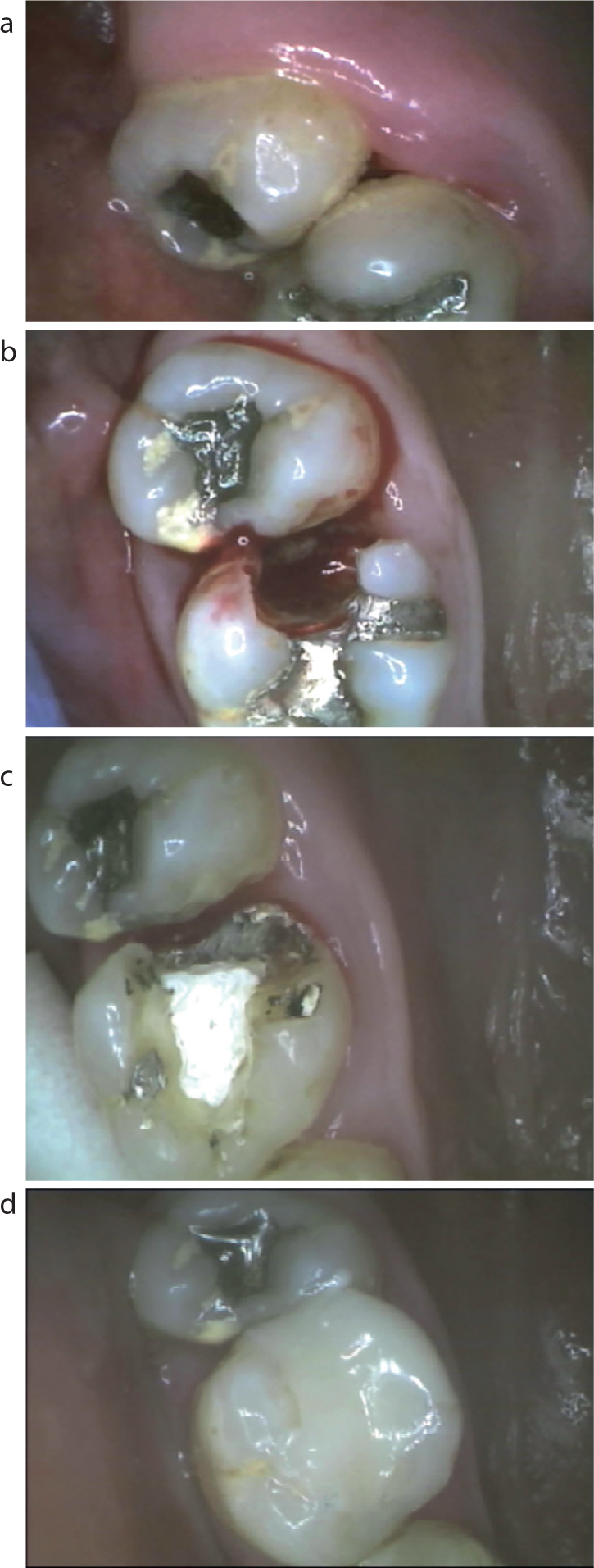
Figure 6.
(a−d) A 68-year-old patient with extensive distal RC in UL6. Risk factors: drug-induced xerostomia; high sugar intake; high plaque index; smoking; poor interdental cleaning. Class II cavity unsuitable for tunnel restoration (TR); gingival tissue hyperaemia. Preparation: rotary instruments and excavators, electrosurgery used to remove hyperplastic gingiva and control hyperaemia. Dressing placed. Definitive restoration: air abrasion GIC/flowable/bulk composite.Figure 7.
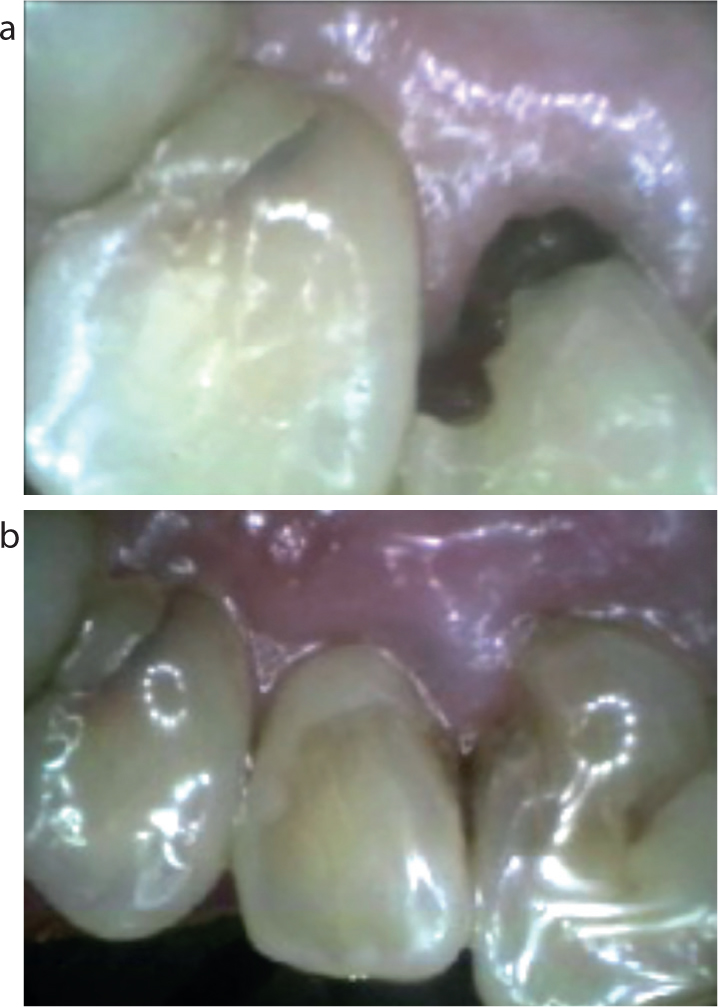
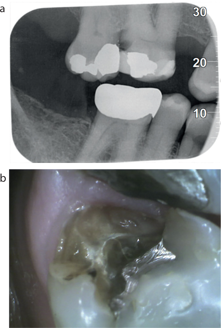
(a) Bitewing radiograph showing Class II distal root caries in UR6 in an 86-year-old patient (b) during treatment by direct access.
Figures 8 and 9 illustrate use of amalgam and composite in TRs.
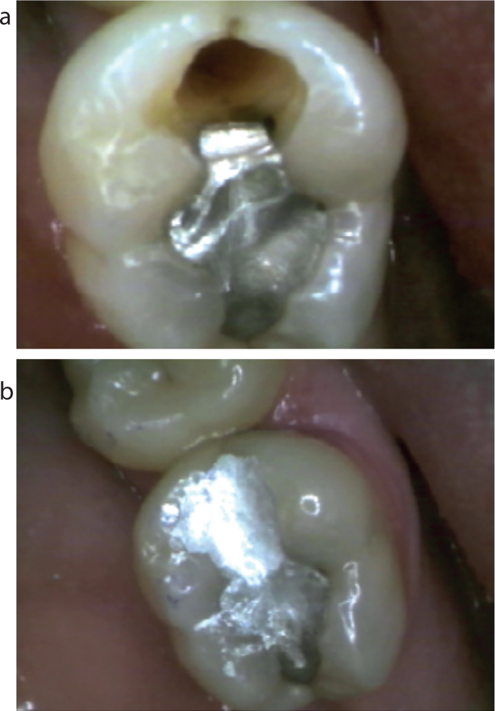
Figure 8. A 63-year-old patient with LL6 distal root caries (a) during caries removal (b) immediately after amalgam placement. Risk factors: xerostomia, high sugar intake, poor interdental cleaning. Tunnel restoration. Rotary instruments and excavators. GIC/amalgam Duraphat 1.1%. Note non-carious ditched amalgam that was subsequently repaired.Figure 9.
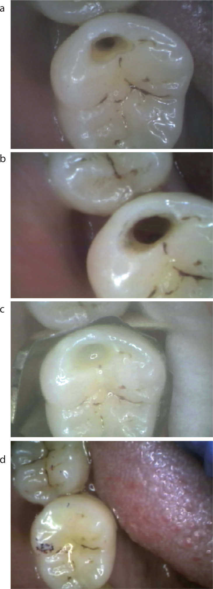
(a, b) A 63-year-old patient with LR6 distal root caries. Risk factors: drug-induced xerostomia; high sugar intake; poor interdental cleaning. Tunnel restoration. Rotary instruments and excavators. GIC/flowable/bulk resin composite Rx OHI, diet advice, Duraphat toothpaste 1.1% and saliva substitutes. (c) Wedged matrix band in place, composite layered on GIC base. (d) Finished TR.
However, there are cases when a minimally invasive cavity preparation, such as the more conservative tunnel restoration (TR) can be used. The TR was first described in 1963 for the restoration of distal proximal surfaces of deciduous second molars70 and was further developed in the 1980s using glass ionomers.71,72 GIC attributes of fluoride release and adhesion to enamel and dentine, as already discussed, are well known and should improve success, provided correct protocols are followed. Proximal carious lesions were accessed and prepared by approaching the lesion from the occlusal aspect, using rotary and hand instruments beneath the marginal ridge, but leaving it intact. Preserving the marginal ridge is the main advantage of the TR over the conventional box preparation.73 However, the TR can be challenging and difficult to prepare, particularly for inexperienced clinicians, often necessitating additional training. Success can therefore be operator-dependent. The marginal ridge can be undermined, resulting in fracture. Due to the limited access, recurrent caries caused by inadequate caries removal can develop and remains a key concern of this technique, as well as inadequacy in filling the prepared cavity.73 Therefore, proper case selection, having the appropriate armamentarium, including magnifying loupes and attention to detail, are imperative to improve success when restoring proximal root caries (Figure 10).
Figure 10.
(a, b) Bitewing radiographs showing amalgam tunnel restorations LR7 and UL7 placed to treat root caries 7 years earlier. Note mesial caries in UL8.
Whilst the 2-year clinical success rate of composite TRs was 96%, there was no significant difference with that of conventional composite box restorations.74 The most common cause of failure of TRs has been reported to be caries found clinically or radiographically adjacent to the TR.75 It was also demonstrated that the only factor significantly associated with failure of TRs was tooth type, with molars five times more likely to fail than premolars. In summary, problems associated with TRs include:75
Undermining the marginal ridge which subsequently fractures;
Insufficient caries removal due to poor access and visibility;
Poor bond to dentine substrate;
Poor restoration seal.
Summary of points to consider when managing RCLs
Whenever possible use non-operative measures, aimed at preventing demineralization and promoting remineralization of active and non-active RCLs.
When to restore? – cavitated lesions with active caries, recurrent caries, when a patient has symptoms from RCLs, when effective plaque control is difficult or impossible, aesthetic concerns.
Access – direct/indirect/TR: correct armamentarium to include the use of magnifying loupes, long rose head burs and a range of excavators with different patterns, especially when restoring proximal root lesions.
Caries detector dyes – are controversial as they stain both infected and affected/demineralized dentine. The present evidence indicates that their use is not recommended.
Caries removal can be subjective – aims: to remove all infected dentine and create a good dentine substrate to optimize restoration bond strength. Tactile feedback is an important factor when using excavators in caries removal. The use of rotary instruments to remove caries contributes to treatment success. Carisolv may be useful when using ART.
Moisture and tissue control: use of rubber dam is not often possible when restoring a subgingival RCL. The use of retraction cord facilitates accurate marginal integrity of restorations placed subgingivally.
Electrosurgery may be useful when restoring subgingival lesions and controlling hyperaemic tissue. Consider interim dressing if conditions for definitive restoration are unfavourable.
Restorative material – GICs give poor results whilst attributes of RMGIs are more favourable. However, incorrect handling of these materials can result in a higher failure rate.
Substrate preparation – orthophosphoric acid for lesions involving enamel or 20% polyacrylic acid tooth conditioner for dentine lesions is recommended.
Use of matrix bands – the best Class V restoration finish is against cellulose matrix strips or metal or metal/cervical formers to minimize ledge and void formation and iatrogenic tissue damage during finishing.
Conclusion
The decision on when and how best to restore active, cavitated root caries, once diagnosed, is fundamental to efficacy. Management of RCLs should, when possible, be non-operative. As discussed in Part 1, being aware of risk factors and educating patients on the importance of reducing the frequency of sugar intake, effective oral and denture hygiene, especially interdental cleaning and the use of high fluoride toothpastes and saliva substitutes, when appropriate, will help to reduce the restorative needs of the elderly which, as illustrated in Figure 11, can be extremely complex. There are a number of factors that can affect restorative treatment success and careful consideration of treatment protocols will improve outcome. Whilst the restorative materials available, especially the RMGIs, have many favourable attributes, further developments are needed to reduce failure. Caries removal is subjective and knowing how to create an effective substrate to ensure good bond strength under difficult conditions of access and moisture control are just some of the challenges clinicians face in restoring RCLs. Frequently updated approaches of the dental team to include teachers of restorative dentistry will be needed to meet future challenges in root caries management of the elderly.
Figure 11.
(a, b) A 68-year-old patient. Risk factors: drug-induced xerostomia, high sugar intake, poor oral hygiene and crowding. Combined extensive coronal and root carious lesions LR2 and LR3 involving sub- and supra-gingival tooth tissue. Tx Rotary burs, excavators and air abrasion, GIC base and composites. Note: good moisture control and absence of tissue trauma. Caries control: OHI, diet advice, Duraphat toothpaste (1.1%), saliva substitutes and 4-month recall. (c, d) Completed restorations.