The Immune System: Basis of so much Health and Disease: 9. Control of Inflammation and Immunity

From Volume 45, Issue 1, January 2018 | Pages 51-56

Authors

Crispian Scully

CBE, DSc, DChD, DMed (HC), Dhc(multi), MD, PhD, PhD (HC), FMedSci, MDS, MRCS, BSc, FDS RCS, FDS RCPS, FFD RCSI, FDS RCSEd, FRCPath, FHEA

Bristol Dental Hospital, Lower Maudlin Street, Bristol BS1 2LY, UK

Articles by Crispian Scully

Eleni A Georgakopoulou

PhD, MD, MSc, DDS

Research Fellow, University of Athens and Dental Practitioner, 4 Fokaias Str, 14232 N Ionia, Greece

Articles by Eleni A Georgakopoulou

Yazan Hassona

BDS, FFD RCSI, PhD

Assistant Professor and Consultant in Oral Medicine and Special Needs Dentistry, The University of Jordan, Amman

Articles by Yazan Hassona

Abstract

The immune system is the body's primary defence mechanism against infections, and disturbances in the system can cause disease if the system fails in defence functions (in immunocompromised people), or if the activity is detrimental to the host (as in auto-immune and auto-inflammatory states). A healthy immune system is also essential to normal health of dental and oral tissues. This series presents the basics for the understanding of the immune system, this article covering control of immunity and inflammation.

Clinical Relevance: Modern dental clinicians need a basic understanding of the immune system as it underlies health and disease.

Article

Control of inflammation

The cardinal features of inflammation are:

  • Redness (rubor);
  • Swelling (tumour);
  • Pain (dolor);
  • Heat (calor);
  • Altered function (functio laesa).
  • Mediators of inflammation are shown in Tables 1 and 2 and involve the following:

  • Inflammation in most instances begins to resolve within the first few hours after it begins;
  • Inflammation is perpetuated and amplified in an ongoing imbalance between pro- and anti-inflammatory factors – the latter also are mainly cytokines;
  • Anti-inflammatory cytokines include the interleukins and lymphokines, and cell signal molecules such as TNF, and the interferons, which regulate the immune response intensity or duration by modulating immunocyte proliferation, or their secretion of other cytokines or antibodies;
  • Inflammation control is not a mere passive regression of pro-inflammatory products but is also an active process which includes:
  • Granulocytes, which inhibit proinflammatory molecules such as leukotrienes;
  • Prostaglandins and leukotriene production from arachidonic acid switches to lipoxins, which initiate a sequence leading to resolution of inflammation;
  • Lipo-oxygenases (LO) in mucosae, leukocytes and platelets convert arachidonic acid to anti-inflammatory lipoxin LXA4 which inhibits chemotaxis, superoxide production and release of pro-inflammatory cytokines, and modulates macrophages to develop anti-inflammatory actions;
  • Apoptosis (programmed cell death) is initiated by the synthesis from omega-3 polyunsaturated fatty acids (PUFA), of resolvins and protectins, which critically shortens the period of neutrophil infiltration. Omega 3 polyunsaturated fatty acids give rise to lipid mediators which inhibit the conversion of arachidonic acid to pro-inflammatory eicosanoids, and include:
  • Alpha linolenic acid (ALA)
  • Eicosapentaenoic acid (EPA) gives rise mainly to resolvins (resolution phase interactive products), which act mainly on phagocytes to inhibit production and release of inflammatory mediators;
  • Docosahexaenoic acid (DHA) gives rise to protectins.
  • Apoptotic neutrophils are removed by macrophage phagocytosis;
  • Macrophages release anti-inflammatory and reparative cytokines such as transforming growth factor TGF-β1, IL-10, IL-13, and IL-1ra and then leave the area via lymphatics;
  • Neutrophil recruitment gradually subsides and ceases;
  • The inflamed area returns to normal.
  • Inflammation, if not resolving, may give rise to a number of untoward sequelae and may, if chronic, injure tissues;
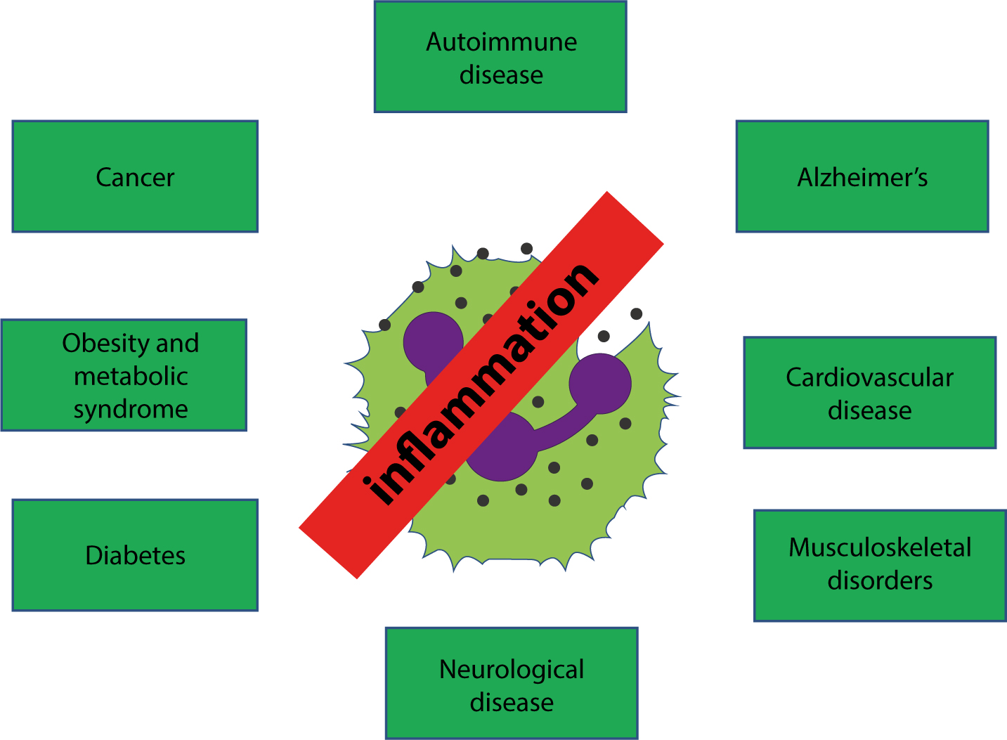
  • The control of inflammation is thus important not only for resolution of damage from trauma and infection but is involved in the pathogenesis of many systemic diseases including cancer, cardiovascular diseases, age-related degenerative conditions like Alzheimer's disease, metabolic diseases such as diabetes (Figure 1) and possibly periodontitis. Thus many of the molecules involved in inflammation (Table 3) are also mediators of systemic diseases.

  • Mediators Source Action
    Chemokines (eg CCL 1-28, CXC1-17) Endothelial cellsMonocytesLymphocytes Directs immune cells from blood into the affected tissue
    Inflammatory cytokines (eg interleukins and tumour necrosis factor TNF) MonocytesLymphocytes Directs immune cells from blood into the affected tissue
    Pain mediators (eg bradykinin) Damaged tissuesMonocytes/macrophages Vasodilation increase the permeability of capillaries
    Clotting mediators (eg fibrin peptides) Damaged tissue Mediates clotting process
    Complement components (eg C5a, C3A, C4a) Monocytes macrophagesCascade-activated Attack and destroy and opsonize bacteria

    Vasodilation Immediate action Delayed action Chemotaxis Opsonins Pain
    Histamine * *** _ _ _ _
    Serotonin (5 hydroxy tryptamine) * * _ _ _ _
    Bradykinin * * _ _ _ ***
    Complement C3a _ * _ _ _ _
    C3b _ _ _ _ *** _
    C5a _ * _ *** _ _
    Prostaglandins *** * * *** _ _
    Leukotrienes _ *** * *** _ _
    Lysosomal proteases _ _ ** _ _ _
    Oxygen free radicals _ _ ** _ _ _

    Key:

    mild mediator;

    moderate mediator;

    important mediator.


    Acronym Full meaning
    AP-1 Activator Protein 1
    AA Arachidonic Acid
    CARD15 Caspase Recruitment Domain Family, Member 15 (also known as NOD2)
    CRP C Reactive Protein
    ESR Erythrocyte Sedimentation Rate
    Fas Fast antigen (also termed APO-1, APT1 and CD95, and now known as tumour necrosis factor receptor superfamily 6 [TNFRSF6])
    FADD Fas Associated Death Domain
    Fas L Fas Ligand
    IFN Interferon-γ
    IL Interleukin
    JNK Jun-n-terminal Kinase
    LPS Lipopolysaccharide
    MAPK Mitogen-Activated Protein Kinase
    NFκB Nuclear Factor-κB
    NOD2 Nucleotide-Binding Oligomerization Domain Containing 2 (also known as the caspase recruitment domain family, member 15; CARD15)
    NSAID Non-Steroidal Anti-inflammatory Drugs
    PAMP Pathogen Associated Molecular Pattern
    PG Prostaglandin
    ROS Reactive Oxygen Species (chemically reactive molecules containing oxygen, such as peroxides, superoxide, hydroxyl radical and singlet oxygen)
    SAA Serum Amyloid A protein
    TNF Tumour Necrosis Factor-α
    TNFR TNF Receptor
    TNFRI TNF Receptor Type I (also termed p55 or CD120a)
    TNFRII TNF Receptor Type II
    TNFRSF TNF Receptor Super Families
    TNFSF1A The gene encoding TNF Receptor Type I
    TRADD TNF Receptor Type 1-Associated DEATH Domain protein
    TRAF TNF Receptor-Associated Factor
    Figure 1. Inflammation is involved in the pathogenesis of many diseases.

    Control of the immune system

    Antigen recognition can be influenced by:

  • Dose and route of antigen exposure;
  • Absence of co-stimulation;
  • Engagement of B7 on activated antigen-presenting cells (APC);
  • Presence of antibodies;
  • Presence of immune complexes;
  • Some MHC types.
  • T-cell activation is influenced by cytokines:

  • Th1 cells and Th2 cells respond to different sets of chemokines;
  • CD4 T-cells can deviate immune responses to Th1 or Th2 type helper T cell responses;
  • Selective migration of lymphocyte subsets to different sites can modulate the local type of immune response.
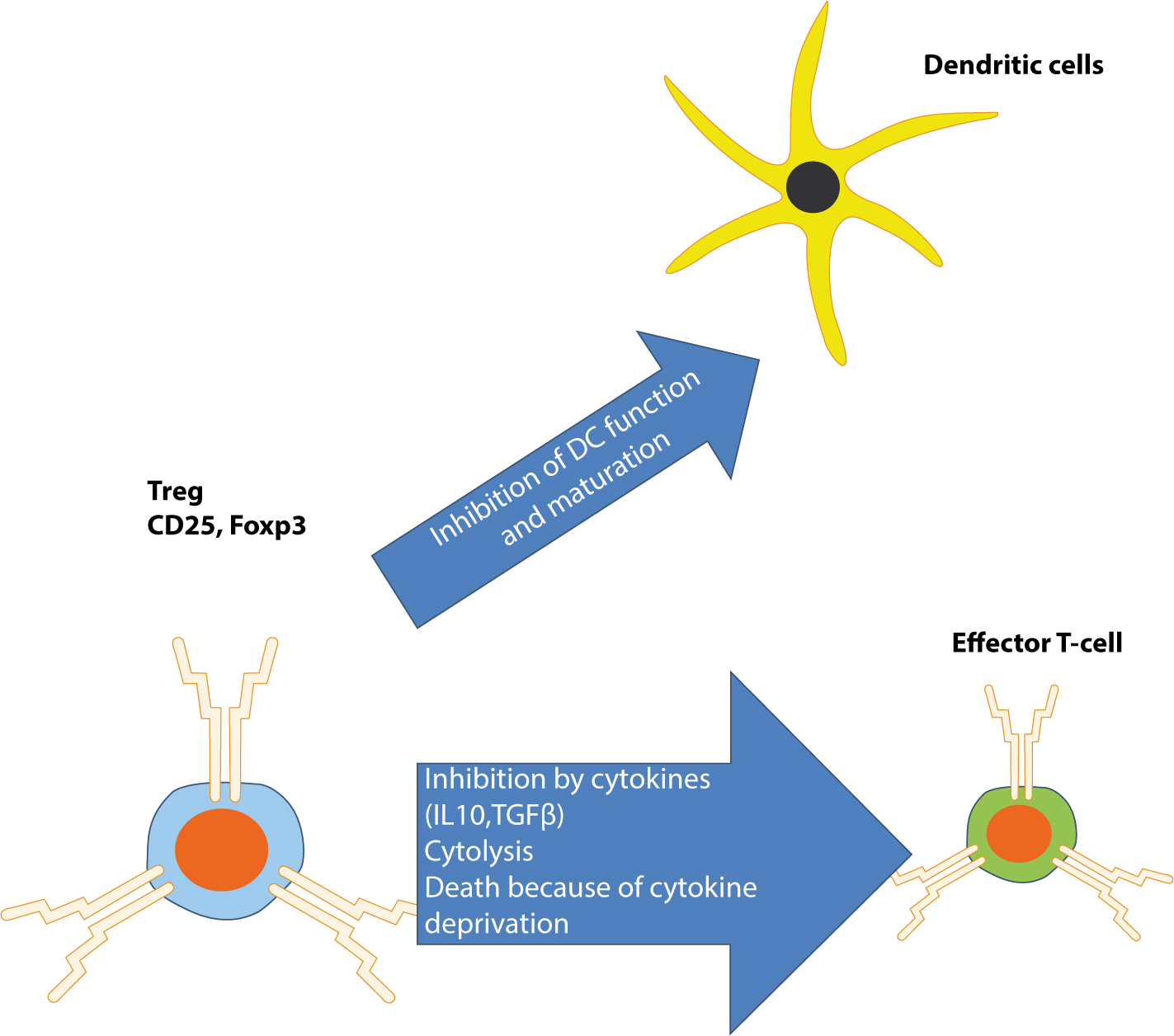
  • APCs produce CCL4 (chemokine [C-C motif ] ligand 4), a chemokine which attracts regulatory T cells (Tregs) to the area:

  • Tregs are usually CD4 T cells (CD25+, CD4+) which limit and suppress the immune system (and may also control aberrant immune responses to self-antigens);
  • Tregs can inhibit immune responses by producing suppressive cytokines, eg interleukin 10 (IL-10) and transforming growth factor-P (TGFP)
  • Tregs are under the control of a specific transcription factor (FoxP3). Figure 2 shows the effects of Tregs on T-cells and dendritic cells.
  • Figure 2. The regulatory effects of T-regs.

    Cytokine release becomes suppressed; for example, IL-1β and TNF-alpha fall under the control of the AUF1 (AU-rich element RNA-binding protein gene) also known as heterogeneous nuclear ribonucleoprotein D (HNRNPD).

    Immunoglobulins can influence the immune response positively in several ways, as anti-idiotypes or through immune complex formation or negatively by reducing antigenic challenge or by feedback inhibition of B-cells.

    Genetic factors which influence the immune system include both MHC-linked and non-MHC-linked genes.

    Tolerance mechanisms are needed because the immune system randomly generates a vast diversity of antigen-specific receptors and some of these will be self-reactive. Self-reactive T-cells share the following features:

  • May ignore self antigens, for example when antigens are in tissues sequestered from the circulation;
  • Response to a self antigen may be suppressed if the antigen is present in a privileged site;
  • May, under certain conditions, be deleted or rendered anergic and unable to respond;
  • May be maintained by immune regulation in a state of tolerance to self antigens;
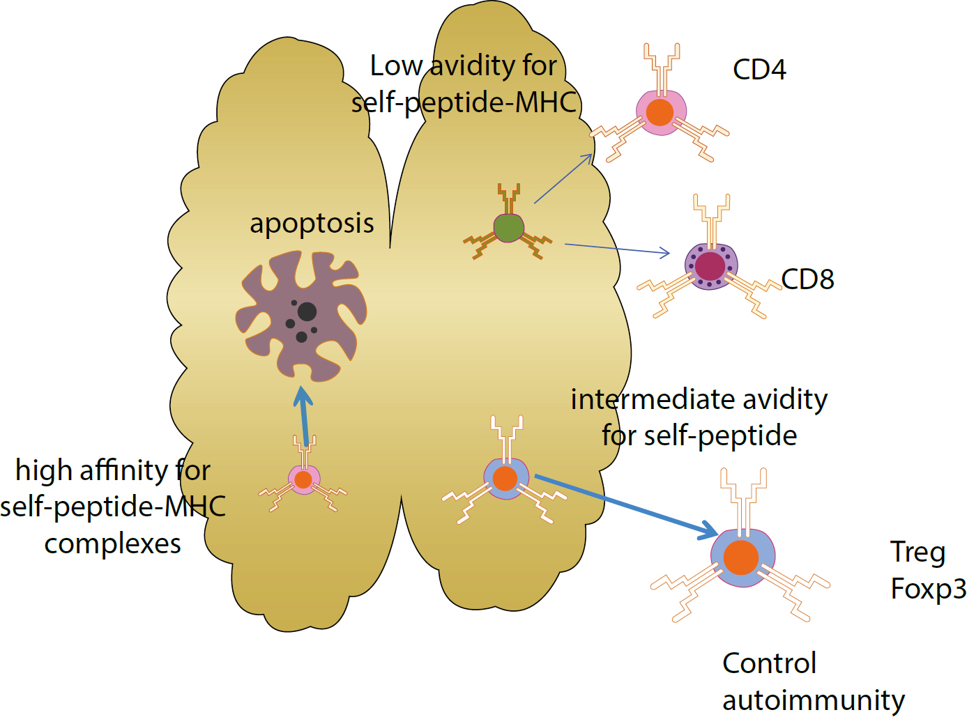
  • Central thymic tolerance to self antigens (auto-antigens) results from the deletion of differentiating T-cells that express antigen-specific receptors and thereby have high binding affinity for intra-thymic self antigens;
  • Low-affinity self-reactive T-cells, and T-cells with receptors specific for antigens that are not represented intra-thymically, mature and join the peripheral T-cell pool.
  • The fates of T-cells, as derived from thymocytes, are depicted in Figure 3.

    Figure 3. Thymic regulation of auto-reactive T-cell control.

    B-cell deletion takes place in both bone marrow and peripheral lymphoid organs. Differentiating B-cells that express surface immunoglobulin receptors with high binding affinity for self-membrane-bound antigens will be deleted soon after their generation in the bone marrow. A high proportion of short-lived, low-avidity, auto-reactive B-cells appear in peripheral lymphoid organs, and may be recruited to fight infection.

    Immune system responsiveness

    Immune system responsiveness is affected by the following:

  • Enhanced by sleep and rest;
  • Enhanced by a diet rich in fresh fruits and vegetables, and by polyunsaturated fatty acids;
  • Impaired by stress;
  • Modulated by:
  • Female sex hormones stimulate innate and acquired immune responses;
  • Male sex hormones suppress the immune system;
  • Vitamin D (a hormone) may suppress the immune system;
  • The neuroendocrines prolactin and growth hormone can modulate immune responses;
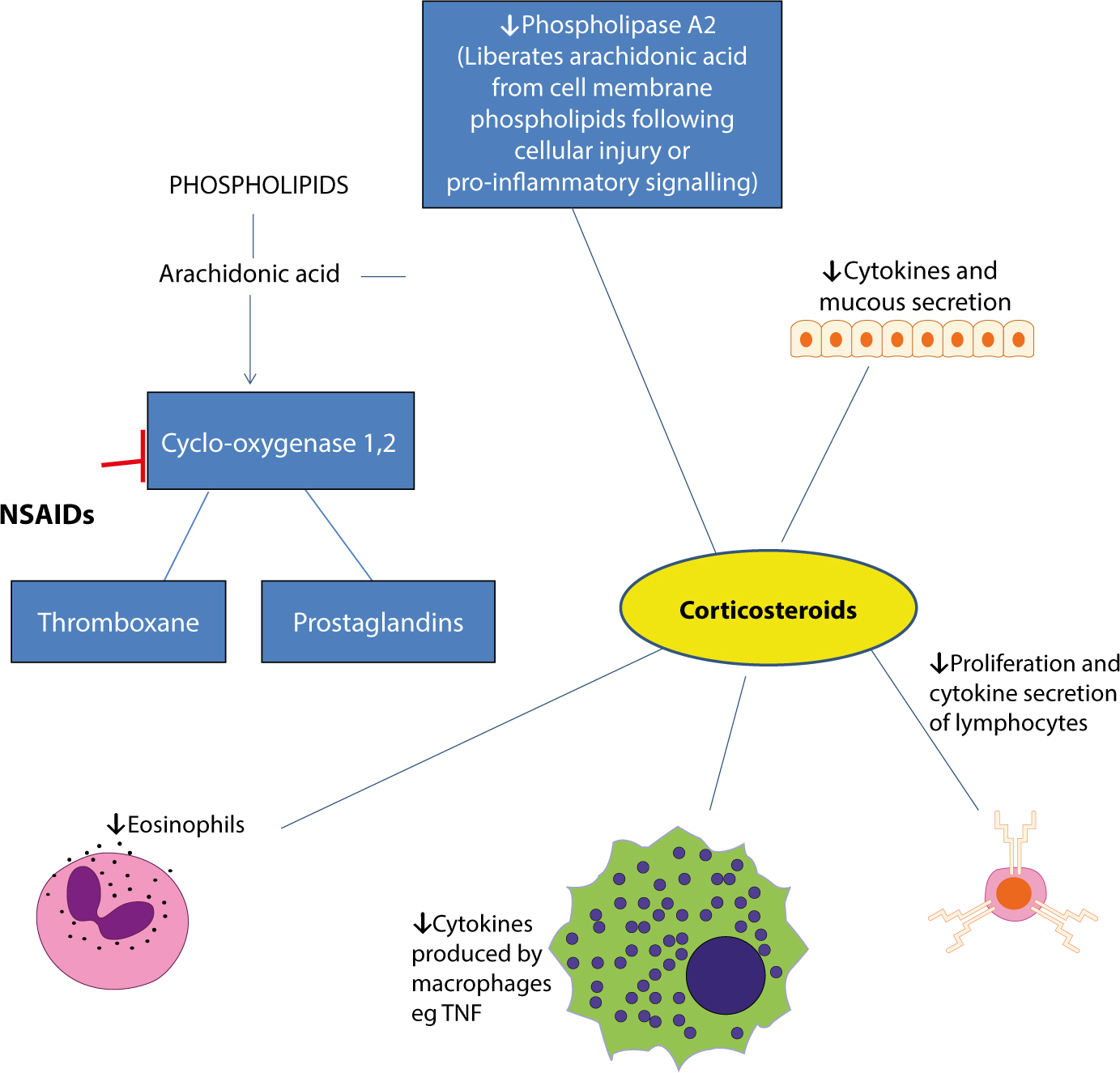
  • Corticosteroids in particular downregulate immune responses and macrophage activation;
  • Prostaglandins (PG) and thromboxanes (TX) belong to the group of biologically active lipid-termed eicosanoids synthesized by most nucleated cells. PGs and TXs are mediators of pain, and inflammation – by vasodilatation and increased vascular permeability. They are inhibited by corticosteroids and non-steroidal anti-inflammatory drugs (NSAIDs).
  • The main actions of corticosteroids and NSAIDs are depicted in Figure 4.

    Figure 4. The main actions of corticosteroids and NSAIDs.

    Conclusion

    Inflammation is beneficial to the organism as it consists of a primary barrier against external and internal detrimental processes. Failure of the mechanism that controls the process of inflammation may result in chronic inflammatory conditions. Uncontrollable inflammation is also involved in the pathogenesis of degenerative diseases such as cancer, diabetes, autoimmune and cardiovascular diseases.