Kalan A, Tariq M. Foreign bodies in the nasal cavities: a comprehensive review of the aetiology, diagnostic pointers, and therapeutic measures. Postgrad Med J. 2000; 76:(898)484-487
Ranalli DN, McWilliams BJ, Garrett WS Tooth and foreign object in the nasal fossa of a child with a cleft: case report. Pediatr Dent. 1990; 12:183-184
Tolhurst DE. The ubiquitous foreign body. Cleft Palate J. 1974; 11:237-239
Hussain K, Brown AJ, Chavda D. Case report: foreign body in the palate of an infant. Br Dent J. 2008; 205:23-25
Soubhia AM, Ribeiro AC, Martins LD, Silva AR, Lopes MA. Unusual wooden foreign body in the palate. Br Dent J. 2007; 203:573-574
de Jong AL, Moola F, Kramer D, Forte V. Foreign bodies of the hard palate. Int J Pediatr Otorhinolaryngol. 1998; 43:27-31
Deniz Y, Zengin AZ, Karli R. An unusual foreign body in the maxillary sinus: dental impression material. Niger J Clin Pract. 2016; 19:298-300
Jones SD, Drake DJ. Case series of undetected intranasal impression material in patients with clefts. Br J Oral Maxillofac Surg. 2013; 51
Lownie JF, Lemmer J, Sykes L. Chronic maxillary sinusitis resulting from displacement of impression material into the maxillary antrum: a case report. J Dent Assoc S Afr. 1989; 44:341-342
Rodrigues MT, Munhoz ED, Cardoso CL, de Freitas CA, Damante JH. Chronic maxillary sinusitis associated with dental impression material. Med Oral Patol Oral Cir Bucal. 2009; 14:E163-E166
Wright RA, Bidra AS. Retrieval of residual alginate impression material from a small oral-nasal communication defect in a maxillectomy patient. J Prosthet Dent. 2015; 113:253-254
Owen M, Macansh J. Foreign body (impression material) in the maxillary antrum. Clin Radiol. 1965; 16:284-288
Rathee M. Single visit feeding appliance for 1-day-old neonate with cleft palate using safe dental putty-gauze hybrid impression technique for maxillary impression. J Surg Tech Case Rep. 2015; 7:7-11
Jacobson BN, Rosenstein SW. Cleft lip and palate: the orthodontist's youngest patient. Am J Orthod Dentofacial Orthop. 1986; 90:63-66
Grayson BH, Maull D. Naso alveolar moulding for infants born with clefts of the lip, alveolus and palate. Semin Plast Surg. 2005; 19:294-301
Robinson HE, Zerlin GK, Passy V. Maxillary sinus development in patients with cleft palates as compared to those with normal palates. Laryngoscope. 1982; 92:183-187
Hardwicke JT1, Landini G, Richard BM. Fistula incidence after primary cleft palate repair: a systematic review of the literature. Plast Reconstr Surg. 2014; 134:618e-627e
Ravikumar N, GunaShekhar M, Prasad SR, Lalitha N, Raju PR, Natesh YA. Unusual foreign body in the nasal cavity of an adult with repaired cleft lip and palate. Cleft Palate Craniofac J. 2015; 52:219-222
Chate RA. A report on the hazards encountered when taking neonatal cleft palate impressions (1983–1992). Br J Orthod. 1995; 22:299-307
Akay C, Karakis D, Yalug S. An alternative impression technique for an infant with cleft palate. Int Dent Res. 2015; 5:38-41
Alani A, Bishop K. Contemporary issues in the provision of restorative dentistry. Br Dent J. 2012; 213:163-170
Locke M, Bishop K. An assessment of the contribution of UK specialists in restorative dentistry to cleft lip and palate services. Br Dent J. 2011; 210
Fonseco RB, Branco CA, Haiter-Neto F Radiodensity evaluation of dental impression materials in comparison to tooth structures. J Appl Oral Sci. 2010; 18:467-476
What's left in the cleft? a rare complication following displacement of dental impression material into a palatal cleft Srishti Datta Abhishek Agarwal Dapo Akintola Aws Alani Dental Update 2024 44:10, 707-709.
Authors
SrishtiDatta
BDS(Hons), BSc(Hons), MFDS RCS(Ed)
Dental Core Trainee, Department of Oral Surgery, King's College Hospital, Denmark Hill, London SE5 9RS, UK
Discoveries of foreign bodies lodged in the nose, palate and maxillary sinuses have been well documented. A rare, iatrogenic cause is displacement of dental impression material which, if left undetected at these sites, may lead to acute respiratory obstruction or chronic problems, such as nasal discharge and chronic sinusitis. This article reports the case of acute complications following displacement of dental impression material into a palatal cleft, discusses immediate surgical management, and considers restorative techniques that should be adopted to prevent such complications in patients with cleft palates.
CPD/Clinical Relevance: Impression-taking in patients with cleft palate carries significant risks and appropriate referral to a multidisciplinary team is appropiate in order to avoid potentially life-threatening complications.
Article
The discovery of foreign bodies in the nose, palate and maxillary sinuses has been well reported in the literature with children under the age of five being most frequently affected.1,2,3,4,5,6,7 Embedded inanimate objects such as erasers, pebbles, beads and coins may simulate pathological lesions and this, coupled with the limited co-operation of children, can make diagnosis extremely challenging.4,5,6 Loose foreign objects in the postnasal space should raise particular concern due to the risk of accidental aspiration and acute respiratory obstruction, which is a life-threatening emergency.1 Chronic complications may include halitosis, nasal discomfort, discharge and sinusitis.7,8 Management of these patients and retrieval of lodged foreign objects therefore requires great skill.1,5,6
One major cause of foreign bodies arising from dental clinical practice is the retention of dental impression material in the oral, nasal or paranasal cavities7,8,9,10,11,12 and this is a significant risk in a particular cohort of patients with orofacial clefts.
Orofacial clefts
Oral clefts are one of the commonest congenital craniofacial malformations in humans and may involve the lip, hard palate or soft palate.13 Classification of oral clefts is based on the phenotype, extent (unilateral or bilateral) and associated features (syndromic or non-syndromic).14 Depending on the site and extent of the defect, patients with such anomalies undergo multiple surgical procedures and prosthetic rehabilitation from infancy to adulthood, under the care of a multidisciplinary team, in order to restore their anatomy, aesthetics and function.13,14
Newborns with abnormal oro-nasal communications are at immediate risk of airway obstruction due to lack of separation between the oral and nasal cavities, which results in regurgitation of fluids and food through the nose and inability to feed.15 These infants require urgent construction of a feeding appliance called an obturator, which relies on taking an accurate impression of the cleft and associated structures using dental impression materials.16 The presence of a surgeon during this process has been recommended to manage an airway emergency, should one arise.17
Patients with orofacial clefts require multiple impressions throughout their lifetime in order to ensure optimal fit of obturators for mastication and speech, as well as prevention of nasal regurgitation and associated chronic sinus infections.18 A systematic review reported an incidence of 8.6% of residual oro-nasal communication in the form of fistulae in patients after primary cleft palate closure.19 These fistulae may allow displacement of foreign bodies into the nasal cavity following dental impressions.8,20 Similarly, foreign bodies may displace into the maxillary sinuses in patients with oro-antral fistulae.7,9,10,12
Acute complications due to dislodged dental impression fragment(s) when taking impressions in patients with orofacial clefts may include:21
Respiratory obstruction;
Cyanotic events including asphyxiation and risk of fatality;
-Patient anxiety/frustration with unknown and delayed diagnosis.
There are three reported cases of nasal foreign bodies arising from dislodged dental impression material in adults with repaired cleft lip and palate but no current literature highlighting acute complications associated with taking a maxillary impression in such patients.8,20 This article reports the case of acute complications following displacement of dental impression material into a palatal cleft, discusses immediate surgical management, and considers restorative dentistry techniques that should be adopted in order to prevent such complications in patients with cleft palates.
Case report
A distressed 66-year-old man attended the dental emergency department complaining of unilateral nasal obstruction and difficulty breathing. He was referred by his dentist following an attempted dental impression for a new obturator earlier that day. He explained that his current obturator had worn away and reported that his symptoms began shortly after completion of the impression procedure. His medical history revealed that he had undergone corrective surgery for cleft lip and palate in his early childhood and that he was otherwise fit and well.
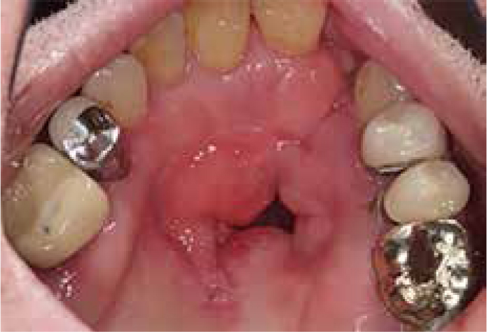
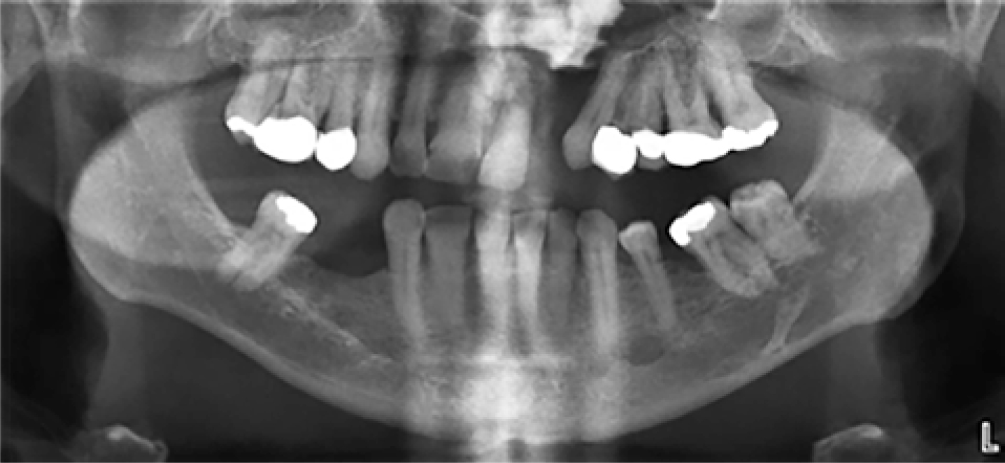
Clinical examination revealed evidence of repaired left unilateral cleft lip and palate with the presence of a mid-palatal fistula communicating with the nasal cavity (Figure 1). The contents of the nasal cavity could not be assessed clinically. Radiographic examination revealed the presence of a large, well-defined, radio-opaque lesion in the left nasal cavity (Figure 2), confirming suspicions of dislodged dental impression material.
Figure 1. Maxillary occlusal view revealed absent UL2, evidence of repaired left unilateral cleft palate and a mid-palatal fistula.Figure 2. DPT radiograph confirmed presence of radio-opaque mass in the left nasal cavity, suggestive of lodged foreign body.
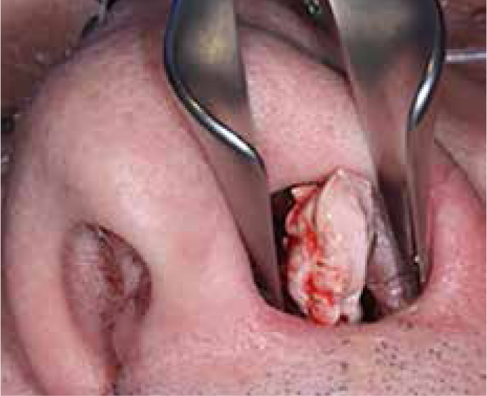
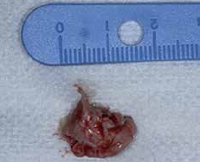
Topical local anaesthetic spray was used to anaesthetize the nasal mucosa and a nasal speculum was used to gain visual access (Figure 3). A large mass measuring 1.5 cm x 1 cm and several smaller fragments of zinc oxide eugenol impression material were carefully removed from the nasal cavity using a pair of college forceps (Figure 4). The patient reported immediate resolution of symptoms following the procedure and was subsequently referred to a specialist in the restorative dentistry department for construction of a new obturator.
Figure 3. A nasal speculum was used to dilate the left nostril to permit retrieval of the foreign body.Figure 4. A large mass measuring 1.5 cm x 1 cm of impression material was successfully removed from the patient's left nasal cavity.
Discussion
This case report highlights a rare occurrence of displacement of dental impression material into the nasal cavity in an adult patient with repaired cleft palate, which may have resulted in potentially life-threatening complications in the absence of appropriate surgical management. To the authors’ knowledge, there are no such reports of acute complications following impression-taking in adults with cleft palate. Chronic complications due to dislodged nasal foreign bodies (later confirmed as dental impression material) in adults have been reported in a few cases.8,20
It is important for clinicians to be aware of the risks posed by the impression-taking procedure in patients with orofacial clefts and to adopt strategies to maximize patient safety. Procedural complications that may be experienced include:8,15
Inadequate coverage of the defect;
Difficulty in withdrawal of the impression due to the presence of cleft undercuts that lock the material;
Distortion or tears in the impression, for instance due to the use of excessive force during withdrawal;
Dislodging of fragments into the nasal and/or paranasal spaces due to the presence of residual oro-nasal and/or oro-antral communication, respectively.
These complications may be avoided by adapting the impression-taking technique to include varying viscosities of the impression material for the cleft and surrounding maxillary sites (Figure 5). We recommend the use of a two-phase impression technique as described below, for a different patient from that described above:
Initial impression of the cleft defect (Figure 5c) – A low viscosity silicone-based impression material, such as silicone putty, is used to produce an impression of the defect. The low viscosity and increased tear strength of silicones reduces the risk of airway obstruction and overflow of material deep into the cleft, thereby preventing tear and displacement of the material into the nasal/paranasal cavities.22
Over impression of remaining anatomy (Figure 5d) – A further silicone material with higher viscosity is used to overlay the initial impression in order to gain greater detail of the fit surface. This produces excellent detail and enables multiple casts to be created from the same impression, if required.
Figure 5. The two-phase, two-stage, impression-taking technique recommended for patients with orofacial clefts. (a) Maxillary occlusal view revealed absent UR1, UR2 and UR3, evidence of repaired right unilateral cleft palate and a mid-palatal fistula. (b) Maxillary anterior view to highlight the labial extent of cleft defect. (c) Initial impression of cleft defect using silicone putty enables control of impression material. (d) Final impression (blue) made using two-phase putty-wash technique where initial impression of cleft is highlighted in yellow silicone. (e) Removable prosthesis fabricated to replace the UR1, UR2 and UR3 with good adaptation to cleft defect on the labial aspect.
The clinician must always check the impression to confirm its integrity and any suspicions of retained impression material must be voiced to the patient with the advice to seek urgent medical care should symptoms develop.8 Fragments of dislodged impression material may act as a nidus for calcification in the nasal and/or paranasal spaces, resulting in the formation of rhinoliths or antroliths, respectively.7,12 If left undetected, these may enlarge with time and cause chronic nasal discharge and sinusitis.10 Construction of obturators in such patients usually constitutes high treatment complexity due to the different factors involved and requires referral to a specialist restorative unit.
Patients with congenital defects, such as cleft lip and palate, are a priority for NHS funded treatment and present a plethora of challenges to the dental team, including maintenance of restorations, increased incidence of periodontal disease and structural tooth defects, lack of bone for implant-retained prostheses and possible failed surgical and orthodontic treatment.23 As these patients transition to adulthood (having likely received multidisciplinary care at specialist cleft centres), they may be unable to access the appropriate dental care because general dental practitioners have limited experience in treating such patients.24 Therefore, restorative specialist input retains a crucial place in cleft care and this patient cohort is best managed in a multidisciplinary team.23,24
A detailed history and radiographic examination are essential in order to aid diagnosis of dislodged foreign bodies and a degree of clinical suspicion is required when faced with patients complaining of chronic sinusitis and/or nasal symptoms.8 In the case presented above, the patient's history, acute presentation and positive radiographic findings enabled prompt diagnosis. However, diagnosis of dislodged foreign bodies can be extremely challenging in cases of delayed presentation (nearly 20 years since the precipitating event was reported in two cases) and inconclusive findings of investigations, such as nasendoscopy and CBCT radiographs due to the presence of anatomical and ghost shadows in the posterior and superior regions of the nasal cavity (where retained dental impression material is often located).8 The radiolucent appearance of several impression materials may further hinder detection on radiographs. This can be overcome by using impression materials with greater incorporation of radio-opaque chemical elements, such as zinc, strontium, zirconium, barium and lanthanum, to enable easier detection of retained fragments.25 Examination under anaesthesia (EUA) has been recommended in cases with no significant clinical and radiographic findings.8
It is important for all clinicians to recognize the risks and challenges involved in an albeit simple clinical procedure such as taking dental impressions in patients with orofacial clefts and the potential life-threatening complications in the case of airway obstruction. The complex and often multidisciplinary treatment needs of patients with orofacial clefts are therefore most appropriately managed via referral to specialists in secondary care.24