Holroyd J. Measurement of scattered and transmitted x-rays from intra-oral and panoramic dental x-ray equipment. J Radiol Prot. 2018; 38:793-806 https://doi.org/10.1088/1361-6498/aabce3
Ludlow JB, Davies-Ludlow LE, White SC. Patient risk related to common dental radiographic examinations: the impact of 2007 International Commission on Radiological Protection recommendations regarding dose calculation. J Am Dent Assoc. 2008; 139:1237-1243 https://doi.org/10.14219/jada.archive.2008.0339
Public Health England (PHE) and Faculty of General Dental Practice (UK) (FGDP(UK)) guidance notes for dental practitioners on the safe use of X-ray equipment 2020: an update for the dental team
Public Health England (PHE) and Faculty of General Dental Practice (UK) (FGDP(UK)) guidance notes for dental practitioners on the safe use of X-ray equipment 2020: an update for the dental team Mark Gribben Dental Update 2025 48:9, 719-724.
PHE and FGDP(UK) co-published Guidance Notes for Dental Practitioners on the Safe Use of X-ray Equipment in 2020 to encompass all relevant information to safely use and maintain radiographic equipment by dental practitioners. This guidance supersedes the first edition, previously published in 2001 by the Department of Health, and there are a number of notable changes that should be highlighted to the dental team. The document is freely available and accessible on the FGDP(UK) website and should be an essential tool to ensure the safety of the dental team, patients and the public.
CPD/Clinical Relevance: Members of the dental team must acknowledge the updated guidance notes and look to incorporate these changes into their working routine to ensure good clinical practice.
Article
Guidance Notes for Dental Practitioners on the Safe Use of X-ray Equipment was first published in 2001 by the Department of Health to summarize the UK regulations relating to the use of X-rays in dentistry.1 These regulations have since been updated through the Ionising Radiations Regulations 2017 (IRR17) and the Ionising Radiation (Medical exposure) Regulations 2017 (IRMER17)2,3 In response to these updates, Public Health England (PHE) and the Faculty of Dental Practitioners (UK) (FGDP(UK)) co-published a second edition of this guidance in 2020 (Figure 1).4 The document is freely available on the FGDP(UK) website and was produced through a working party of PHE, which included regulatory bodies, professional bodies representative of dentistry and radiation protection, consultant dental radiologists and general dental practitioners. It is important to highlight changes that will impact the dental team to ensure safety and optimize clinical practice.
Figure 1. Cover page for Guidance Notes for Dental Practitioners on the Safe Use of X-Ray Equipment
In line with the General Dental Council's (GDC) ‘Standards for the Dental Team’, principle 7.1.1 states that ‘You must find out about current evidence and best practice which affect your work, premises, equipment and business and follow them’.5 Therefore, the obligation for the dental team to be aware of these changes is both a professional and legal requirement.
Key aspects of the updated guidance notes
With new regulations set out by both IRR17 and IRMER 17, updated guidance has been provided to help navigate these changes and ensure safe clinical practice during the use of dental X-ray equipment. There are a number of important changes in the updated guidance notes that should be highlighted to all dental practitioners:
Registering ‘Work with a radiation generator’
All dental practices in England, Scotland and Wales are now required to register their ‘work with a radiation generator’ with the Health and Safety Executive (HSE), prior to working with dental X-ray equipment for the first time. It is advised that employers who have yet to register under IRR17 should do so as soon as possible, because it is an offence to work with X-ray generators without doing this. Registration is via the following website: https://services.hse.gov.uk/bssd/. The HSE sees no distinction between the use of dental radiography equipment for diagnostic, non-medical imaging or research use. Those in Northern Ireland can register with the Health and Safety Executive in Northern Ireland (HSENI) (www.secure.hseni.gov.uk/Forms/IonisingRadiationNotification.aspx).
Quality assurance of images and viewing screens
Quality assurance (QA) is essential to continually maintain the best clinical care provided to patients. In a world of technological innovation, as seen in new digital workflow systems, it is increasingly important to uphold standards through the correct quality assurance practises. This will help to provide the best clinical care to patients and to ensure their safety.
With increased use of digital radiography equipment, superseding film-based systems, a major source of reduced image quality through wet chemical processing has been removed. However, it is important to develop a quality assurance programme to ensure that the images produced, and the screens available for viewing are to a satisfactory standard. Since digital sensors and phosphor plates are reusable, they can easily become damaged resulting in a reduced image quality. As a part of this programme, the guidance notes recommend three types of tests to be carried out every 3 months, or when damage is suspected:
Visual inspection: the receptor should be removed from its protective packaging, inspected for damage and cleaned if required.
Image uniformity: the receptor should be exposed to a low dose, which should be recorded and used for future tests. The image should show uniform density across all of the exposed area of the receptor, with any evidence of deterioration of image quality should result in a failed test.
Subjective check of image quality: this can be done using a step-wedge or commercially available phantom, with the same object being used for each test. Any evidence of deterioration from the baseline image should result in a failed test.
Before carrying out these tests, it is recommended that the viewing screens are checked regularly and adjusted as required. This will ensure the best possible conditions for displaying and assessing radiographic images.
Quality assurance rating system
Previously, the National Radiological Protection Board (NRPB) set out performance targets in the FGDP guidance notes published in 2001, based on a subjective quality rating system:
Grade 1: excellent;
Grade 2: diagnostically acceptable;
Grade 3: unacceptable.
The targets expected for each grade were to be no less than 70% (grade 1), 20% (grade 2) and 10% (grade 3).
An important update to the guidance outlines a new two-tier QA system, with images being rated as either ‘diagnostically acceptable (A)’ or ‘diagnostically not acceptable (N)’. These are summarized in Table 1 along with the expected target percentage of dental radiographs or CBCT image quality to be achieved. It should be that no less than 95% of digital images are diagnostically acceptable (no less than 90% for film images), and no greater than 5% of digital images are diagnostically not acceptable (no greater than 10% for film images). It is recommended that if any practice struggles to achieve these expected targets, then auditing of radiographic protocols would be beneficial to determine any failings so that changes can be implemented accordingly.
Quality rating
Basis
Digital image target (%)
Film image target (%)
Diagnostically acceptable (‘A’)
‘No errors or minimal errors in either patient preparation, exposure, positioning, image receptor processing or image construction, and of sufficient quality to answer the clinical question.’
≥95%
≥90%
Diagnostically not acceptable (‘N’)
‘Errors in either patient preparation, exposure, positioning, image (receptor) processing or image reconstructions which render the image diagnostically unacceptable.’
≤5%
≤10%
Working with dental radiography equipment on another employer's premises
Any other employer carrying out work on your dental radiography equipment (eg radiation protection advisor (RPA)/medical physics expert (MPE), service engineers, etc) must complete a handover of the controlled area from and back to the dental practice. This should generally advise that you have released control and they have given back control once any appropriate maintenance has been completed and the equipment is working safely. These handover forms should be signed by both parties and copies retained for records. Appendix C in the Guidance Notes document provides a template of the handover form to be used to document this process.
Changes to the controlled area
A controlled area is designated to assist with the control and restriction of exposure to radiation. A change to the rules regarding the controlled area is the recommendation to monitor the level of radiation within the area, including any transmitted radiation or scatter at its boundaries. This can be done by measuring the radiation dose at the operators' positions and any other areas outwith the controlled area, in a representative clinical situation. Transmitted radiation and scatter can be measured by leaving dosimeters in the operators' positions for a maximum of 3 months. Occupied areas outwith the controlled area should also be monitored, and radiation levels calculated based on the image receptor position, using empirical methods described by Holroyd.6
Dosimetry
Dosimetry can be described as the measurement, calculation and assessment of ionizing radiation dose absorbed by the environment, an object or the human body. In line with the requirements of IRR17, the Health and Safety Executive (HSE) advise that they no longer accept ongoing ‘calculated’ assessment of doses to operators and any non-classified persons who may enter the controlled areas in the dental practice. This is to confirm that the surgery/room local rules are effective in restricting radiation exposure, with the expectation to undertake measurements through personal dosimeters, or other suitable means, such as environmental dosimeters.
It is a legal requirement to assess dose in those who enter a controlled area. A way this can be achieved is by providing staff who enter controlled areas with a personal dosimeter. The assessed dose can be reported to the employer through an approved dosimetry service. Direct electronic personal dosimeters are also available and can keep a log of doses locally. The annual dose can also be estimated based on the operator's position, and is a part of controlled area monitoring. The decision on the most appropriate measurement of personal doses should be made in consultation with the RPA as part of the risk-assessment process.
Dosimetry in dental practices does not need to be long term, but should be considered when using hand-held dental X-ray units, opening new dental practices, using new dental X-ray equipment, in the presence of pregnant staff, and when there are changes in practice routines.
Warning signs and lights
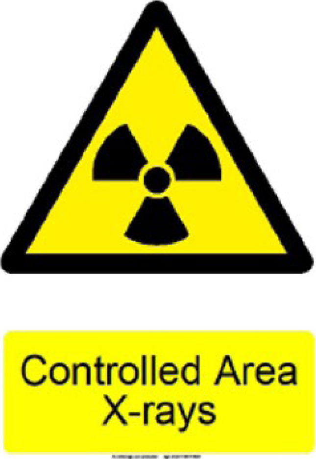
Any designated controlled area must now have warning signs that must be visible at each area entrance (eg dental surgery room), including any entrance in which the operator stands. These signs should include the ionizing radiation trefoil symbol and wording to include ‘X-rays’ and ‘controlled area’ designated when the X-ray unit is switched on. An example is shown in Figure 2.
Figure 2. Example X-ray trefoil with clearly marked ‘controlled area’ signage.
Contingency plans
A radiation risk assessment is required to be carried out and recorded prior to starting any new radiography activity within a practice. As part of this risk assessment, a contingency plan must be in place in the event of any foreseeable radiation accidents to prevent them from happening or, if they do happen, to limit their consequences. The new guidance states a requirement to formally investigate, in consultation with the RPA, any circumstances when these contingency plans are activated to determine the underlying cause and take any action to prevent recurrence. There is also a need to rehearse these contingency plans and the guidance suggests that this is done annually, or when the contingency plans are subject to change. This will ensure that staff are aware of the requirements and provide a safe environment for workers, patients and the public.
Optimizing patient dose
Rectangular collimation has been shown to reduce the effective dose to patients, and radiation scatter to staff, by approximately 50%.7 It is, therefore, essential to use collimation when taking a peri-apical, bitewing or occlusal radiograph, unless there are sound clinical reasons for not doing so. The use of a circular collimator must be justified and documented in the patient's clinical notes.
The new guidance also states that rectangular collimation should be provided as standard with all new intra-oral dental X-ray units, and in any units still equipped with a circular collimator, this should be adapted or replaced by a rectangular one. The size of the collimator should be no greater than 40 x 50 mm, and preferably less than 35 x 45 mm, which should improve image quality and reduce the number of repeat X-ray exposures for the patient.
Training
IRR17 and IRMER17 both state that the responsibility to ensure that those involved in dental radiation exposure should be appropriately trained in the theoretical and practical aspects of their role. There should also be continuing education and training in radiation protection, in keeping with the continuing professional development (CPD) requirements set out by the GDC in each 5-year cycle.8
Training in the use of CBCT units is divided into two levels. Level 1 training relates to dentists and dental nurses who use the equipment, or referrers requesting CBCT scans. Level 2 relates to those able to diagnose and report on CBCT images. For CBCT trained staff, it is recommended that at least 1 out of the 5 hours of verifiable CPD in radiation protection/radiology should be dedicated to CBCT.
Expectations for service engineers
Service engineers typically assist dental practices, providing the RPA with relevant information including room plans and equipment details, to allow installation or relocation of radiography units. They also carry out installation, undertake test exposures as a part of the critical examination and have a duty to pass on any information regarding safe use, testing and maintenance to the appropriate persons. To comply with IRR17, service engineers must:
Register their work with radiation generators HSE;
Appoint a suitable RPA;
Draft a radiation risk assessment that should identify their conditions to designate and demarcate controlled areas when working on clients' sites, how access will be restricted and working instructions when co-operating with other employers, including handover arrangements;
Draft local rules and contingency plans;
Appoint and train suitable staff as radiation protection supervisors (RPS) adequately;
Consult with the RPA about conducting critical examinations on the equipment that they install and how to treat the results, as well as discussing arrangements for co-operation between employers;
Ensure that they receive continued further training regarding how to conduct critical examinations in accordance with protocols, as advised by the RPA.
Disposal or selling on of X-ray equipment
When X-ray equipment is deemed no longer fit for use, consideration should be given to ensure that it is disposed of safely. The unit must be rendered inoperable by cutting the main power cable after being switched off and disconnected from the mains supply. Any warning signs should be removed to avoid alarm following disposal. It is recommended that any contractor providing this service for redundant radiographic equipment should provide evidence of its appropriate and safe disposal, due to the tube head containing hazardous materials including lead, beryllium and mineral oil.
The transfer of an X-ray unit to another person must include adequate information regarding the safe use, testing and maintenance of the equipment, as expected of the original supplier.
Retention times for records
Records are required to be kept for specific timescales, outlined by IRR17. Investigation of contingency plans, reports on dose investigation levels, monitoring of controlled areas, monitoring and maintenance of engineering controls, and safety and warning feature checks are required to be kept for at least 2 years. Some records are not included within IRR17, however the guidance notes have provided recommendations on retention periods. These include:
Record of registering with HSE: until registration is changed or cancelled;
Radiation risk assessment: until this is replaced by an updated version;
Record of critical examinations: for the working life of the equipment;
Personal dose monitoring in non-classified employees: at least 2 years;
Local rules: until these are replaced by an updated version;
Examination of PPE: at least 2 years after date of test;
Record of staff training in radiation safety: at least 5 years;
Controlled area handover forms: at least 3 years.
Conclusion
PHE and FGDP have provided extensive and essential guidance on the safe use of dental radiographic and CBCT equipment through its recently updated document. The new guidance notes clarify radiation protection for the dental team, patients and the public, aid compliance and should be a valuable resource for dental practices for many years to come.