Sutures are routinely used to achieve haemostasis and to approximate soft tissues after extractions, periodontal procedures, implant surgery and soft tissue biopsies. This paper provides the general dental practitioner with the knowledge and understanding of sutures to select the most appropriate for use. Common suturing techniques are also discussed.
Clinical Relevance: Suturing is an important aspect of dental practice, the selection of an appropriate suture and technique is integral to obtaining an optimal outcome.
Article
The history of surgical suturing has been traced back to ancient Egyptian times and the first written record of suture materials and techniques can be found in the texts of the Indian physician Sushruta from the sixth century BC.1 Well known suture materials, such as catgut and silk, have existed since ancient times; no great advances in suture materials were made until after World War II, when synthetic non-resorbable and resorbable fibres were developed.2
The purpose of a suture is to hold the tissues in place to permit healing by primary intention and to control bleeding.3 Suturing is undoubtedly integral to the practice of dentistry and every dentist develops the skill as an undergraduate. In dentistry, sutures are routinely used to achieve haemostasis and to approximate soft tissues after extractions, periodontal procedures, implant surgery and soft tissue biopsies. Other uses for sutures are to approximate oro-facial skin lacerations and to ligate blood vessels. There are numerous suturing techniques, but the common techniques that the general dental practitioner should be familiar with are:
Interrupted suturing;
Continuous suturing;
Vertical mattress suturing; and
Horizontal mattress suturing.
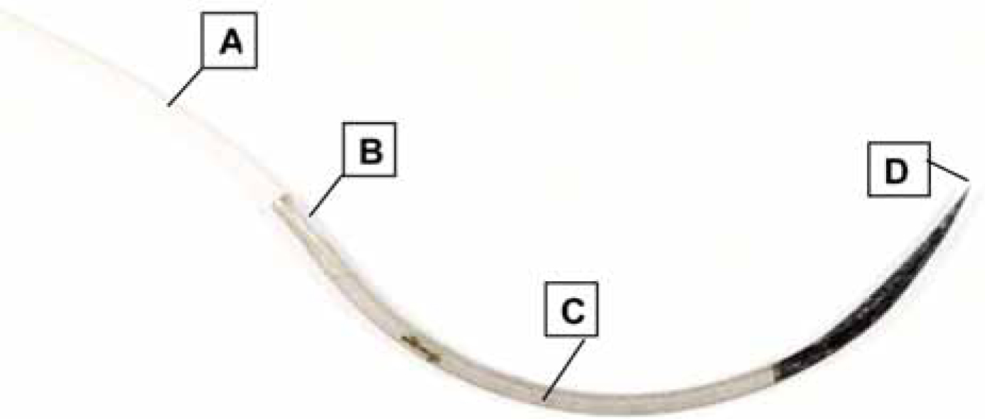
A suture consists of a thread material that is attached to a needle (Figure 1). The thread material may be natural or synthetic, absorbable or non-absorbable. A suitable thread diameter for use in dentistry is 3–0 to 6–0. The most commonly used suture needles are made from stainless steel and vary in shape of curvature, as well as cutting characteristics. When choosing a suture for a given situation, the general dental practitioner is faced with a huge array of options and descriptions found in routinely used dental catalogues. The aim of this paper is to provide the general dental practitioner with the knowledge and understanding of sutures to select the most appropriate for use. In addition, we also describe common suturing techniques.
Figure 1. A suture needle and thread. A - suture thread; B - swaged end; C - body; D - cutting point.
Suture thread material
The suture thread material can be natural or synthetic, absorbable or non-absorbable, mono-filament or multi-filament. Absorbable sutures are eliminated by different types of degradation, according to the type. Catgut is eliminated by a foreign body reaction with enzymatic involvement, whereas Vicryl (synthetic) is absorbed by hydrolysis.4
Monofilament threads consist of one thread and have a closed interior; they are smooth and display no capillarity. Monofilaments generally form thin suture threads as the thicker they are the more their structure is impaired; this has an adverse effect on the quality of the suture. Multifilament threads are produced by twisting or braiding many thin threads together. Twisted threads display high capillarity, which means that these materials can act as bacterial reservoirs. Braided sutures, on the other hand, impede the passage of fluid as the filaments are aligned obliquely to the longitudinal axis of thread. Owing to the process of production, multifilament suture threads tend to be rough, and this makes their passage through tissues more difficult, but conversely, this results in superior knot tying characteristics. Suture threads are often coated to reduce the friction when passing through tissues and to reduce the capillarity.5
Commonly used sutures in dentistry are coated Vicryl and Vicryl Rapide. Both are absorbable, with Coated Vicryl offering an effective wound support for 28 days, with complete absorption at 56–70 days and Vicryl Rapide offering wound support for 10 days, with total absorption by 42 days. With Vicryl Rapide wound support is 50% at 5 days.6Table 1 shows commonly used sutures in dentistry.
Recently, Vicryl Plus has come on to the market. It offers a suture with antibacterial properties, however, its use for intra-oral wound closure has not been demonstrated as yet.
Suture thread size
Suture thread diameter is described by gauge, which varies from 1–0 to 11–0, the greater the gauge, the thinner the suture thread, and also the lower the tensile strength. For dental surgery, we select the smallest appropriate thread size with sufficient strength to approximate the tissue in question effectively. Suture thread gauge 3–0 and 4–0 are commonly used.
Suture needle shape and cutting characteristics
Suture needles vary in shape of curvature, the most commonly used shapes in dentistry are 3/8 circle, 1/2 circle and 5/8 circle. The 3/8 circle is the least curved, but a needle with a greater curvature is required when access to an area with restricted space is needed.
Needle types may generally be described as taper (‘smooth’) or cutting. Taper needles gradually taper to the point and are used for tissues that are easy to penetrate, such as blood vessels or bowel. In dentistry, for closure of mucosa and mucoperiosteal flaps we use cutting needles. These are triangular in shape with the apex forming the cutting surface permitting ready penetration of tougher tissues. Taper needles should not be used for tougher tissues, and especially not for skin, because of the trauma caused by difficulty of tissue penetration. Reverse cutting needles are similar to conventional cutting needles except that the cutting edge faces down rather than up, which reduces the chance of the suture pulling through the tissue.
Needle holders and forceps
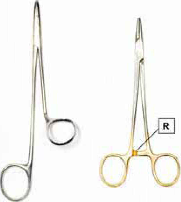
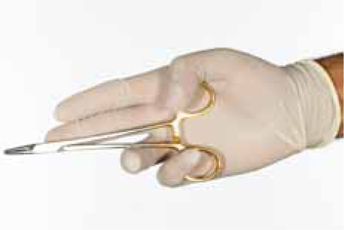
For intra-oral suturing it is usual to use a needle holder to grip the needle. There are many designs of holders and some have a ratchet so that they maintain the grip (Figure 2). The needle holder is held with a palm grip to allow mobility of the wrist and the needle is grasped about two-thirds back from the needle tip. Holding the needle too close to where the suture material is swaged to the needle may weaken or bend it. The thumb and ring finger should not be placed far into rings of the needle holder and some simply grasp the body of the instrument in the palm of the hand to increase mobility (Figure 3).
Figure 2. Two different types of needle holders, one with (right) and one without (left) a ratchet (R).Figure 3. Needle holders in a palm grip.
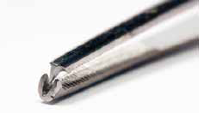
Toothed tissue forceps (Figure 4) should be used to position the edge of the tissue gently and facilitate perpendicular passage of the needle. The forceps are rested on the web space between the thumb and index finger so that the arms of the forceps act as extensions to the thumb and index finger. The forceps can also be used to grasp the needle when repositioning it in the needle holders to avoid having to touch the needle with the fingers.
Figure 4. The ‘toothed’ end of toothed tissue forceps.
Suturing techniques
The ideal suture should form a rectangle, penetrating the tissue perpendicular to its surface, then turning at a right angle across the depth of the wound, then turning again to emerge on the other side again, perpendicular to the tissue surface. Whilst this is usually achievable with mucoperiosteal flaps, it is not possible when closing mucosa bound down to bone in the gingival region when a less than perpendicular angulation is accepted. This is described as a square knot. The needle should be passed through tissue at a distance of at least 3 mm from the free edge and suction should be adequate so that good visibility is maintained.
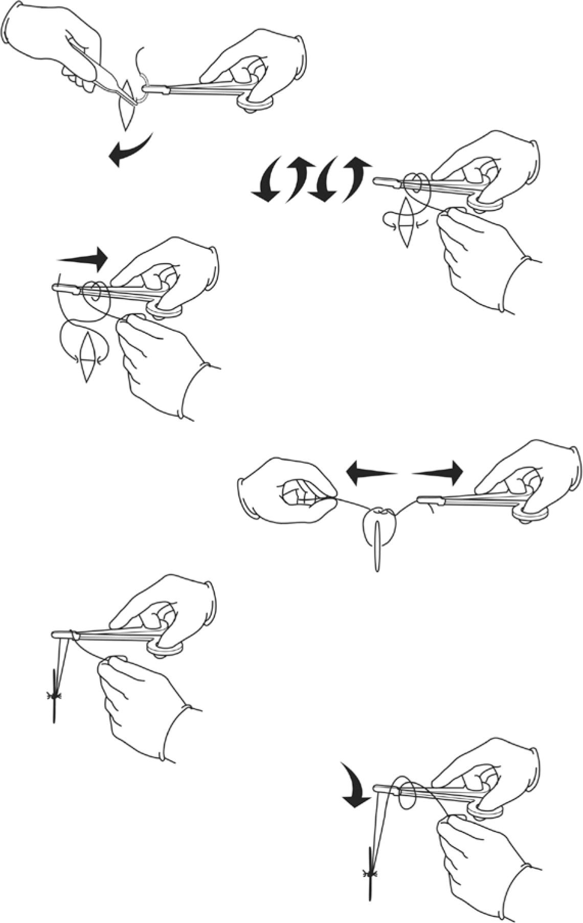
Knot tying is achieved by wrapping (throwing) the long end of the suture thread around the tip of the closed needle holder twice before grasping the short end with the needle holder. This first double knot is then pulled tight; following this a further single throw of the suture thread is made; this throw is made and pulled in the opposite direction. Figure 5 shows this process in diagrammatic form. Sutures should not be too tight as they will cause oedematous swelling of the soft tissues. The suture is cut with scissors to leave about 3–4mm of thread. If too short then the knot may untie, but if too long then this is uncomfortable for the patient, as he/she will be more aware of thread in the mouth. Non-resorbable sutures used for skin wound closure may be left a little longer to facilitate their removal. The scissors are held with the thumb slightly in one ring and the ring finger in the other. The index finger rests on the shaft of the scissors to stabilize them and is particularly important to avoid accidently injuring the lips when entering the mouth. Iris type scissors or similar are suitable for cutting intra-oral sutures.
Figure 5. Knot tying technique.
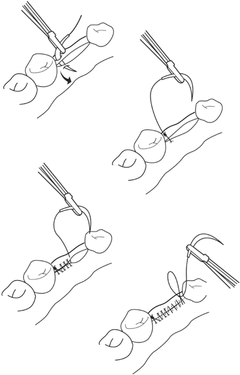
Simple interrupted suture
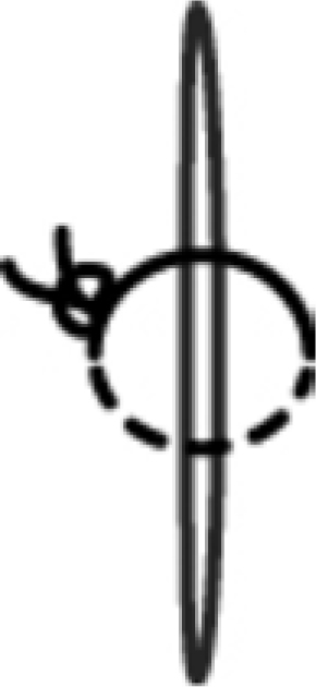
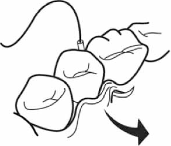
With this type of suture, the wound edges are held with toothed forceps and the needle enters the tissues on one side of the wound and leaves on the other, as described above. Each suture is secured with a knot (Figure 6). When closing buccal soft tissue papillae by suturing to palatal or lingual papillae when teeth are present, the needle must be carefully passed below the contact area of the adjacent teeth (Figure 7). Alternatively, a sling suture may be used to secure adjacent papillae without involving the papillae on the opposite side (Figure 8).
Figure 6. Simple interrupted suture.Figure 7. Suture needle passing below the contact area of adjacent teeth.Figure 8. Sling suture technique.
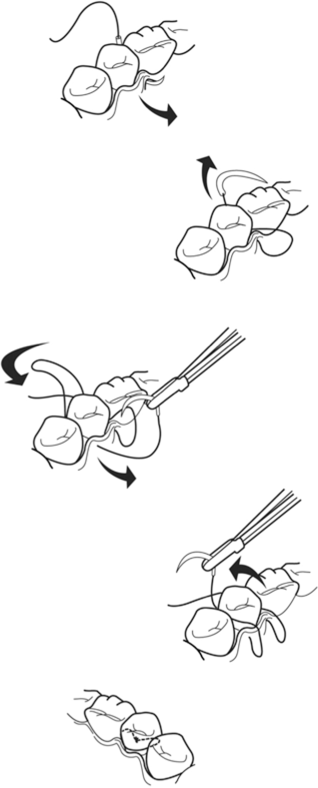
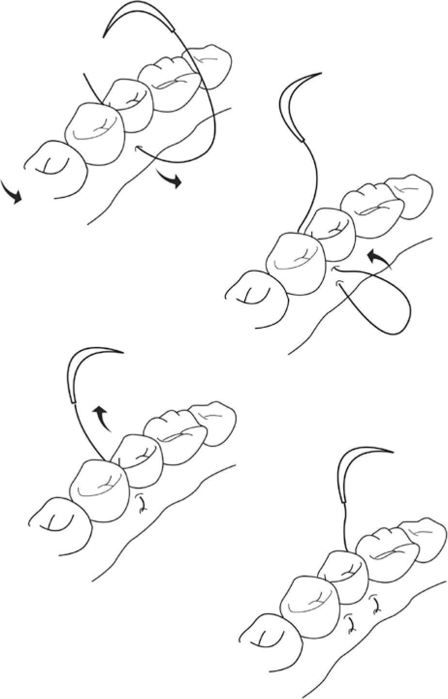
Continuous suture
This type is faster to place but relies on fewer knots so is more vulnerable. An interrupted suture is undertaken and then the free end is cut before the needle is reintroduced and directed diagonally across the wound, to leave the tissue on the other side. The suture is then brought across perpendicular to the wound edge and reintroduced on the first side again with each bite. After the entire wound is closed, a loop is made with the last pass of suture and this loop is grasped with the needle holder to tie the knot (Figure 9).
Figure 9. Continuous suture technique.
Vertical mattress suture
This suture is used to add additional wound edge eversion and decrease dead space within the wound. The needle is introduced about 5–6 mm from the wound edge and a deep bite of tissue is taken before exiting in the tissue in the same position on the opposite side of the wound edge. The needle position is then reversed in the needle holder and reintroduced about 1–3 mm from the second side of the wound, and a smaller bite of tissue is taken before leaving the first side of the wound; a knot is then placed (Figure 10).
Figure 10. Vertical mattress suture technique.
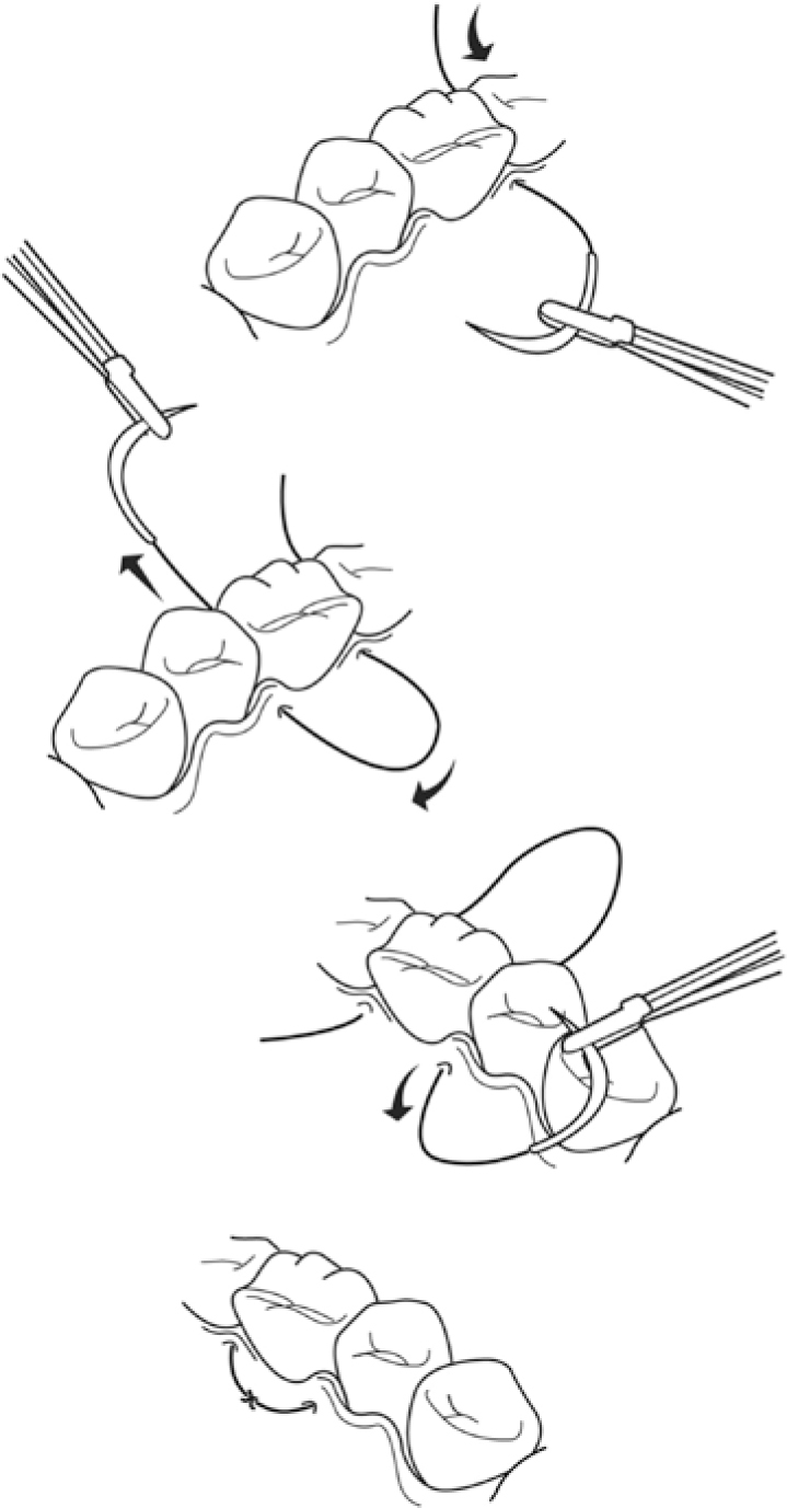
Horizontal mattress suture
When it is important to distribute wound tension across larger wounds, even for just the first few sutures, then the horizontal mattress suture can be useful. The needle is introduced about 5–6 mm from the wound edge and leaves the opposite side tissue. The needle is then introduced on the second side of the wound but 3–5 mm along the wound from the exit point. The suture exits in the same position on the first side of the wound and the suture is tied (Figure 11).
Figure 11. Horizontal mattress suture technique.
Summary
Simple interrupted sutures are the most commonly used type of suture for intra-oral wound closure and arrest of haemorrhage from dental extraction sockets, but other techniques should be used too, when appropriate.
Vicryl Rapide is a commonly used material but sometimes, when the wound requires support for a period longer than around 7 days, then Vicryl should be used. A vestibular wound in the anterior buccal sulcus, such as when providing access for anterior mandibular bone harvesting, may be more quickly closed with a continuous suture. However, this wound is under significant tension with the movement of the lower lip, and some may prefer to use multiple simple interrupted or a series of mattress sutures. A horizontal mattress suture is ideal for apposing the mentalis muscle during this procedure, with a resorbable suture with tie left deep in the wound.
Surgical wounds for endodontic surgery may also provide situations when increased wound support is required and so regular Vicryl rather than Rapide thread, and mattress rather than simple interrupted technique, is appropriate. This is the case when the incision is placed in the vestibule and is under greater tension with the movement of the lip.
A longer period of wound support using Coated Vicryl rather than Vicryl Rapide may also be required when healing is compromised by the patient's age, low body weight or a general health condition that reduces the immune response. Poor local blood supply and local infection may also necessitate using a suture offering wound support for a longer period of time.
In conclusion, dentists undertaking surgery should be familiar with the different suturing techniques so that they can use the most appropriate for the patient and the wound.