Kolokotronis A, Doumas S Herpes simplex virus infection, with particular reference to the progression and complications of primary herpetic gingivostomatitis. Clin Microbiol Infect. 2006; 12:(3)202-211
Arduino PG, Porter SR Herpes Simplex Virus Type 1 infection: overview on relevant clinico-pathological features. J Oral Pathol Med. 2008; 37:(2)107-121
Wittek M, Doerr HW, Allwinn R Varicella and herpes zoster. Part 2: therapy and prevention. Med Klin (Munich). 2010; 105:(6)399-403
Oxman MN Zoster vaccine: current status and future prospects. Clin Infect Dis. 2010; 51:(2)197-213
Mustafa MB, Arduino PG, Porter SR Varicella zoster virus: review of its management. J Oral Pathol Med. 2009; 38:(9)673-688
Bravender T Epstein-Barr virus, cytomegalovirus, and infectious mononucleosis. Adolesc Med State Art Rev. 2010; 21:(5)251-264
Aberle SW, Mandl CW, Kunz C, Popow-Kraupp T Presence of human herpesvirus 6 variants A and B in saliva and peripheral blood mononuclear cells of healthy adults. J Clin Microbiol. 1996; 34:(12)3223-3225
Nikkels AF, Pierard GE Herpesvirus 6. What attention does it deserve in general practice?. Rev Med Liege. 2006; 61:(5-6)317-321
Stoeckle MY The spectrum of human herpesvirus 6 infection: from roseola infantum to adult disease. Annu Rev Med. 2000; 51:423-430
Braun DK, Dominguez G, Pellett PE Human herpesvirus 6. Clin Microbiol Rev. 1997; 10:(3)521-567
Ongradi J, Kovesdi V, Kovats E Human herpesvirus 7. Orv Hetil. 2010; 151:(16)645-651
Caselli E, Di Luca D Molecular biology and clinical associations of Roseoloviruses human herpesvirus 6 and human herpesvirus 7. New Microbiol. 2007; 30:(3)173-187
Ongradi J, Kovesdi V, Medveczky GP Human herpesvirus 6. Orv Hetil. 2010; 151:(13)523-532
Shiboski CH, Patton LL, Webster-Cyriaque JY The Oral HIV/AIDS Research Alliance: updated case definitions of oral disease endpoints. J Oral Pathol Med. 2009; 38:(6)481-488
Kabani S, Greenspan D, deSouza Y, Greenspan JS, Cataldo E Oral hairy leukoplakia with extensive oral mucosal involvement. Report of two cases. Oral Surg Oral Med Oral Pathol. 1989; 67:(4)411-415
Mendoza N, Diamantis M, Arora A Mucocutaneous manifestations of Epstein-Barr virus infection. Am J Clin Dermatol. 2008; 9:(5)295-305
Mesri EA, Cesarman E, Boshoff C Kaposi's sarcoma and its associated herpesvirus. Nat Rev Cancer. 2010; 10:(10)707-719
Taylor GS, Blackbourn DJ Infectious agents in human cancers: lessons in immunity and immunomodulation from gammaherpesviruses EBV and KSHV. Cancer Lett. 2011; 305:(2)263-278
Cleveland JL, Junger ML, Saraiya M, Markowitz LE, Dunne EF, Epstein JB The connection between human papillomavirus and oropharyngeal squamous cell carcinomas in the United States: implications for dentistry. J Am Dent Assoc. 2011; 142:(8)915-924
Syrjanen S, Lodi G, von Bultzingslowen I Human papillomaviruses in oral carcinoma and oral potentially malignant disorders: a systematic review. Oral Dis. 2011; 17:58-72
D'Souza G, Fakhry C, Sugar EA Six-month natural history of oral versus cervical human papillomavirus infection. Int J Cancer. 2007; 121:(1)143-150
D'Souza G, Kreimer AR, Viscidi R Case-control study of human papillomavirus and oropharyngeal cancer. N Engl J Med. 2007; 356:(19)1944-1956
Mannarini L, Kratochvil V, Calabrese L Human Papilloma Virus (HPV) in head and neck region: review of literature. Acta Otorhinolaryngol Ital. 2009; 29:(3)119-126
D'Souza G, Dempsey A The role of HPV in head and neck cancer and review of the HPV vaccine. Prev Med. 2011; 53:S5-S11
Leao JC, Ribeiro CMB, Carvalho AAT, Frezzini C, Porter S Oral complications of HIV disease. Clinics (Sao Paulo). 2009; 64:(5)459-470
Scott LA, Stone MS Viral exanthems. Dermatol Online J. 2003; 9:(3)
Moss WJ, Griffin DE Measles. Lancet. 2012; 379:(9811)153-164
Sabella C Measles: not just a childhood rash. Cleve Clin J Med. 2010; 77:(3)207-213
Coloe J, Burkhart CN, Morrell DS Molluscum contagiosum: what's new and true?. Pediatr Ann. 2009; 38:(6)321-325
Hille JJ, Webster-Cyriaque J, Palefski JM, Raab-Traub N Mechanisms of expression of HHV8, EBV and HPV in selected HIV-associated oral lesions. Oral Dis. 2002; 8:161-168
Purgina B, Pantanowitz L, Seethala RR A review of carcinomas arising in the head and neck region in HIV-positive patients. Patholog Res Int. 2011; 2011 https://doi.org/10.4061/2011/469150
Orofacial viral infections – an update for clinicians Raj G Nair Ali Salajegheh Anut Itthagarun Sahar Pakneshan Michael T Brennan Lakshman P Samaranayake Dental Update 2024 41:6, 707-709.
Authors
Raj GNair
MSc, PhD, MRACDS(Oral Med)
Oral Medicine, School of Dentistry and Oral Health, Centre for Medicine and Oral Health, Griffith Health Institute, Griffith University and Department of Haematology and Oncology, Gold Coast Hospital
Orofacial viral infections may be less common but appear in different clinical forms. Often these infections get initially treated by antibiotics which obviously will have limited or no effect. The authors review the current concepts of orofacial viral infections, causative agents, their classification and clinical manifestations and a basis for treatment.
Clinical Relevance: Most viral infections do not require any specific treatment except in patients who are immunosuppressed or immunodeficient. Appropriate diagnosis and timely management of orofacial viral lesions are important irrespective of whether it is localized or a manifestation of a systemic infection.
Article
Patients often present with orofacial infections in general medical and dental practice. These infections may be caused by bacterial, fungal or viral pathogens. Viral infections may manifest in different clinical forms and affect all age groups. The most common viral infection affecting the orofacial region is caused by the herpes simplex virus (HSV). Infection from a virus follows a different aetiopathogenic pathway compared with bacteria, fungi and other organisms, as a virus metabolism is dependent on host cells. In general, most of the viral infections of the orofacial region are self-limiting in an otherwise healthy individual, whilst compromised individuals may present with a myriad of local and systemic complications of viral infections.
General dental practitioners should be aware of the wide range of clinical manifestations of viral infections, which may affect the orofacial region as a localized disease or a manifestation of a systemic viral infection, such as the human immunodeficiency virus (HIV) disease. The aim of this short review is to provide a state-of-the-art, concise account of orofacial viral infections of humans and their management.
Viruses causing orofacial infections
There are a number of viruses that may produce a subclinical or an overt infection of the peri-oral, oral, and oropharyngeal region, the most common being a group belonging to the Herpesvirida and the Papillomaviridae family.
Herpes group of viruses
The herpes group of the Herpesviridae family include eight viruses in three subclasses as Alpha, Beta and Gamma herpes viruses. Alpha herpes viruses include the herpes simplex virus-1 (HSV-1), herpes simplex virus-2 (HSV-2) and varicella zoster virus (VZV, HHV-3). Beta herpes viruses are grouped as cytomegalovirus (CMV, HHV-5), human herpesvirus-6 (HHV-6) and human herpesvirus-7 (HHV-7). Gamma herpes viruses consist of Epstein-Barr virus (EBV, HHV-4) and human herpesvirus-8 (HHV-8).
Interestingly, most of the viruses in the Herpesviridae family are known to cause oral and peri-oral infections, although there is controversy as to the true causative agent of some of these orofacial infections.1 Orofacial viral infections are common among immunocompromised patients; the most common being those caused by the herpes simplex virus (HSV). Human herpesvirus-6 has been proposed as an aetiologic factor in recurrent aphthous stomatitis.2 Human herpesvirus-8 (HHV-8) is the aetiopathogenesis of Kaposi's sarcoma.3 Varicella-zoster virus is less common but occurs in severe forms, as in herpes zoster.4 Epstein-Barr virus in oral hairy leukoplakia (OHL)5 and many of the herpes viruses and certain human papilloma viruses (HPV) are known for their association in malignant neoplasms.6
Alpha herpes viruses
Herpes simplex virus
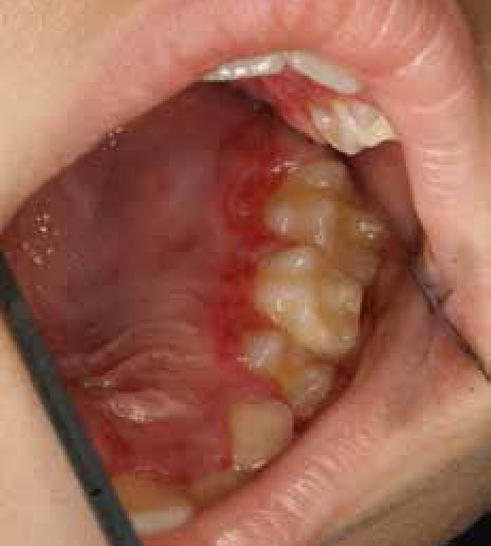
As a general rule, herpes simplex virus-1 (HSV-1) affects the areas above the waistline, and HSV-2 mainly causes genital lesions or lesions below the waistline, although HSV-1 and -2 can affect either area. Primary infection of HSV-1 occurs either during childhood as gingivostomatitis or, if not exposed, then as pharyngotonsilitis in an adult. Latent reactivation of HSV-1 most commonly manifests as herpes labialis (or the common cold sore) or as atypical forms in an immunocompromised individual7 (Figures 1 and 2). The virus is shed into saliva and the main mode of transmission of HSV-1 is through contaminated saliva and transmission may occur via kissing a child, for example.8 Both primary and secondary or recrudescent infections are self-limiting. Immunosuppression, ultraviolet sun exposure, stress, changes in weather, especially colder months, could all attribute to the initiation of herpes labialis.9 Primary infection is common among children and young adults, either asymptomatic or in the form of gingivostomatitis, following a usual course of fever, headache, irritability, loss of appetite, lethargy, hypersalivation and cervical lymphadenopathy.10 Most of the individuals who suffer from recurrent herpes labialis will experience a prodromal phase of tingling, burning sensation, itching, mild pain and/or fever. Upper respiratory tract infection may precede the onset of the disease. Clinically, single or multiple small erythematous papules that develop and form vesicles appear on either upper or lower lip, which may or may not coalesce. They will either rupture or may heal by crusting, leaving no noticeable scar in most circumstances. An atypical clinical presentation may occur in immunocompromised patients. The healing may be prolonged with pain in the immunocompromised patient. There may be widespread vesicles with ulcerations involving large areas of the lip mucosa and adjacent cutaneous surfaces of the peri-oral region.10 Diagnosis is clinical but laboratory confirmations may be needed in atypical cases and immunocompromised patients.10 Antiviral treatment is often recommended for the immunocompromised and in moderate to severe infection in otherwise healthy individuals. Systemic acyclovir, 200 mg, 5 times daily for 7 days or topical application of 5% acyclovir cream every four hours for 5 days is the recommended dose. Valacyclovir 1–2 milligrams twice daily may be used as a prophylactic treatment, which is most effective when initiated early during the prodrome phase.10
Figure 1. Generalized gingival involvement in primary herpetic stomatitis in an immunocompetent child.Figure 2. Recurrent HSV-1 infections.
Varicella-zoster virus
Chickenpox is the primary infection in childhood due to VZV as a droplet infection from the nasopharynx. If a child is not exposed during childhood, an overt infection may occur during adulthood. Chickenpox can have orofacial lesions such as vesicles, especially over the facial skin and the oral mucosa, in addition to the cutaneous lesions of the trunk. If not secondarily infected, usually the lesions on the facial skin will heal without scarring.11
After chickenpox, VZV remains latent in sensory ganglia until reactivation and replication, resulting in herpes zoster (shingles). Herpes zoster affects those above the age of 50 years or the immunocompromised and is characterized by a unilateral, distinctive painful vesicular rash over a dermatome, corresponding to the sensory ganglion where the VZV was latent. Orofacial manifestations are within the ophthalmic, maxillary and mandibular nerve distribution of the trigeminal nerve, with maxillary and mandibular causing intra-oral vesicles and painful ulcerations. Preventive therapy includes vaccination against VZV (HHV-3).12
Anti-viral therapy is not indicated for chickenpox in otherwise healthy individuals but may be considered in children 12 years or older, patients with chronic cutaneous or pulmonary disease, patients on short to intermittent courses of aerosol corticosteroids and those on long-term salicylates.13 Treatment is based on symptomatic relief and antiviral drugs. In general, antiviral therapy in varicella zoster reduces the acute symptoms of pain and malaise, limits the spread and duration of the skin lesions and may prevent the development of post-herpetic neuralgia and reduce ophthalmic complications.13
Beta herpes viruses
Cytomegalovirus
Cytomegalovirus is known to cause mononucleosis-like disease characterized by fever, pharyngitis, lymphadenopathy and fever in healthy immunocompetent individuals requiring no specific therapy.14 In the orofacial region, the mononucleosis-like disease can present with palatal petechiae and submandibular lymphadenopathy. This virus also has been implicated to non-specific oral ulcers and salivary gland disease, especially in the human immunodeficiency virus (HIV) infected patients.6
Human herpesvirus-6
Human herpesvirus-6 is one of the first, so called ‘ancient’ human herpes viruses identified by molecular characterization. The main mode of viral transmission is through contaminated saliva. It has been demonstrated that HHV-6 is present in the saliva of a large proportion of the healthy adult population.15 The primary infection is usually asymptomatic and commonly occurs during childhood by age 2 years.16 The clinical form is called exanthema subitum or roseala infantum or ‘sixth disease’. This biphasic disorder usually runs a benign course, causing fever, then a maculopapular rash on subsidence of fever at the end of the fourth febrile day. Uvulo-palatoglossal junction ulcers are useful early signs. The condition requires no antiviral treatment.17 Recent reports have emphasized the critical role of HHV-6 in the aetiology of human oral squamous cell carcinoma. It is, however, unclear whether the virus acts in combination with other carcinogens as a so-called co-carcinogen.18
Human herpesvirus-7
Human herpesvirus-7 was first identified in 1990 and is closely related to HHV-6. It establishes latency in macrophages and T-lymphocytes and reactivates frequently with asymptomatic virus shedding through saliva. Most children acquire infection by the age of 3–4 years, and seronegative individuals are at risk of infection at any age. The spectrum of diseases caused by primary HHV-7 infection is similar to HHV-6, with milder clinical presentation.19,20 Severe complications due to HHV-6 and 7 are treated with ganciclovir and its derivatives or foscarnet and cidofovir.21
Gamma herpes viruses
Epstein-Barr virus
Epstein-Barr virus has been known to cause both local and systemic infections and benign and malignant diseases of the orofacial region. They include infectious mononucleosis or glandular fever, OHL and malignancies such as lymphomas (non-Hodgkins and Burkitt's) and nasopharyngeal carcinoma.22
Clinical features of infectious mononucleosis are pharyngitis, cervical lymphadenopathy, generalized arthromyalgia and associated fever and malaise. Symptomatic treatment is indicated, such as anti-pyretics, analgesics and anti-inflammmatories, with no specific anti-viral drug treatment. Oral hairy leukoplakia is a classic feature of immunosuppression, HIV disease and iatrogenic immunosuppression, such as cancer therapy. Clinically, lesions appear as white corrugated patches commonly on the lateral border of the tongue and gingiva.23
Lymphomas may present as a swelling and/or an ulcer in the oral cavity and orofacial region. Lymphomas and nasopharyngeal carcinomas obviously need more aggressive cancer therapies, depending on the type and stage of the disease.24
Human herpesvirus-8
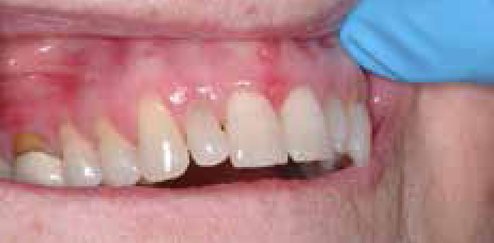
Human herpesvirus-8 is associated with Kaposi's sarcoma (KS), especially in patients with HIV disease or acquired immune deficiency syndrome (AIDS).25 HHV-8 has been found in all the different types of KS affecting humans, hence is known as Kaposi's sarcoma herpes virus or KSHV. Kaposi's sarcoma, seen in HIV disease or AIDS-associated KS, is usually asymptomatic with either purple or bluish macules or swellings affecting the orofacial skin and oral mucosae (Figure 3). In the oral cavity, the hard palate is the most common site, though it could be on other areas of the oral cavity.22,26 Management may include intra-lesional injections of cytotoxic drugs and surgery is warranted only to restore aesthetics, such as the labial gingivae, for example. Anti-retroviral therapy or highly active anti-retroviral therapy (ART/HAART) has significantly improved the management of orofacial KS associated with AIDS.22
Figure 3. HIV disease or AIDS-associated KS affecting the oral mucosae.
Human papilloma virus (HPV)
The Papillomaviridae family are a group of double-stranded circular DNA viruses commonly found in the oral and oropharyngeal mucosa, tracheo-bronchial mucosa and ano-genital region. They are grouped into more than a hundred types and HPV type 16 and 18 have been implicated in oral, oropharyngeal and tonsillar carcinomas.27 More recently, there has been an increasing understanding of the risk factors of HPV in oral cancers, especially the risk of oro-genital sexual activity.28,29,30
Orofacial manifestations of HPV are:
Verruca vulgaris or the common wart on the peri-oral skin;
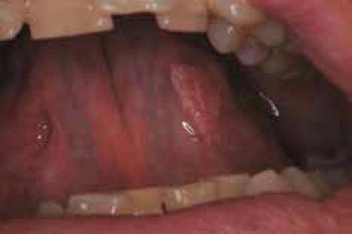
Oral papilloma (squamous cell papilloma) of the oral mucosa (Figure 4);
Focal epithelial hyperplasia; and
Condyloma accuminatum, a sexually transmitted disease.31
Figure 4. Oral papilloma (squamous cell papilloma) of the oral mucosa (HPV).
Management of HPV infection depends on the clinical presentation, such as papilloma, usually using complete surgical excision and/or topical drug therapy. Laser and cryotherapy are not recommended owing to lack of a tissue for histopathological evaluation and a possible seeding of the lesion to the surrounding area in the process.22,23 Two HPV vaccines are currently available and have a clear role in preventing many ano-genital cancers and conditions related to HPV infection. The effectiveness of HPV vaccines in preventing oral HPV infection and cancer is unknown. Studies are underway to evaluate the long-term efficacy of the vaccine against both ano-genital and non-ano-genital endpoints.32
Human immunodeficiency virus
Human immunodeficiency virus infection is pandemic and the disease has become more of a chronic viral infection due to the advent of multiple drug therapy using ART or HAART. The disease may progress to a serious and debilitating stage of AIDS with increased viral load and a significant decrease in the CD4 cells, leading to several opportunistic infections.33
There are several orofacial manifestations associated with HIV disease and these are among the earliest manifestations and considered important indicators of HIV infection, with some carrying a prognostic value. Oral manifestations of HIV/AIDS have been classified into three groups, based on the clinical features and intensity. Viral infections relevant to this review are as follows:
Lesions that are ‘strongly associated’ with HIV infection such as hairy leukoplakia, Kaposi's sarcoma and Non-Hodgkin's lymphoma;
Lesions that are ‘less commonly associated’, such as HSV, HPV and VZV; and
CMV and molluscum contagiosum, which are classified as ‘lesions that are seen’ in HIV infection.33
Coxsackie virus
Coxsackie virus causes hand, foot and mouth disease (strain A16) and herpangina. These viruses can pass through the oral mucosa and small intestine and the regional lymph nodes. Clinical features of hand, foot and mouth disease include, a mild prodrome followed by sparse distribution of vesicles with an erythematous halo affecting the oral mucosa, hands and feet. Painful ulcerative lesions occur anywhere in the oral cavity, but are commonly found on the hard palate, tongue and buccal mucosa. The exanthem (mucosal lesions) begins as 2–8 mm erythematous papules, a short vesicular stage and yellow-grey ulcers with an erythematous halo. Lesions may coalesce, the tongue may become red and oedematous and painful, interfering with oral intake. Oral lesions heal without treatment within 5–7 days.34 No specific treatment is necessary except for isolation of the patient, especially children, to avoid spread of the disease in a community.34
Herpangina is also a disease of the early life, with an incubation period of 4 days followed by an abrupt onset of fever with malaise, headache, neck or back pain. The oral mucosal lesions consist of 1–2 mm grey-white papulo-vesicular lesions that progress to ulcers surrounded by an erythematous halo or rim and the oropharynx may appear diffusely hyperaemic. Lesions are distributed on the anterior tonsillar pillars, soft palate, uvula and tonsils and usually last for a week. Common complaints of affected patients are anorexia, dysphagia and sore throat. No associated cutaneous lesion is typically seen. Only symptomatic treatment is necessary, such as anti-pyretics, analgesics and anti-inflammatories, if necessary.34
Other viruses and their orofacial manifestations
Measles is caused by an RNA virus of the paramyxovirus group of the respiratory tract and skin through droplet infection, mainly affecting infants and young children. With an increase in measles vaccine coverage or a decrease in population density, the age distribution shifts towards older children. In temperate climates, annual measles outbreaks typically occur in the late winter and early spring, while in tropical climates, a combination of high birth rates and variable associations of measles outbreaks with the rainy season creates highly irregular large outbreaks.35Measles is a highly contagious disease. After an incubation period of about 10–12 days, constitutional symptoms such as fever, malaise, conjunctivitis, coryza and cough start and last for a week, followed by hyperpyrexia. Pathognomonic Koplic spots are punctate blue-grey lesions surrounded by an erythematous ring (so-called grains of sand on a red background) on the buccal mucosa. They appear 1–2 days prior to the onset of the rash and remain for 2–3 days.34 Pinpoint raised red lesions on the soft palate may coalesce and the entire oropharynx turns red, lasting for 6–7 days. Other features include Herman spots on tonsils as bluish-grey areas. Treatment is mainly based on supportive measures such as fluids and anti-pyretics. Current active immunization is two doses of live-virus measles vaccine for all healthy children before they begin school.36
Molluscum contagiosum is a disease due to a large DNA virus. Patients who are immunocompromised or deficient, such as the HIV infected, are more prone to this disease entity. The clinical presentation includes asymptomatic, multiple, flesh-coloured, dome-shaped papules of the skin with a central depression.37
Viruses and other head and neck malignancies
Viruses are a recognized risk factor for cancer of the head and neck, as mentioned earlier. The known viruses implicated in malignancies affecting the head and neck region are HPV, EBV and HHV-8.38 More recently, observational studies have found that HPV is a strong risk factor for the development of other head and neck cancers, in particular HPV subtype 16 (HPV-16) associated with oral squamous cell carcinoma, oropharyngeal carcinomas, tonsillar carcinomas and squamous cell carcinoma of the larynx and conjunctiva. Epstein-Barr virus (EBV) has been implicated in lymphomas, nasopharyngeal carcinoma, salivary gland lympho-epithelial carcinoma and HHV-8 in Kaposi's sarcoma.39,40
Conclusions
Viral infections are unique in the sense that they may appear as localized lesions in the absence of constitutional symptoms, unlike bacterial or other infections. Knowledge of such viral infections, their clinical manifestations and lesions with a virus as an aetiological agent or in association is important while making decisions on management (Table 1). The majority of systemic viral infections require no specific treatment in many cases, except in situations such as in immunosuppression. The treatment of virus-induced oral lesions is also important since recurrence is common if treated inappropriately. Some of the virus-induced oral lesions may regress with systemic therapy without the need for surgical intervention. Viral infections remain an important differential of oral and orofacial lesions, which warrants appropriate and timely diagnosis with appropriate management, if required.