Senapati S, Samal SC, Kumar R, Patra S. Necrotizing sialometaplasia: manifestation of a localized unclassified vasculitis. Indian J Pathol Microbiol. 2016; 59:232-234 https://doi.org/10.4103/0377-4929.182018
Nah KS, Cho BH, Jung YH. Necrotizing sialometaplasia: report of two cases. Korean J Oral Maxillofac Radiol. 2006; 36:207-209
Daudia A, Murty GE. First case of full-thickness palatal necrotizing sialometaplasia. J Laryngol Otol. 2002; 116:219-220 https://doi.org/10.1258/0022215021910384
Rajendran R, Sivapathasundharam B. Tumors of salivary glands, 6th edn. In: Shafer WG, Hine MK, Levy BM (eds). India: Elsevier; 2009
Renehan A, Lowry JC. The oral tumours of two American presidents: what if they were alive today?. J R Soc Med. 1995; 88:377-383
Cooper PH. President Cleveland's palatal tumor. Arch Dermatol. 1986; 122:747-748
Anneroth G, Hansen LS. Necrotizing sialometaplasia. The relationship of its pathogenesis to its clinical characteristics. Int J Oral Surg. 1982; 11:283-291 https://doi.org/10.1016/s0300-9785(82)80027-6
In this case of a 45-year-old woman, who attended the primary care service at Birmingham Dental Hospital with widespread ulceration affecting the hard palate, histopathological, haematological and immunological testing confirmed a diagnosis of necrotizing sialometaplasia (NS), a rare benign minor salivary gland condition. The aetiology of NS is unknown; however, it is thought to represent necrosis of the mucoserous acini due to ischaemia. The clinical features of NS are similar to of squamous cell carcinoma and histopathological investigations may be required to exclude malignancy.
CPD/Clinical Relevance: The article highlights the importance of recognizing necrotizing sialometaplasia to prevent misdiagnosis and ensure correct management is undertaken.
Article
Necrotizing sialometaplasia (NS) is a rare, rapidly developing benign minor salivary gland disease of inflammatory origin. The term necrotizing sialometaplasia is used to describe the most consistent feature of the condition: necrosis of the salivary glands causing mucosal breakdown. It is thought to be caused by trauma to the mucoserous glands.
The clinical features of NS are similar to neoplastic changes seen in oral squamous cell carcinoma and muco-epidermoid carcinoma, hence prompt histopathology may be warranted to eliminate malignancy. The case of a patient who presented with necrotizing sialometaplasia and was treated with debridement and antimicrobial mouthwash is presented.
Case report
A 45-year-old female attended the primary care service at Birmingham Dental Hospital for urgent care, with a complaint of acute pain in the palate for the previous 2 weeks. The patient initially experienced symptoms of a sore throat, persistent cough and sought medical attention (from her general medical practitioner and out of hours A&E) on numerous occasions, where she was prescribed morphine and discharged on oral antibiotics (doxycycline). She reported no history of chemical, thermal or physical trauma to the palate. She developed an extensive ulcer affecting the palate, and progressive worsening of symptoms and distress. The patient's medical and social history revealed she had a gastric band placed several years previously, suffered from gastro-oesophageal reflux and was being monitored for hypertension; however, she was not taking any medications. In addition to this, she reported smoking around 10 cigarettes a day.
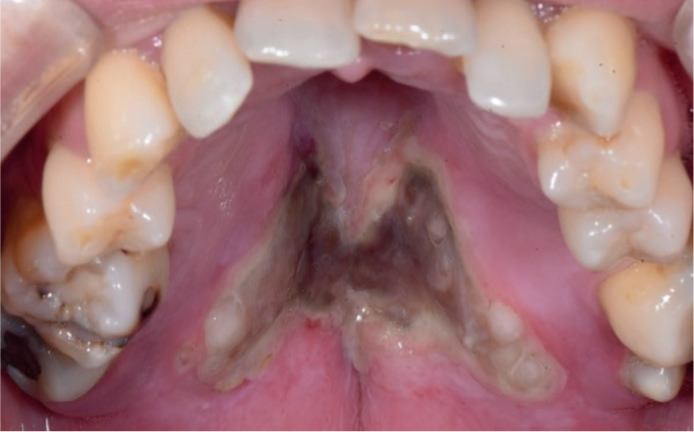
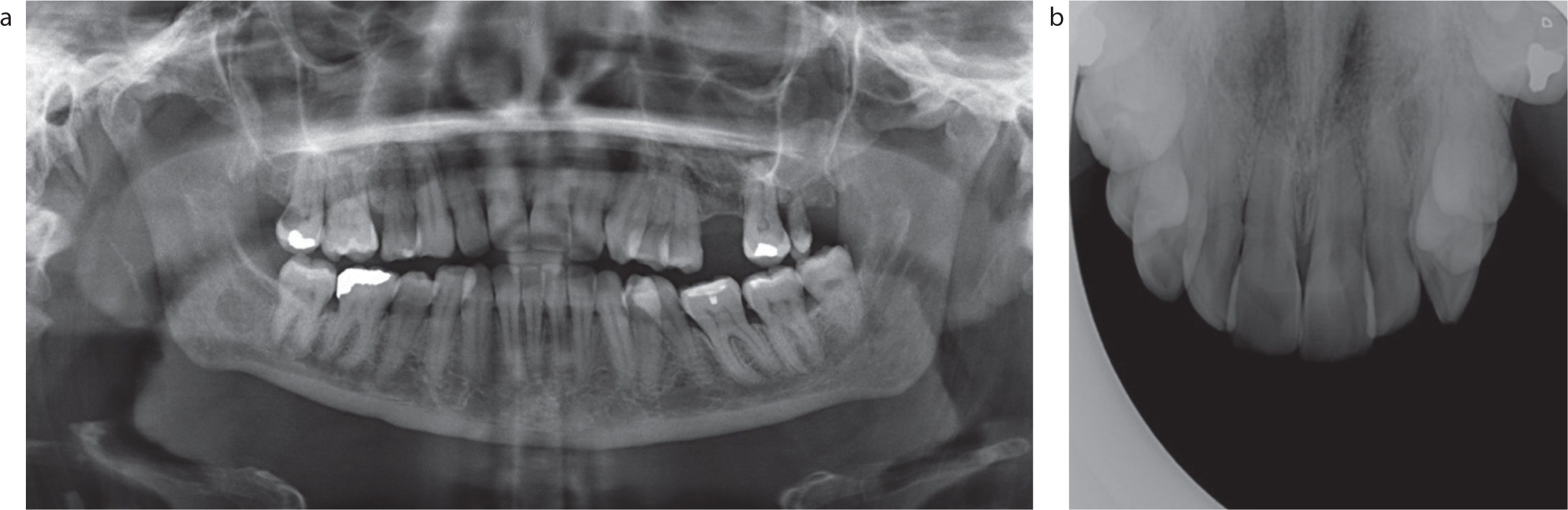
On extra-oral examination, there was no lymphadenopathy or other abnormal signs. Intra-oral examination revealed extensive necrotic tissue measuring 30 mm in diameter, located at the border of the hard and soft palate. There was sloughing of the soft palate mucosa, extending onto the posterior pharyngeal wall causing gagging and difficulties swallowing (Figure 1). Further clinical and radiographic investigations were undertaken. The patient's vital signs were nothing of concern (temperature 36.5°C, blood pressure 135/99mmHg and pulse rate 72bpm). The orthopantomogram showed no bony abnormalities (Figure 2).
Figure 1. Intra-oral image of the hard palate on initial presentation.Figure 2.
(a, b) A full orthopantomogram and maxillary occlusal radiograph.
Haematological investigations including FBC, LFT and U&E were undertaken. The haematology report showed all the values were within normal range, with the exception of slightly elevated white blood cells (11.4 x 10⁹/L) and neutrophils (8.9 x 10⁹/L). Immunological studies showed slightly raised IgM (2.69 g/L), positive p-ANCA but low MPO and PR3 antibodies, not regarded as clinically significant. A biopsy was completed, and the histopathology revealed mixed inflammatory infiltrate, rich in neutrophils, with a mixture of small sheets of suppuration, admixed eosinophils and foci of fat necrosis. The minor salivary gland parenchyma was difficult to identify. Focally, towards the deep aspect, residual atrophic acinar units were identified, deep within sheets of granulation tissue. There was no evidence of dysplasia or malignancy. A plasma cell-rich infiltrate, as expected in syphilis, was negative.
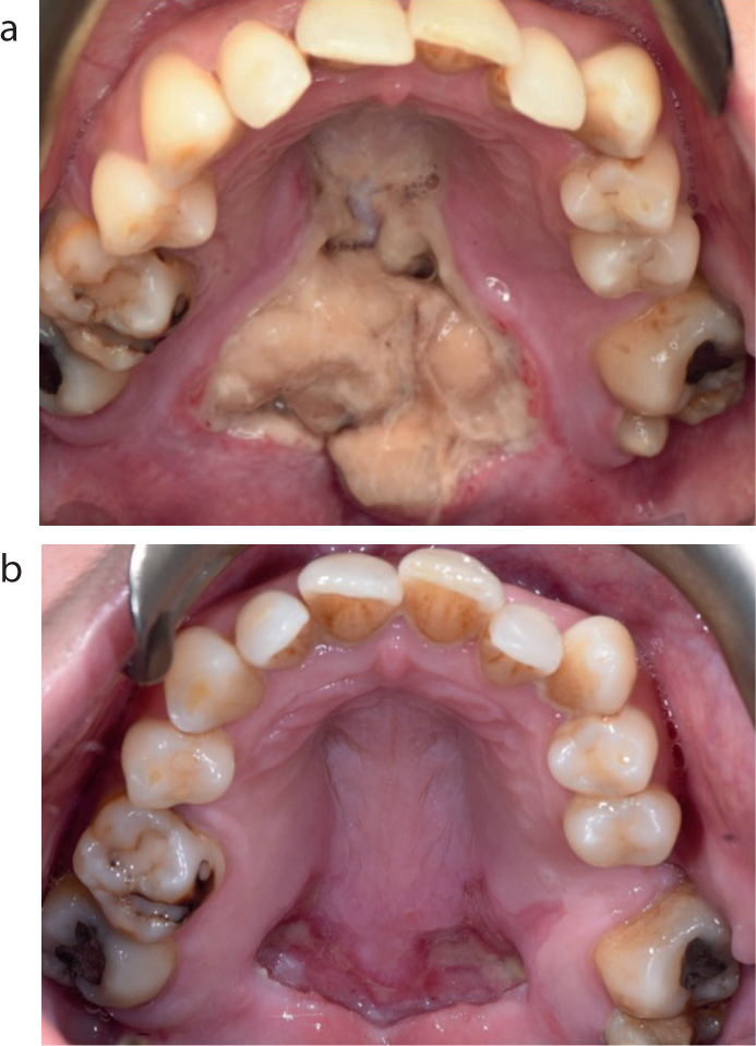
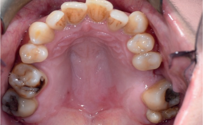
The area was debrided under local anaesthesia, and clean margins were achieved. The patient was advised to maintain good oral hygiene and carry out mouth rinses with 2% hydrogen peroxide mouthwash four times per day. Regular review showed progressive healing (Figure 3). She reported an improvement in her symptoms with reduced pain, increased ability to tolerate a range of foods and improved taste sensation. There was complete remission of the lesion at 7 weeks post debridement, with no signs of palatal defects (Figure 4).
Figure 3.
(a) Intra-oral image of the hard palate, evidence of ulceration and sloughing 1 week after the initial presentation. (b) Intra-oral image of the hard palate 3 weeks post-surgical debridement, showing healing.Figure 4. Intra-oral view of the hard palate 7 weeks after surgical debridement, showing complete closure.
Discussion
Necrotizing sialometaplasia is a benign condition of the minor salivary glands, most commonly affecting the hard palate. The aetiology of the condition is unclear; however, it is thought to be caused by trauma to the mucoserous glands of the hard palate causing ischaemia and necrosis of soft tissues.
Predisposing factors are thought to include smoking, alcohol abuse, local anaesthetic administration, mechanical irritation from ill-fitting dentures, gastric reflux, bulimia and diabetes. It is hypothesized that the most likely cause in this patient was her long-term cigarette smoking, which might have led to thermal trauma and dehydration of the palatal mucosa, making it more susceptible to trauma. Gastro-oesophageal reflux may have contributed to trauma to the mucosa.
Necrotizing sialometaplasia was first described in the literature by Abrams 1973,1 most frequently affecting the hard palate; however, there have been reported cases affecting the soft palate, buccal mucosa, tongue, retromolar trigone, tonsils, nasal cavity, maxillary sinus and other seromucinous glands.2 The area of ulceration reportedly ranges from 0.7 cm to 5.0 cm.3 In the case of this patient, it presented as a 3-cm ulcer affecting the majority of the hard and soft palate. Two-thirds of cases present unilaterally, bilateral involvement is a rarity.4 There is reportedly a higher incidence in Caucasian men than women, with a ratio of 2:1 and it usually affects people in their fifth decade.5
The clinical presentation raises the possibility of several differential diagnoses including squamous cell carcinoma, muco-epidermoid carcinoma, primary adenocarcinoma, B cell lymphoma, secondary syphilis and tuberculous ulcer.3 Diagnosis can be difficult and is based on a thorough clinical history and histology as necessary. Prompt investigations are essential to eliminate malignant neoplasm. Supplementary investigations with immunohistochemistry and haematology may eliminate infectious and other inflammatory processes.
Patients can present with minor pain or even painless ulcerations and may have prodromal symptoms of fever, malaise or a swelling, with intact mucosa, at early presentation. As the condition progresses, the swelling becomes ulcerated, and perforation of the palate may ensue, somewhat similar to a muco-epidermoid carcinoma.
The histological features of necrotizing sialometaplasia consist of necrosis of glandular acini and squamous metaplasia of the ducts without cytological atypia. There can also be mixed inflammatory infiltrate consisting of neutrophils, eosinophils, lymphocytes and plasma cells. The mucosal surface is often ulcerated with pseudo-epitheliomatous hyperplasia, hence mimicking a neoplastic growth, which may be mistaken for muco-epidermoid carcinoma and squamous cell carcinoma. Hence a careful clinical history is paramount.5
NS has been misdiagnosed for a century. In 1893, President Grover Cleveland presented with a painless ulcer on the hard palate, which was treated as a malignancy.6 Surgical intervention was undertaken with an intra-oral partial maxillectomy. However, there was some mystery around the pathology report of the collected specimen. Clinicians questioned the diagnosis of a malignancy nature as the President survived an additional 15 years with no radiotherapy. In the 1970s, NS was added to the list of possible diagnoses.7
Treatment is usually not required, although healing and patient comfort maybe facilitated with surgical debridement of the site. Anneroth et al described the histopathogenesis in five stages: infarction of the blood vessels; bony sequestration leading to perforation; ulceration; repair; and healing. These stages cannot be explained by histology alone due to its admixed natures, and therefore regular clinical review is required.8 The area usually takes around 4–10 weeks to heal by secondary intention.
Conclusion
Necrotizing sialometaplasia is a rarity and is usually self-limiting. It may be mistaken clinically and microscopically as a malignant neoplasm, leading to the incorrect aggressive management. Histopathological analysis may need to be supplemented by haematology and immunohistochemistry to establish the diagnosis.