Lomas A, Leonardi-Bee J, Bath-Hextall F. A systematic review of worldwide incidence of nonmelanoma skin cancer. Br J Dermatol. 2012; 166:(5)1069-1080 https://doi.org/10.1111/j.1365-2133.2012.10830.x
Egart GW. Seborrhoeic keratoses, solar lentigines, and lichenoid keratoses. Dermatoscopic features and correlation to histology and clinical signs. Dermatol Clin. 2001; 19:(2)347-357
Leiter U, Eigentler T, Garbe C. Epidemiology of skin cancer. Adv Exp Med Biol. 2014; 810:120-140
Wall D, Fraher M, O'Connell B, Watson R, Timon C, Stassen LF, Barnes L. Infection of the Beard area. Kerion: a review of 2 cases. Ir Med J. 2014; 107:(7)219-221
Facial skin lesions are common; patients may present with a nodule, crack, ulcer or abnormal discoloration of the skin that is not normally present. Ideally, dentists should include face examination in their routine clinical examination. Any suspicious lesion should be referred to a dermatologist as an early diagnosis and treatment could be life-saving. This article will discuss the diagnosis and treatment of common lesions of the face.
Clinical Relevance: Dentists should recognize facial lesions, understand the differential diagnosis and refer suspicious lesions for treatment.
Article
Dentists are in a good position to observe their patients' faces for any suspicious lesions. Patients may present with a lump, crack, ulcer or abnormal discoloration of the skin that is not normally present. Dentists should include facial examination in their routine clinical examination and should appreciate the differential diagnosis of common skin lesions. This article will discuss the signs, symptoms, diagnosis and treatment of common facial lesions and it will enable the general dental practitioner to differentiate among the various lesions, appropriately evaluate the differential diagnosis and refer any suspicions lesions to a dermatologist or a maxillofacial surgeon, when indicated.
Basal cell carcinoma (BCC)
BCC is the most common type of skin cancer on the sun-exposed areas of face, head and neck. It accounts for about 75% of non-melanoma skin cancers.1,2 BCC presents as an epidermal neoplasm; a malignancy of the basal cells of the skin. It is slow growing, locally invasive and rarely metastases. BCC is capable of extensive local destruction and invasion. This is why early referral is important. The most significant aetiological factor implicated is ultra-violet light.3
BCC has a variety of presentations, the common ones are:
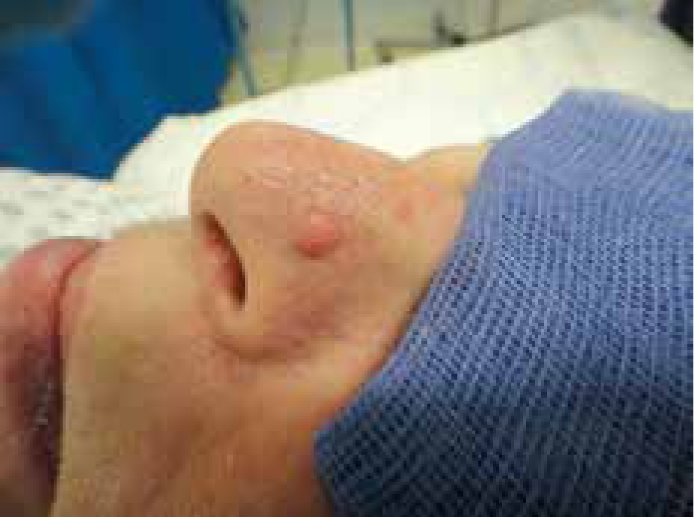
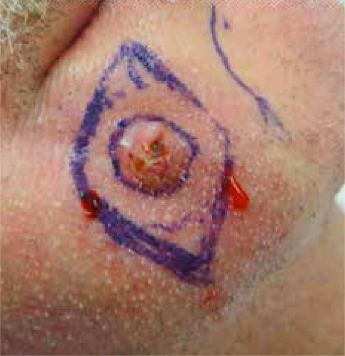
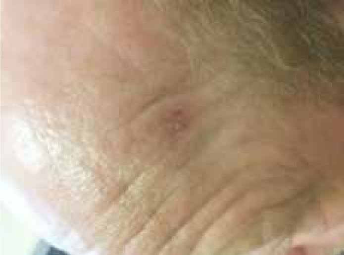
Nodular BCC: most BCCs on the face are the nodular type. These present as a single shiny nodule with telangiectasia and could appear as a pearly translucent nodule, which may ulcerate centrally and could bleed or create a crust (Figures 1, 2).
Morphoeic BCC: This is also known as sclerosing or infiltrative BCC. It appears on the middle of the face as a whitish/yellow, poorly defined patch or plaque. It is usually aggressive and prone to recurrence.
Superficial BCC: This presents as an erythematous patch or plaque, most commonly on the trunk.
Pigmented and basosquamous BCC (uncommon).
Figure 1. Nodular BCC. Note the telangiectasia sign on the nodule head.Figure 2. BCC ulcerated centrally with rolled edge.
Patients who present with features of BCC should be referred to a specialist for treatment.
Squamous cell carcinoma (SCC)
SCC is the second most common skin cancer. It accounts for about 20% of non-melanoma skin cancers.1,2 SCC is a malignant epithelial neoplasm of the keratinocytes of the epidermis. It grows faster than BCC. It is locally invasive and also spreads to distant organs and lymph nodes. Distant metastases can occur in the lung, liver, brain and bone.
Excessive sun exposure is a significant aetiological factor.3 Other risk factors include:
The presence of precursor lesions like actinic keratosis and Bowen's disease;
Smoking;
Long-standing leg ulcers;
Previous burn scars;
Previous radiotherapy;
Previous PUVA treatment;
Previous exposure to arsenic; and
Immunosuppressed patients (Leukaemia and lymphoma patients).3,4
The clinical appearance of SCC is variable. It could present as a non-healing ulcer, as a keratinized nodule, or as crusted plaque on sun-exposed sites which may bleed or ulcerate.
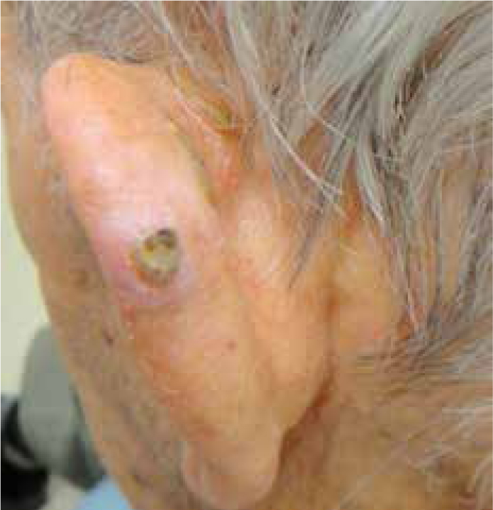
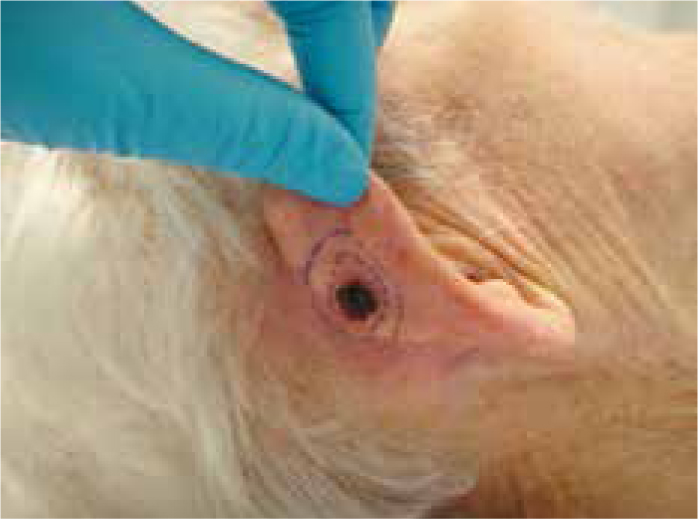
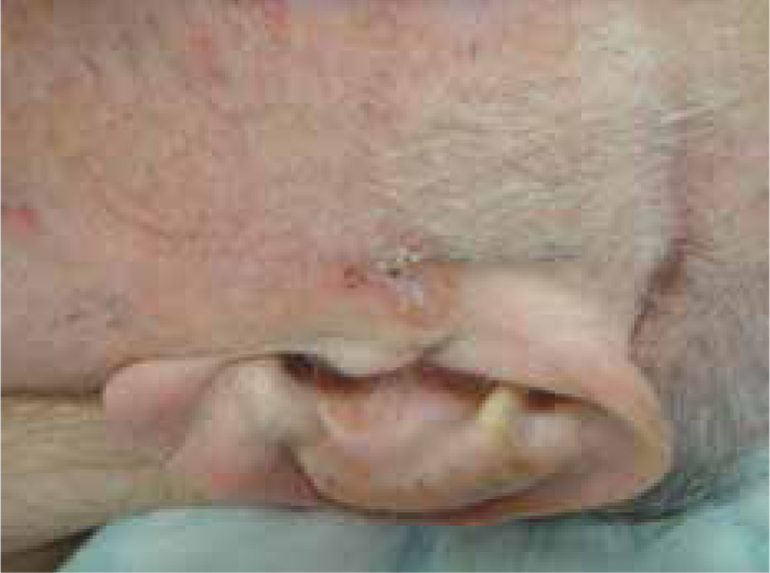
Patients who present with features of SCC should be urgently referred for diagnosis and treatment (Figures 3, 4).
Figure 3. SCC on left ear margins presented as small crack covered with crust.Figure 4. SCC on right ear presented as non-healing ulcer.
Bowen's disease
Bowen's disease (also known as cutaneous squamous cell carcinoma in situ) appears as an asymptomatic, single, slow-growing, scaly, erythematous patch or plaque with definite borders on the face, ear, back of the hand and the lower leg (Figure 5). The surface of the plaque could be flat, scaly, eroded or velvety. The silent nature of the lesion can lead to a large plaque by the time of presentation. It may be difficult to differentiate Bowen's plaque from SCC, BCC, or discoid eczema. This is why patients should be referred to arrange the biopsy as soon as possible.
Figure 5. Bowen's disease presented as single erythematous patch with scaly surface.
Keratoacanthoma
Keratoacanthoma is a benign fast-growing nodule with a central crateriform core filled with a keratinous material. It involutes by itself leaving a scar on sun-exposed sites. Excessive sun exposure is one of the main aetiological factors for keratoacanthoma. Sometimes it may be difficult to differentiate keratocanthoma from squamous cell carcinoma. Patients should be referred to a specialist for biopsy to confirm diagnosis.
Actinic (solar) keratosis
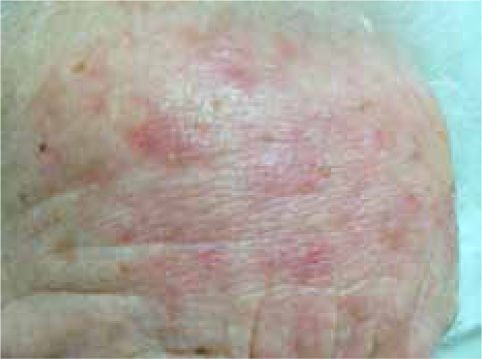
Actinic keratosis is a common lesion present on the sun-exposed skin of fair skinned elderly patients (Figure 6). It is a pre-malignant intra-epidermal skin lesion which may develop into squamous cell carcinoma. (Not all develop into invasive SCC. The estimated risk of developing into SCC ranges from 0.5% to 16%5). Chronic sun exposure is the aetiological factor for developing actinic keratosis. It appears as single or multiple brownish, yellowish or whitish plaques on the face and head, especially nose, cheeks, temples and forehead (Figure 7). These plaques are scaly and have a rough surface.6 They may cause discomfort and irritation. They should be referred to a specialist for either topical treatment or cryotherapy.
Figure 6. Actinic keratosis. Note the yellowish surface and the sun-damaged skin.Figure 7. Multiple actinic keratosis on forehead.
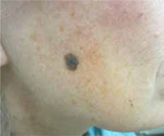
Malignant melanoma
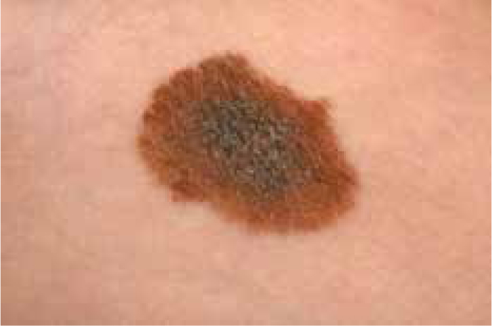
Although malignant melanoma is the least common type of skin cancer, it has the highest mortality rate. It could start either as a small dark patch anywhere on the skin or from an existing mole. It is the cancer of fair-skinned patients who have a long history of sun exposure (Figure 8).3,7
Figure 8. Malignant melanoma. Note the dark irregular margins.
General dental practitioners should ask about any recent changes in size, shape or colour of moles (those changes are known as major changes). Dentists should also ask about other symptoms, such as itching, bleeding, crusting and inflammation (these changes are known as minor changes).7 Dentists should arrange for urgent referrals to specialists if major or minor changes are noted.
The most common presentation of malignant melanoma on the face is the lentigo maligna melanoma which usually presents in patients over 60 on sun-damaged skin of the face (in particular the nose and the cheeks). Lentigo maligna melanoma could be preceded by the presence of a long-term pigmented freckle or lentigines, known as a lentigo maligna, which is an early form of melanoma in situ (the malignant cells are confined to the epidermis). The risk of this transformation is under 5% overall.7
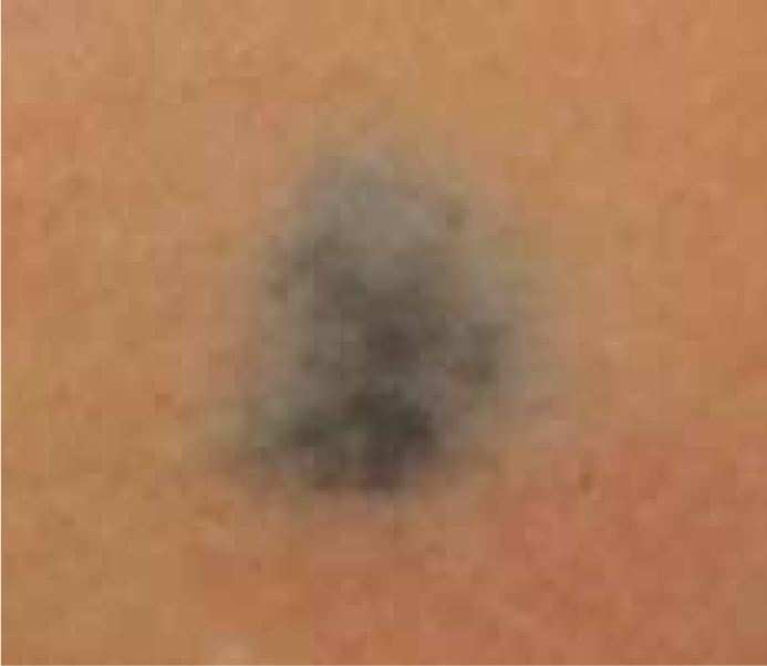
Blue naevi
Blue naevi is a benign mole which presents as a raised nodule or flat patch, usually on the face or scalp (Figure 9). It is usually very small and very dark blue/black in colour (the dark colour is because of the melanocytes located deeper in the skin than in the commoner brown mole). Blue naevi usually appears in teenagers, but could develop at any age. No referral or treatment is needed.
Figure 9. Blue naevi.
Seborrhoiec keratosis
Seborrhoiec keratosis is a common, well-defined benign nodule on the face and the scalp characterized by a ‘stuck-on appearance’ (Figure 10). It has a uniform skin colour or brown colour with a waxy surface. It could be flat, raised or pedunculated. It is most common in the elderly and presents as a single or multiple nodules.6 No referral or treatment is needed for asymptomatic seborrhoiec keratosis. Treatment could be indicated for cosmetic reasons or if symptomatic (itchy or rubbing against clothes).
Figure 10. Seborrhoiec keratosis. Note the waxy surface and the stuck-on appearance.
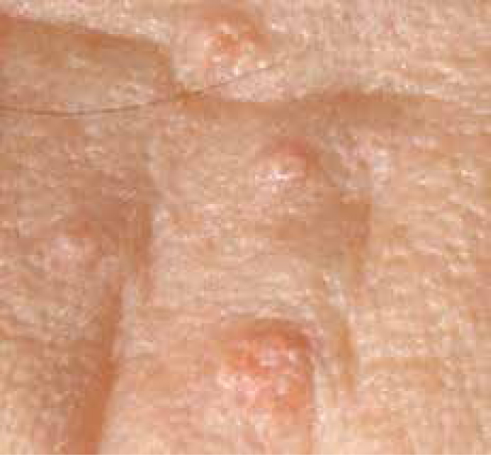
Sebaceous hyperplasia
Sebaceous hyperplasia is a common benign enlargement of sebaceous glands and the sebaceous duct (Figure 11). It presents as single or multiple yellowish soft and asymptomatic papules on the face (particularly on the nose, cheeks and forehead). It is most common in the elderly population. There is no need for referral or treatment of asymptomatic cases. Treatment is sometimes indicated for cosmetic reasons.
Figure 11. Sebaceous hyperplasia.
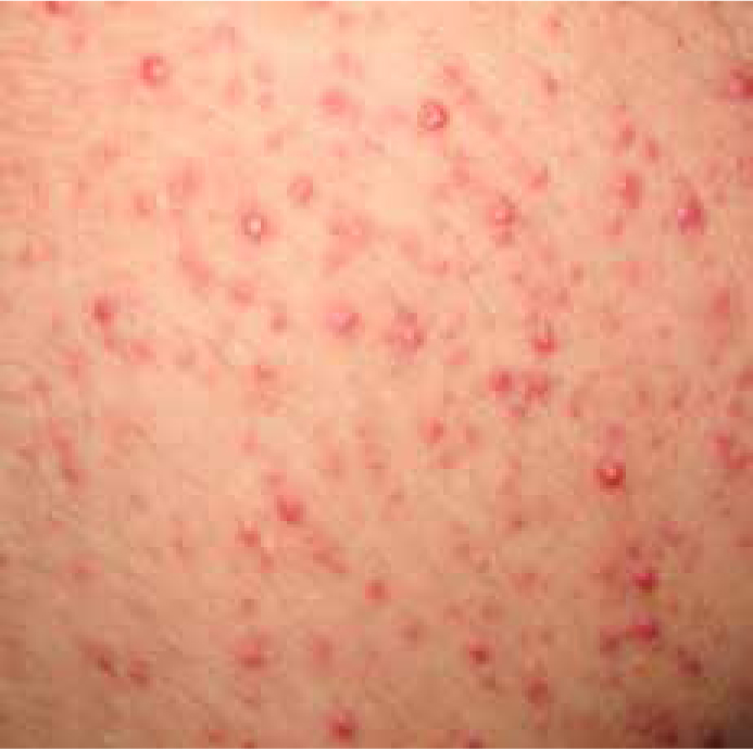
Folliculitis barbae
Folliculitis barbae is a common inflammation of bread hair follicles caused by inflammatory reaction surrounding ingrown facial hair (which can result from shaving) (Figure 12).8 A local infection of Staphylococcus aureus is usually associated with this. It presents as red- or skin-coloured itchy papules. Multiple follicles are usually affected. Patients who present with folliculitis barbae could be referred to their GPs as they sometimes benefit from a short course of antibiotics (topical or systematic).
Figure 12. Folliculitis barbae.
Conclusion
Dentists should recognize common facial lesions, understand the differential diagnosis and refer the suspicious lesions. Early referral is essential to provide the best management and prognosis.