Blau JN. How to take a history of head or facial pain. Br Med J. 1982; 285:1249-1251
Murphy E.London: Quintessence; 2008
Murray H, Locker D, Mock D, Tenenbaum HC. Pain and the quality of life in patients referred to a craniofacial pain unit. J Orofac Pain. 1996; 10:316-323
Svensson P, Baad-Hansen L, Newton-John T, Ng S, Zakrzewska JM. Investigations. In: Zakrzewska J (ed). Oxford: Oxford University Press; 2009
Scott J, Huskinson EC. Graphic representation of pain. Pain. 1976; 2:175-184
Melzack R, Katz J, Jeans ME. The role of compensation in chronic pain; analysis using a new method of scoring The McGill Pain Questionnaire. Pain. 1975; 23:101-112
Zigmond AS, Snaith RP. The Hospital Anxiety and Depression Scale. Acta Psychiatr Scand. 1983; 67:361-370
Pilling S, Anderson I, Goldberg D, Meader N, Taylor C. Br Med J. 2009; 339
Vickers ER, Zakrzewska JM. Dental causes of orofacial pain. In: Zakrzewska JM (ed). Oxford: Oxford University Press; 2009
Forssal H, Ohrbach R. Temporomandibular disorders (TMD). In: Zakrzewska J (ed). Oxford: Oxford University Press; 2009
Bergstrom I, List T, Magnusson T. A follow-up study of subjective symptoms of temporomandibular disorders in patients who received acupuncture and/or interocclusal appliance therapy 18–20 years earlier. Acta Odontol Scand. 2008; 66:88-92
Zakrzewska JM. Facial pain: an update. Curr Opin Support Palliat Care. 2009; 3:125-130
De Boever JA, Nilner M, Orthlieb JD, Steenks MH. Recommendations by the EACD for examination, diagnosis, and management of patients with temporomandibular disorders and orofacial pain by the general dental practitioner. J Orofac Pain. 2008; 22:268-278
Glaros AG, Owais Z, Lausten L. Diurnal variation in pain reports in temporomandibular disorder patients and control subjects. J Orofac Pain. 2008; 22:115-121
Zakrzewska JM. Assessment and treatment of trigeminal neuralgia. Br J Hosp Med. 2010; 71:490-494
Gronseth G, Cruccu G, Alksne J Practice parameter: the diagnostic evaluation and treatment of trigeminal neuralgia (an evidence-based review): report of the Quality Standards Subcommittee of the American Academy of Neurology and the European Federation of Neurological Societies. Neurology. 2008; 71:1183-1190
Jitpimolmard S, Radford SG. Neuropathic pain. In: Zakrzewska JM (ed). : Oxford University Press; 2009
Patton LL, Siegel MA, Benoliel R, De Laat A. Management of burning mouth syndrome: systematic review and management recommendations. Oral Surg Oral Med Oral Pathol Oral Radiol Endod. 2007; 103:S13-S39
Zakrzewska JM, Forssell H, Glenny AM. Interventions for the treatment of burning mouth syndrome. Cochrane Database Syst Rev. 2005;
Chong MS. Other neurological causes of head and face pain. In: Zakrzewska JM, Harrison SD (eds). Oxford: Elsevier; 2002
List A, Feinmann C. Persistent idiopathic facial pain (atypical facial pain). In: Zakrzewska JM (ed). Oxford: Oxford University Press; 2009
Baad-Hansen L. Atypical odontalgia – pathophysiology and clinical management. J Oral Rehab. 2008; 35:1-11
Zakrzewska JM. Diagnosis and management of non-dental pain. Dent Update. 2007; 34:134-136
Loder E, Rizzoli P. Tension-type headache. Br Med J. 2008; 336:88-92
Headache Classification Subcommittee of the International Headache Society. The International Classification of Headache Disorders 2nd Edition. Cephalalgia. 2004; 24:9-160
Differential diagnosis for orofacial pain, including sinusitis, tmd, trigeminal neuralgia Anne M Hegarty Joanna M Zakrzewska Dental Update 2024 38:6, 707-709.
Authors
Anne MHegarty
BDentSc, MSc(OM), MBBS, MFD, RCSI, FDS(OM) RCS
Consultant and Honorary Clinical Lecturer in Oral Medicine, Charles Clifford Dental Hospital, Sheffield S10 2ZS
Professor of Pain in Relation to Oral Medicine, Clinical and Diagnostic Oral Sciences Dental Institute, Barts and the London Queen Mary's School of Medicine and Dentistry, Turner Street, London E1 2AD, UK
Correct diagnosis is the key to managing facial pain of non-dental origin. Acute and chronic facial pain must be differentiated and it is widely accepted that chronic pain refers to pain of 3 months or greater duration. Differentiating the many causes of facial pain can be difficult for busy practitioners, but a logical approach can be beneficial and lead to more rapid diagnoses with effective management. Confirming a diagnosis involves a process of history-taking, clinical examination, appropriate investigations and, at times, response to various therapies.
Clinical Relevance: Although primary care clinicians would not be expected to diagnose rare pain conditions, such as trigeminal autonomic cephalalgias, they should be able to assess the presenting pain complaint to such an extent that, if required, an appropriate referral to secondary or tertiary care can be expedited. The underlying causes of pain of non-dental origin can be complex and management of pain often requires a multidisciplinary approach.
Article
Management of orofacial pain can only be effective if the correct diagnosis is reached and may involve referral to secondary or tertiary care. The focus of this article is differential diagnosis of orofacial pain (Table 1) rather than available therapeutic options.
The underlying cause of the majority of facial pain presentations in primary care will be of a dento-alveolar origin. These will not be discussed further here but their differentiating features are summarized in Table 2.
Diagnosis
Site
Character
Duration
Severity
Triggers
Radiation
Relieving factors
Associated factors
Appropriate referral point
DENTOALVEOLAR
Reversible pulpitis
Tooth
SharpStimulation evoked
Intermittent
Mild to moderate
Thermal Tactile Chemical
Adjacent teeth Upper/lower jaw
Removal of stimulus
Attrition Erosion Caries Cracked tooth
Irreversible pulpitis
Tooth
Sharp Throbbing
Intermittent Continuous
Mild to severe
Heat Chewing Lying supine
Regional Unilateral Upper/lower jaw
Cold
Deep caries
Periapical periodontitis
Tooth/gingival/bone
DeepContinuousBoring
Paroxysmal
Moderate to severe
Biting
RegionalUnilateral
Removal of trauma
Periapical erythemaSwelling Tooth mobility
Acute pericoronitis
Unerupted or partially erupted third molar mainly lower
Ache
Continuous
Moderate to severe
Biting
Ear Unilateral
Removalof traumaIrrigationAntibiotics
FeverMalaiseRegionallymphadenopathy
BONY PATHOLOGY
Alveolar osteitis (Dry socket)
Affected bone
SharpDeep seatedAche
Continuous 4–5 days post-extraction
Moderate to severe
Nil
RegionalUnilateral
IrrigationAntibiotics
Loss of clotExposed bone Halitosis
MUCOSAL DISEASE
Mucosal pathology
Affected mucosa
Sharp Burning Tingling
Intermittent
Mild to severe
SINUSITIS
Maxillary sinusitis
Over affected sinusUnilateral or bilateral
DullAchingBoring
Continuous
Mild to moderate
TouchBendingBiting upper teeth
Rare
DrainageMedication
History of URTI Purulent nasal discharge Fullness over cheek +/- erythema over cheek
SALIVARY GLANDS
Blocked salivary gland
80% Sub-mandibular
Burning Aching
Paroxysmal
Mild to severe
Smell or taste of food/drink
Local or regional if associated infection
Cessation of eating Removal of cause
Swelling Erythema Possible infection with pus from salivary gland duct
Oral Surgery
MUSCULOSKELETAL
TMD
Masticatory muscles TMJs
DullAchingThrobbingSharp
ContinuousorIntermittent
Mild to moderate
ProlongedChewing>Openingwide such as yawningStress
EarsHeadNeck
MedicationWarm compressesAvoidance of triggering factors
ClickingCrepitusLimitation in mouth openingDeviation of mandible on openingEar pain, fullness TinnitusDepressionAnxiety
Facial Pain Centre Oral Surgery
NEUROPATHIC
BMS
TonguePalateLipsPharynx
BurningTinglingTenderItching
Continuous +/-paroxysms
Mild to moderate
StressSpicy, acidic foods
To sites involved
Eating
Altered tasteAbnormalsalivaSensory change
Oral Medicine
Postherpetic neuralgia
Localized to site of herpes zoster infection Intra-oral but more often extra-oral
BurningTinglingShootingTenderItching
Continuous
Mild to moderate
Touch
Nil
MedicationLocal anaesthetic
AllodyniaHyperalgesiaAltered sensation
Oral MedicineFacial Pain Centre
Trigeminal neuropathic pain
Local to widespread Neuroana-tomical
Burning, tinglingAchingThrobbing
Continuous
Mild to severe
Light touchSpontaneous
Regional
MedicationLocal anaesthetic
AllodyniaTrauma historySensory change
Facial Pain Centre
TN
Trigeminal nerve
SharpShootingStabbingElectric-shock like
ParoxysmalSecondsRemits weeks/months
Moderate to severe
Light touchCold airWashing faceSpontaneous
Unilateral
MedicationSurgery
Trigger pointsPossible sensory change
Facial Pain Centre
Glosso-pharyngeal neuralgia
EarTonsilsNeck
SharpShootingStabbing
ParoxysmalSeconds/minutesRemits for weeks/months
Moderate to severe
SwallowingCoughingTouch
RegionalRarely bilateral
Medication
Possible TN
Facial Pain Centre
VASCULAR
Cluster headaches
UnilateralPeriorbitalTemple
BoringThrobbing
RegularRecurring 1–8 attacks per day, lasting 15–180 mins‘Alarm clock’ wakeningCompleter emission for months to years
Moderate to servere
SmokingAlcoholAltitude seasonal
PeriorbitalTemple
Medication
Autonomic features such as nasal congestion, eye redness/injection
Facial Pain CentreNeurology
Paroxysmal hemicrania
UnilateralPeriorbitalTemple
Boring
Paroxysmal 1–40 attacks per day lasting 2–30 mins
Moderate to severe
Neck movement
PeriorbitalTemple
Medication
Autonomic features
Facial Pain CentreNeurology
SUNCT/SUNA
Unilateral mainly first second division trigeminal
Stabbing
Recurring 1–200 attacks per day, 10–250 seconds each
Moderate to severe
Cutaneous triggers
OrbitalTemporal
Medication
TearingConjunctival injection or other autonomic features for SUNA
General MedicalPractitionerNeurology (if complicated)
OTHER
Atypical odontalgia
Tooth bearing area
DullAching, tinglingThrobbingSharp
Continuous
Mild to moderate
Touch
Rare
Nil
Prior dental treatment
Facial Pain Centre
CIFP
Non anatomicalIntra-oralExtra-oral
DullAchingNaggingSharp
ContinuousIntermittentParoxysmal
Mild to moderate
StressFatigueChewing
Deep poorly localizedNo specific radiation site
Rest
Multiple body symptoms Life events
Facial Pain Centre
URTI = upper respiratory tract infection
TMD = temporomandibular disorder
BMS = burning mouth syndrome
TN = trigeminal neuralgia
CIFP = chronic idiopathic facial pain
SUNCT = Short-lasting, Unilateral, Neuralgiform headache with Conjunctival injection and Tearing/SUNA Short-lasting, Unilateral, Neuralgiform headache with Autonomic features
To establish a differential diagnosis for orofacial pain we must first consider the history, examination and relevant investigations.
Although both may co-exist, the more rare non-dental pain must be distinguished from dental pain to avoid unnecessary dental treatment and to organize appropriate referral for the patient. It is essential that patients are referred to the correct departments within secondary or tertiary care to ensure the most efficient management for patients and to maximize use of NHS resources.
Pain history
A thorough pain history is crucial and time needs to be taken when taking it as it should provide sufficient detail to guide clinicians to the most likely diagnosis. It is also important to institute relevant investigations.
In 1936, Ryle's classic analysis of pain highlighted 11 essential questions to be included in the pain history1 and these still apply today and have been further expanded and grouped in more recent years.2 Questions include:
Onset;
Frequency;
Duration;
Site;
Radiation, deep or superficial;
Triggering;
Aggravating or relieving factors;
Quality;
Severity;
Associated symptoms.
This format allows a logical approach to history-taking, which is essential.
Orofacial pain interferes with daily life activities, impacting negatively on quality of life and this impact should therefore be established.3,4
Other aspects of the history of particular relevance when considering chronic orofacial pain aetiologies and determining best therapy include:
Previous management;
Past medical and dental history;
Medications and allergies;
Social and family history, which may disclose psychological factors and aspects of a patient's beliefs of the cause of pain, which may in turn influence the extent and nature of the pain.
Chronic orofacial pain results in decreased quality of life and psychological effects rarely seen in dental pain.
Clinical examination
Clinical examination should include a thorough extra-oral and intra-oral examination to corroborate history findings and assist in reaching a diagnosis. Extra-oral examination should include temporomandibular joints (TMJs), regional lymph nodes, muscles of mastication and cervical muscles, salivary glands and face and eyes for any autonomic signs, such as flushing, tearing, ptosis or sweating. Cranial nerves examination may be required in some cases and, in primary care at least, a gross examination of the facial and trigeminal nerves would be expected to assess any motor or sensory abnormalities. Sensation to light touch and pin prick can easily be elicited by the use of cottonwool and an appropriate sterile pin, respectively, and assessment of the facial nerve should include a patient's ability to raise the eyebrows, close the eyes tightly shut and show his/her teeth whilst observing any facial asymmetry.
Limitation of mouth opening and/or deviation of the mandible on opening, TMJ tenderness, TMJ crepitus and/or click and masticatory muscle pain or tenderness may indicate temporomandibular disorders (TMD) and can be determined by palpation over the TMJs and masticatory muscles. Most patients can open comfortably to 35–45mm, equating to approximately three finger breadths, although some may open to a greater distance. Crepitus and clicking can usually be elicited by palpation over the TMJs and loud clicking will be audible. Facial swelling/asymmetry should be assessed.
The intra-oral examination should include a comprehensive oral examination, including:
Assessing the teeth;
Occlusion;
Salivary glands;
Oral mucosae; and
Oropharyngeal region.
As most primary care practitioners are familiar with examining the oral cavities, these will not be detailed here.
Oral malignancy, in particular gingival or alveolar bone malignancy, may present with facial pain. Clinical features of oral malignancy may also include sensory changes or trismus. Metastatic disease spreading to the jaws must also be considered. Evidence of oral candidosis or xerostomia may be relevant when excluding a diagnosis of burning mouth syndrome (BMS).
Pain associated with the salivary glands may occur as a result of salivary stones blocking the ducts and giving rise to intermittent pain typically associated with eating. There may be tenderness of the glands. Bimanual palpation of the submandibular glands is essential. Infections and tumours of the salivary glands may also give rise to localized pain in the region of the glands and a purulent discharge may be seen from the openings of the ducts within the mouth, indicative of infection. Pain may be relieved by massaging the glands involved.
Investigations
The history and examination alone may lead us to the correct diagnosis. Investigations help exclude or confirm a diagnosis, assist in treatment planning and monitor treatment effects, including adverse effects. Investigations can be broadly classified as haematological, radiological, physiological and psychological. Simple chairside tests to confirm a dental source of pain include vitality testing using heat, cold and electric stimuli, transillumination to highlight proximal caries or a cracked tooth and use of tooth ‘slooth’ to assist localization of cracked cusps. With transillumination, a light beam is reflected where there is a change in the integrity of the tooth structure. The tooth ‘slooth’ is an instrument incorporating an indentation allowing pressure to be directed to a specific cusp to elicit pain suggestive of a cracked cusp.
Measuring erythrocyte sedimentation rate (ESR) in giant cell arteritis (GCA) is important as a high ESR helps to reach the diagnosis. ESR is a simple haematological investigation which measures the rate at which red blood cells fall in a column of blood within an hour and is a non-specific measure of inflammation. In giant cell arteritis, the ESR usually exceeds 50 mm/hr and sometimes 100 mm/hr. GCA can cause a sudden loss of vision due to anterior ischaemic optic neuropathy, constituting a medical emergency, therefore ESR should be checked and treatment commenced as soon as possible if GCA is suspected to prevent possible sight loss.
Baseline blood tests may be useful in eliminating any possible systemic disorders which may cause or exacerbate facial pain. A variety of conditions may lead to burning sensation of the oral mucosae, including vitamin and nutritional deficiencies and, therefore, to diagnose burning mouth syndrome (BMS) correctly, it is essential to check full blood count, haematinics (iron, vitamin B12 and folate levels) and blood glucose before reaching a diagnosis of BMS. Immunological tests help to exclude connective tissue disorders which can cause trigeminal neuropathy.
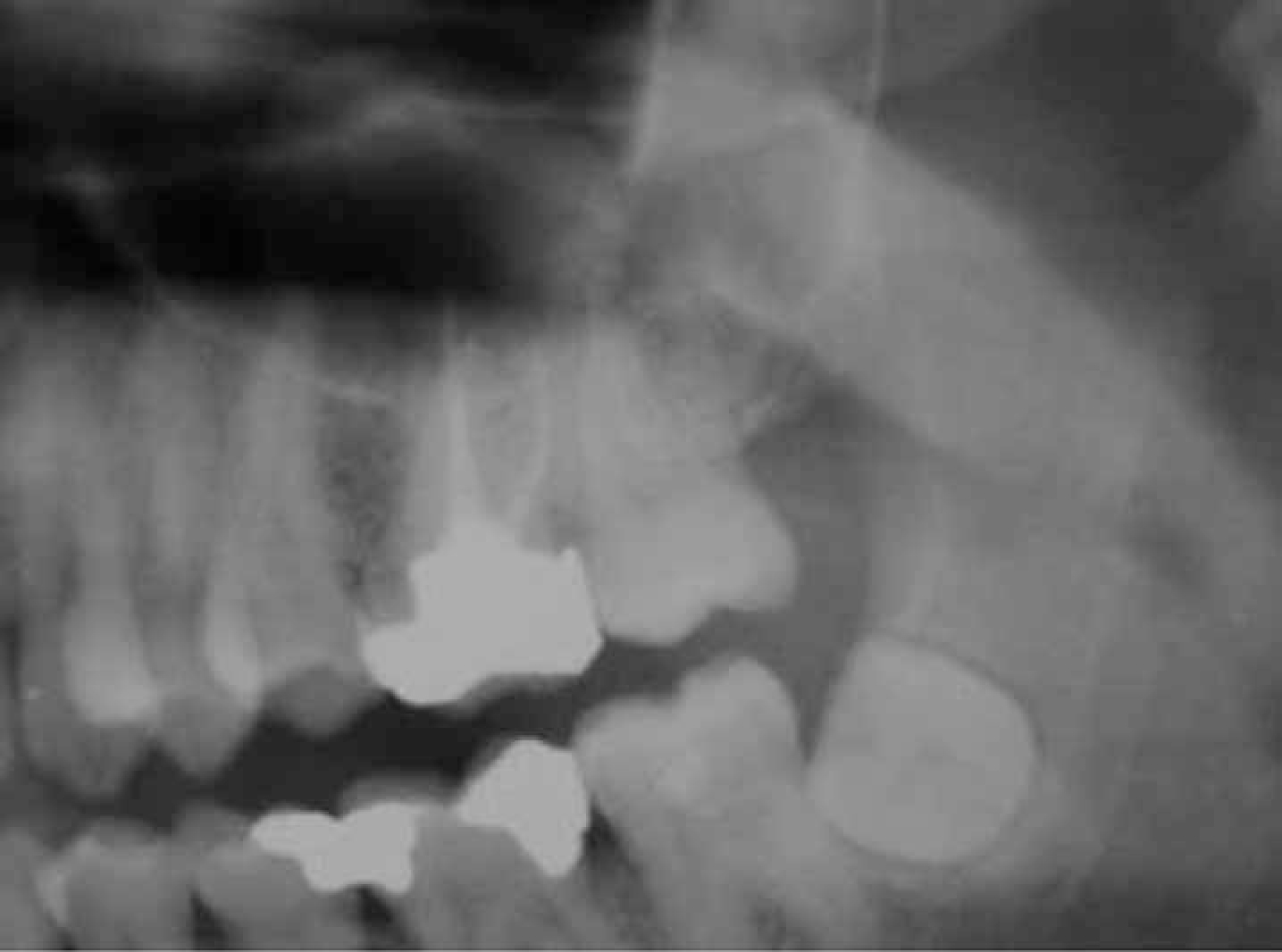
Intra-oral radiographs may involve periapical, bitewing and occlusal views, while extra-oral radiographs include dental panoramic tomography (DPT) and oblique laterals. Intra-oral radiographs are the recommended first line investigation for diagnosing caries, periodontal and periapical pathology (Figure 1), with DPT being reserved for bony lesions and when views of the condyles are required. Other forms of radiological studies, such as CT, MRI, bone scintigraphy, ultrasound scan, sialography and arthrography, may be useful in diagnosing salivary gland disease, malignancies, bony pathology and temporomandibular joint disease, but should only be carried out if there are clinical indications. MRI will demonstrate secondary causes of trigeminal neuralgia.
More complex investigations, such as quantitative sensory testing (QST) which may be useful when investigating trigeminal neuropathic pain, should only be arranged by specialists who can interpret the results. Quantitative sensory testing is a psychophysical test and is a reliable way of assessing large and small sensory nerve fibre function. Results should always be interpreted in light of the patient's clinical presentation. Psychological investigations are generally not arranged by the primary dental practitioner but should be encouraged. There are a wide variety of validated questionnaires available that assess all aspects of pain experience including:
Pain intensity;
Mood; and
Disability.
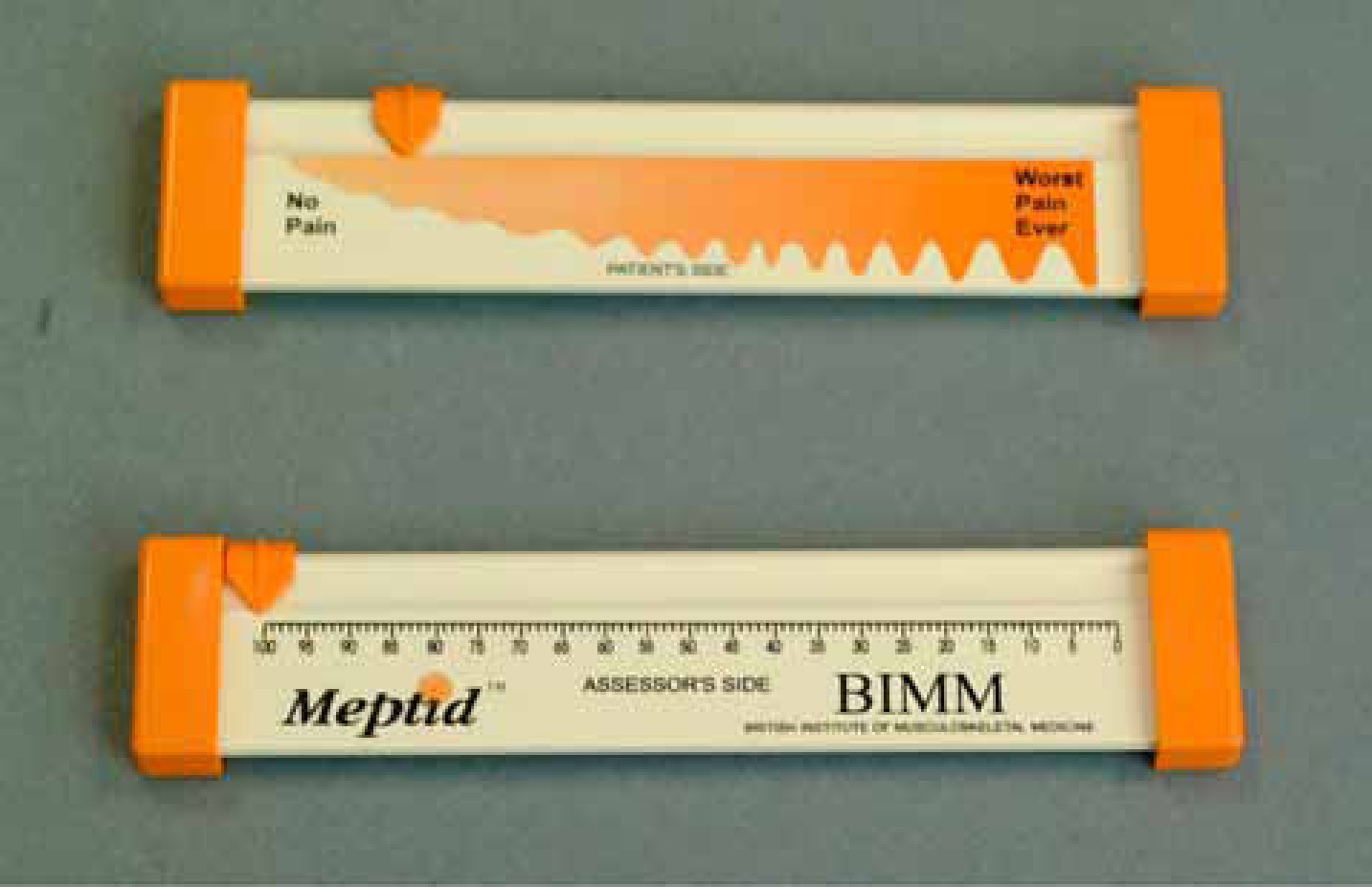
Their use should be mandatory in patients with chronic orofacial pain.5 The Visual Analogue Scale, VAS (Figure 2), is useful for assessing pain intensity and the McGill Pain Questionnaire useful for assessing pain quality.
Figure 2. Visual Analogue Scale (VAS).
The VAS is a simple, reproducible instrument that allows the severity of pain to be expressed as a numerical value.6 The VAS is represented as a plain horizontal 100 mm line (Figure 2) with 0 representing no pain and 100 representing worst possible pain. Patients mark on this line the point which they feel best represents their perception of their facial pain. This can also be used as a verbal rating scale asking patients to express their pain as a range from 0 to 10. Another commonly used pain assessment tool is the McGill Pain Questionnaire which allows the patient to indicate easily the quality of his/her pain using such descriptors as throbbing, shooting, distressing, excruciating.7
A number of psychological tests are useful in patients with chronic facial pain as it has been reported that depression may be co-existent in up to 50% of patients and anxiety in 15%.5 The Hospital Anxiety and Depression Scale (HADS) is a screening tool which involves the patient completing a questionnaire composed of statements (Table 3), with a choice of answers relevant to either generalized anxiety or depression. The answers are scored and, when the final score is compared to a set of normal values, anxiety or depression is suggested, triggering a referral to either a psychologist or psychiatrist.8
I still enjoy the things I used to enjoy:
Definitely as much
0
Not quite so much
1
Only a little
2
Hardly at all
3
Recent NICE guidelines on depression advocate the use of two questions in primary care settings to determine patients at risk of being depressed:
During the last month, have you often been bothered by ‘feeling down,’ depressed, or hopeless?
During the last month, have you often been bothered by having little interest or pleasure in doing things?
9
Non-dental causes of facial pain
Maxillary sinusitis
Maxillary sinusitis causes a constant boring pain with zygomatic and dental tenderness from the inflammation of the maxillary sinus.10 Acute maxillary sinusitis is defined by the International Association for the Study of Pain (IASP) as constant burning pain with zygomatic and dental tenderness from the inflammation of the maxillary sinus.10 In chronic cases there may be no pain or just occasional mild diffuse discomfort. Diagnostic criteria for maxillary sinusitis include:
Purulence in the nasal cavity;
Simultaneous onset of headache and sinusitis;
Pain over the antral area which may radiate to the upper teeth or forehead; and
Headache disappearing after treatment of acute sinusitis.10
The character of the pain of maxillary sinusitis is dull, aching, boring and tender, of mild to moderate severity, is usually continuous and may be either unilateral or bilateral commencing after an upper respiratory tract infection. The pain is triggered by bending forward, touching the area or biting on the upper teeth. Headache is located over the antral area. In the presence of the key diagnostic symptoms, investigations are not required but confirmation of the diagnosis can be confirmed by maxillary sinus radiographic examination (although this is not generally advised as, apart from showing possible fluid levels in acute sinusitis, it is not of great benefit), computerized tomography (CT) or magnetic resonance imaging (MRI).
Temporomandibular disorders
Temporomandibular disorders encompass pain affecting the masticatory muscles and/or temporomandibular joints (TMJs). They consist of muscular pain MSK (referred to by some as myofascial pain), TMJ disc interference disorders and TMJ degenerative joint disease; this latter rarely causes pain but results in limitation of opening.11 In the case of trauma, the pain is usually self-limiting but psychological aspects may contribute to chronicity of the pain, therefore it is important to manage it early. TMD (MSK) is more prevalent in females and the natural history is that of intermittent pain with continuation for many years.12 Tension type headaches can be mistaken for TMD. There is increasing evidence that TMD is linked to many other chronic pain conditions, such as headaches, migraine, post-traumatic stress disorder and fibromyalgia.13 The relationship between TMD pain and clenching habit or bruxism is far from simple5,13,14, and daily variations in pain do not correlate with self-reports of clenching or grinding.15
Trigeminal neuralgia
Trigeminal neuralgia (TN) is defined by the International Association for the Study of Pain (ISAP) as a ‘sudden and usually unilateral severe brief stabbing recurrent pain in the distribution of one or more branches of the fifth cranial nerve’.16 Idiopathic and secondary forms are recognized and conditions such as multiple sclerosis, benign or malignant lesions being contributory factors. Categorization of TN into classical and atypical forms is based on symptoms and not aetiology.17 TN is being increasingly recognized with its annual incidence now being estimated around 12.8 per 100,000, with a peak incidence in 50–60-year-olds. TN symptoms arising in younger patients should alert the clinician to the possibility of an underlying cause, such as multiple sclerosis.
Classical TN presents with shooting, sharp, unbearable pain in the distribution of one or more branches of the trigeminal nerve, of moderate to intense severity, lasting seconds.17,18 The right side of the face is affected in 60% of sufferers, it is unilateral in 97% of cases and rarely in first division only. It is precipitated by light touch, but may be spontaneous, and there are often associated trigger points. Patients may have periods of remission lasting days, weeks or longer.
Glossopharyngeal neuralgia (GPN)
Defined by the IASP as sudden severe recurrent pain in the distribution of the glossopharyngeal nerve, GPN is very rare, with an incidence of 0.7 per 100,000, and is more common in females and those aged over 50 years.19 Classic and secondary forms are recognized. Classic GPN is severe recurrent stabbing pain in the ear, base of tongue, tonsillar fossa or below the angle of the mandible. It is precipitated by swallowing, talking or coughing.
Secondary GPN presents with an additional ache that may persist between attacks and is secondary to a cranial lesion demonstrable by investigations or surgery. The pain is unilateral in location and there are no obvious motor neurological defects. Episodes of pain may last from weeks to months.
Although also rare, a syndrome known as Eagles syndrome should be considered in patients presenting with classical symptoms of GPN. Eagles syndrome describes symptoms related to an elongated styloid process impinging on adjacent anatomical structures and is associated with pain and dysphagia on chewing and on turning the head to the affected side.
Burning mouth syndrome (BMS)
BMS is characterized by continuous burning pain of the oral mucosa in the absence of any contributing local or systemic pathology.20,21 There is an increasing number of studies suggesting that this is not just a psychological condition but is probably neuropathic.13
The symptoms of BMS include:
Burning sensation affecting tongue, palate, gingiva, lips and pharynx;
Tingling sensation;
Altered taste;
Perceived dry mouth; and
Altered tactile sensations.
Most patients have continuous pain but it can vary throughout the day. Most patients do not associate food or drink with the onset of pain but some will describe the pain as being exacerbated by certain foods, such as spicy or acidic food, whereas others find feeding relieves their pain. Many BMS patients score high in tests for depression or anxiety, possibly because the condition is not recognized, patients are not believed and they are not given an adequate explanation.
When excluding an organic cause for BMS, a thorough, systematic soft tissue examination is important and recommended investigations include:
Haematological and biochemical investigations to assess if anaemic, low in iron, folate or vitamin B12, or if there is a raised level of glucose;
Microbiological tests for candidosis;
Baseline saliva flow rate if there is any question of hyposalivation;
Sensory testing;
Allergy testing; and
Immunological testing for conditions such as Sjögren's syndrome or systemic lupus erythematosus. A detailed drug history will highlight any drugs that may be associated with burning oral pain.
Trigeminal neuropathic pain
Trigeminal neuropathic pain or traumatic induced neuralgia is a form of chronic facial pain arising as secondary to injury to the trigeminal nerve, such as facial trauma or a dental procedure. It is rare but increasingly recognized and the pain is described as a continuous burning sensation localized to the injured area, but may be described as constant, dull, burning with or without intermittent sharp stabbing pain. Numbness and tingling may also be present due to nerve dysfunction. The pain symptoms may be classed under the following:
Dysaesthesia (abnormal perception of pain);
Allodynia (due to a stimulus which does not normally provoke pain); or
Hyperalgesia (an increased sensitivity to pain).
Proposed mechanisms for trigeminal neuropathy include peripheral or central sensitization, beta fibre reorganization and sympathetically maintained pain due to alpha receptor sprouting.19
Trigeminal neuropathy, with and without pain, is associated with a number of connective tissue disorders including:
Scleroderma;
Sjögren's syndrome;
Mixed connective tissue disease;
Systemic lupus erythematosus;
Rheumatoid arthritis; and
Dermatomyositis.
The underlying pathology for trigeminal nerve dysfunction in these patients is unknown but could be related to a form of vasculitis.22
Atypical odontalgia (AO)
Considered by most as a form of trigeminal neuropathic pain, atypical odontalgia may in fact have both psychological and neuropathic origins. There is limited evidence on the incidence and prevalence of AO. Clinical features include persistent pain, often commencing in conjunction with some form of dental treatment, particularly root canal therapy or extraction. In fact, over 80% of patients relate the onset of their pain to dental treatment, including local anaesthesia.13 The most common site of pain is the molar and premolar region. The pain is intra-oral, well localized and not associated with radiation to adjacent areas or extra-orally. Several studies have reported associated features of hyperaesthesia and allodynia at the pain site, with a prolonged response to ethyl chloride.23
AO often results in repeated, and possibly unnecessary, dental treatment such as extractions, root canal therapy and apicectomies in the pursuit of pain relief.21 A patient presenting with such pain and giving a history of multiple extractions possibly preceded by root canal therapies should raise suspicions of AO. Diagnosis and management as early as possible is vital24 to avoid unnecessary invasive treatments.
Post-herpetic neuralgia (PHN)
PHN is persistent burning pain but can be an excruciating/severe pain accompanied by intermittent shooting sensation localized to the site of previous herpes zoster infection, occurring 3–6 months after resolution of the infection.19 Allodynia, hyperalgesia and numbness in the affected area have been reported. The neuralgia is dermatomal in location. Ramsay Hunt syndrome, also caused by herpes zoster infection, specifically of the geniculate ganglion of the VIIth cranial nerve,22 presents with facial pain, lower motor neuron palsy and ipsilateral vesicles, affecting the skin of the ear canal, auricle and/or mucous membrane of the oropharynx. The pain is usually localized, paroxysmal, deep within the ear, but can radiate externally and may become more dull and diffuse in nature. Unlike PHN, the onset of pain usually precedes the rash by several hours or days and the disease is self-limiting.
PHN is thought to affect 40% of patients and most patients are more than 70 years of age. Following initial exposure to the herpes zoster virus from chicken pox, the virus lies dormant in the trigeminal ganglion and, when activated, gives rise to the rash of shingles. PHN is the result of damage to the nerve by the virus.
Chronic idiopathic facial pain
Chronic (persistent) idiopathic facial pain (CIFP), previously atypical facial pain, is persistent facial pain which is poorly understood, but its persistence is likely to result in psychological distress. The pain is described as aching, heavy, nagging, sometimes throbbing or stabbing.25 It does not follow anatomical pathways and can be local or very extensive, radiating into the head and neck. The pain is often constant but with varying intensity.
Psychological stresses or fatigue may worsen the symptoms and it is therefore important to take a relevant psychosocial history and record associated stress-related factors. Information regarding marital status, family medical history, employment status, history of depression or anxiety and sleep problems are all relevant. Exploring patients' beliefs about their pain can be particularly enlightening. There are no specific relieving factors and the patient may also suffer irritable bowel syndrome, back and neck pain and poor sleep.
Migraine
Migraine affects one in four women and one in 12 men in the UK.26 It is a chronic headache disorder affecting the frontotemporal region. It often co-exists with TMD, which may exacerbate migraines. The headache is typically unilateral, severely disabling, usually lasting 4–72 hours and is associated with photophobia, phonophobia, nausea and/or vomiting. It may be preceded by an aura in 15% of cases, with visual disturbances being most common.3 It will not be discussed further here but the salient features of the condition are highlighted in Table 2. Changes in frequency, intensity and location are often found in women whose migraines are hormonally driven.
Tension type headache
Episodic and chronic forms of tension-type headache are recognized. The episodic form lasts from 30 minutes to days, with a pressing quality, of mild to moderate intensity, is bilateral, with less than 15 attacks per month and no aggravating factors or associated symptoms, unlike the chronic form which, although of similar character and location, occurs more than 15 times per month for at least 6 months with associated nausea, photophobia or phonophobia. The pathophysiology of this form of headache is not fully understood, its prevalence is quoted as 2.2% and is more common in females.27 It can mimic TMD MSK.
Giant (temporal) cell arteritis (GCA)
GCA is a form of vasculitis involving cell-mediated immune damage to blood vessel walls and mainly affects blood vessels in the head and neck region. It is rare under the age of 50 years and females are about 3 times more likely than men to develop this disease. The temporal artery is commonly affected giving rise to temporal arteritis. Symptoms include unilateral or bilateral headache of aching or throbbing quality, often intense and continuous. Patients may have features of scalp tenderness, visual changes and/or neurological changes.
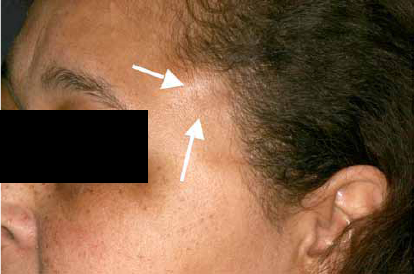
Criteria stipulated by the International Headache Society (IHS) for a diagnosis of temporal arteritis is any new persistent headache in the temporal region, with either swollen tender scalp artery (Figure 3) and raised ESR or CRP, or temporal artery biopsy demonstrating giant cell arteritis.19 Major improvement or resolution of headache within 3 days of high dose steroid treatment also helps confirm the diagnosis.
Figure 3. Temporal arteritis with swollen temporal artery highlighted.
GCA may be associated with polymyalgia rheumatica, jaw claudication, weight loss, altered sensation or loss of vision. Owing to the high risk of early visual loss as a result of anterior ischaemic optic neuropathy, prompt diagnosis and management is essential.
Trigeminal autonomic cephalalgias (TACs)
TACs are a group of headache syndromes incorporating short lasting severe unilateral headache attacks, accompanied by cranial autonomic symptoms. TACs is included in the International Headache Society classification of headaches28 and includes cluster headache, paroxysmal hemicrania and short-lasting unilateral neuralgiform headache attacks with conjunctival injection and tearing (SUNCT), all of which display trigeminal distribution pain and ipsilateral cranial autonomic features. The primary site of pain is in the distribution of the first division of the trigeminal nerve and autonomic features present. They are rare, not expected to be diagnosed in primary care and the main features are highlighted in Table 2.
Conclusions
Differentiating the many causes of facial pain can be difficult for busy practitioners, but a logical approach to history-taking is important and will aid more rapid diagnoses with effective management. Although primary care clinicians would not be expected to diagnose rare pain conditions, they should be able to assess the presenting pain complaint and refer to the appropriate secondary or tertiary care centre. It is important that primary care practitioners provide sufficient detailed information of history, examination and investigation findings in their referral letters to ensure appropriate direction of the referral within the secondary/tertiary care institution.
Underlying causes of orofacial pain are wide ranging and complex, but a greater understanding of a patient's facial pain symptoms, towards establishing a diagnosis or differential diagnosis, can be achieved by obtaining a good pain history, carrying out a good clinical examination and instituting relevant investigations or referring to secondary or tertiary care when appropriate.