
Article


Specialist referral may be indicated if the Practitioner feels:
Truly white oral lesions appear white usually because they are keratotic (composed of thickened keratin, which looks white when wet) or may consist of collections of debris (materia alba), or necrotic epithelium (such as after a burn), or fungi (such as candidosis). These can typically be wiped off the mucosa with a gauze swab.
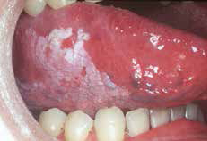
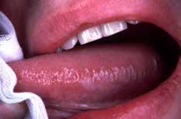
Other lesions, which cannot be wiped off, also appear white usually because they are composed of thickened keratin (Figure 1). A few rare conditions that are congenital, such as white sponge naevus (Figure 2), present in this way, but most white lesions are acquired and many were formerly known as ‘leukoplakia’, a term causing misunderstanding and confusion. The World Health Organization originally defined leukoplakia as a ‘white patch or plaque that cannot be characterized clinically or pathologically as any other disease’, therefore specifically excluding defined clinicopathologic entities such as candidosis, lichen planus (LP) and white sponge naevus, but still incorporating white lesions caused by friction or other trauma, and offering no comment on the presence of dysplasia. A subsequent international seminar defined leukoplakia more precisely as:
‘…a whitish patch or plaque that cannot be characterized clinically or pathologically as any other disease and which is not associated with any physical or chemical causative agent except the use of tobacco’.
There is a range of causes of white lesions (Table 1), but morphological features and site may also give a guide to the diagnosis.
|
Local causes
|
For example, focal lesions are often caused by keratoses; multifocal lesions are common in thrush (pseudomembranous candidosis) and in LP; striated lesions are typical of LP; and diffuse white areas are seen in the buccal mucosa in leukoedema and some LP, in the palate in stomatitis nicotina and at any site in keratoses. White lesions are usually painless but this may not be the case in burns, candidosis, LP, or lupus erythematosus.
Local causes of white lesions
Debris, burns (from heat, chemicals such as mouthwashes), grafts and scars may appear pale or white. Materia alba can usually easily be wiped off with a gauze.
Furred tongue
Tongue coating is common, particularly in edentulous adults on a soft, non-abrasive diet, people with poor oral hygiene, and those who are fasting or have febrile diseases. The coating appears more obvious in xerostomia. The coating consists of epithelial, food and microbial debris and the tongue is the main reservoir of some micro-organisms, such as Candida albicans and some Streptococci, and the various anaerobes implicated in oral malodour (Article 5).
Diagnosis
The history is important to exclude a congenital or hereditary cause of a white lesion. The clinical appearances usually strongly suggest the diagnosis: biopsy is only required if the white lesion does not scrape away from the mucosa with a gauze.
Management
Treatment is of the underlying cause where this can be identified.
Congenital causes of white lesions
Fordyce spots
Some common whitish conditions, notably Fordyce granules (ectopic sebaceous glands), are really yellowish, but may cause diagnostic confusion (Figure 3). This condition is entirely benign and does not require any further intervention, though there is some evidence they may become more prominent in hereditary nonpolyposis colorectal cancer.
Leukoedema
Leukoedema is a common benign congenital whitish-grey filmy appearance of the mucosa, seen especially in the buccal mucosae bilaterally in people of African or Asian descent. Diagnosis is clinical; the white appearance disappears if the mucosa is stretched. No treatment is available or required.
Inherited dyskeratoses
Inherited disorders of keratin are rare, but may be diagnosed, especially if there is a positive family history or other associated features, such as lesions on other mucosae, or skin appendages such as the nails.
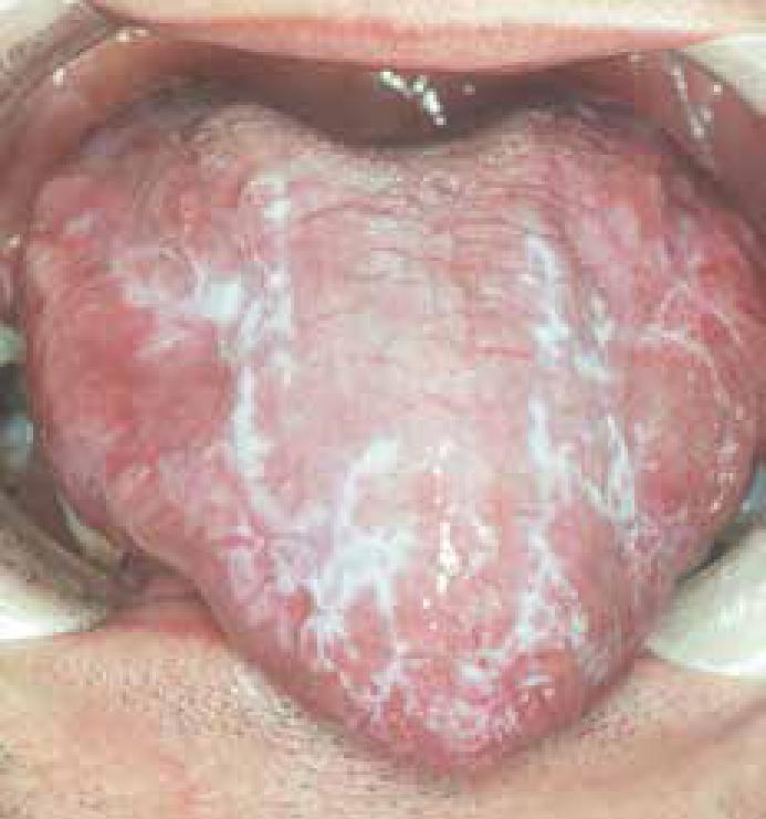
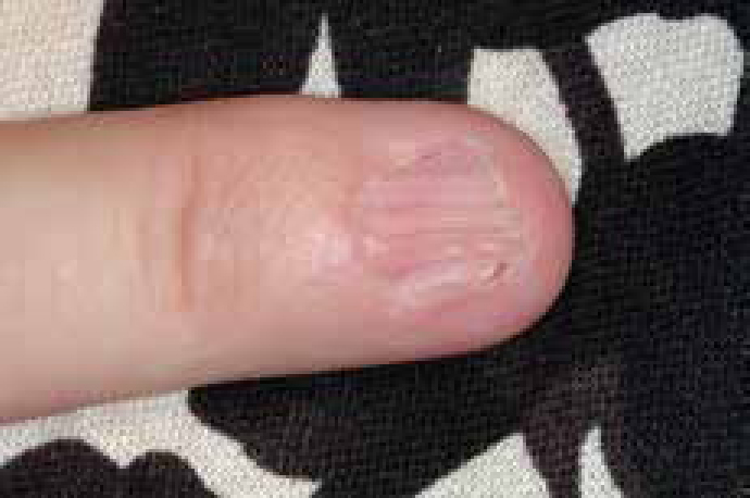
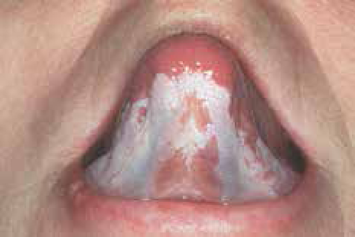
White sponge naevus, the commonest of the inherited dyskeratoses, is an autosomal dominant condition with variable expression and a high degree of penetrance. It generally presents during childhood and is characterized by thickened, folded white patches, most commonly affecting the buccal mucosae. Other mucosal sites in the mouth may be involved and some patients may have similar lesions affecting genital and rectal mucosa. Since the other dyskeratoses may have wider implications and, in particular the risk of malignant transformation, specialist care is indicated.
Inflammatory causes of white lesions
Infections
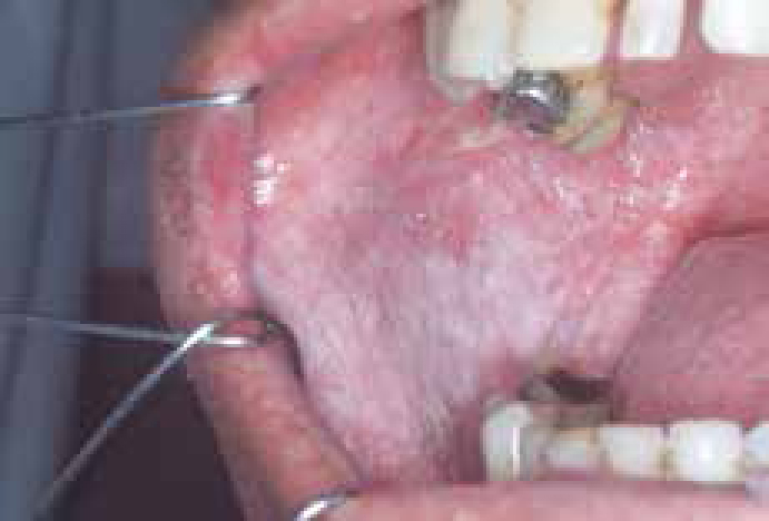
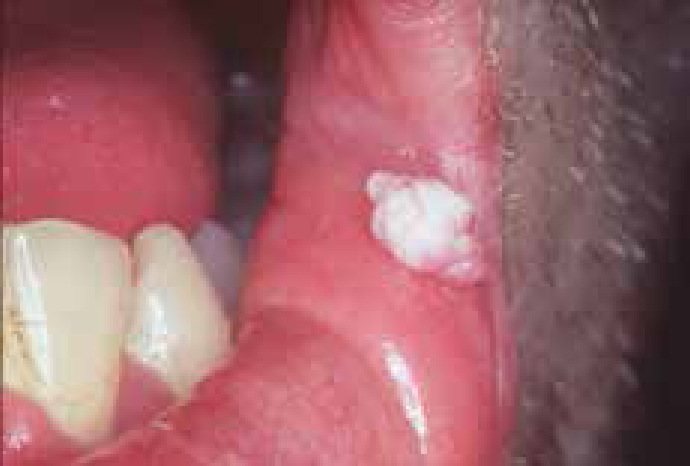
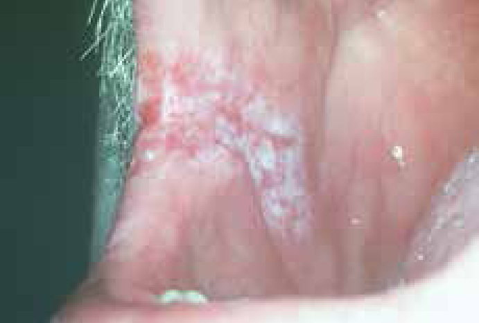
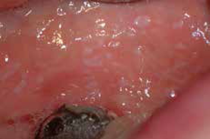
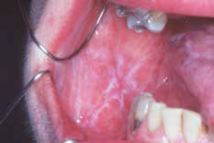
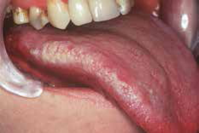
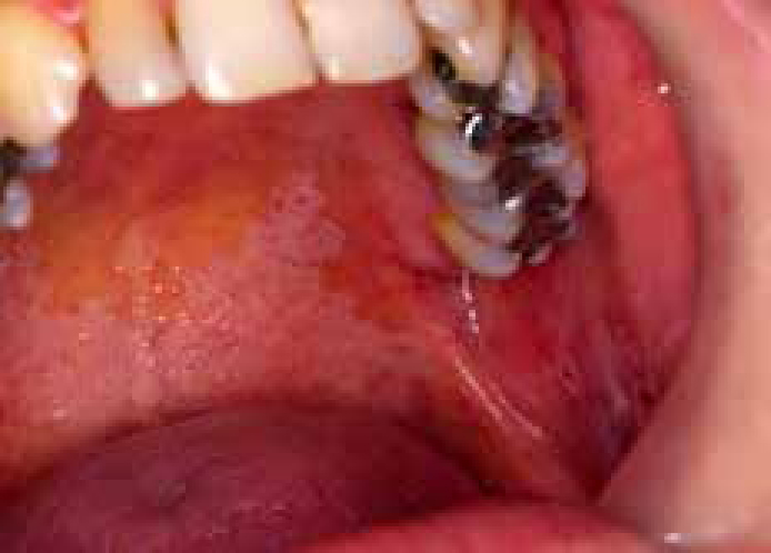
White lesions which can result from infections include candidosis (Figure 4), hairy leukoplakia caused by Epstein-Barr virus (Figure 5), warts and papillomas (caused by human papillomaviruses) (Figure 6), and the mucous patches and leukoplakia of syphilis. Specialist care is usually indicated.
Candidosis (candidiasis; moniliasis)
The importance of Candida has increased greatly, particularly as the HIV pandemic extends, since this common commensal can become opportunistic if local ecology changes, or the host immune defences fail. Candida albicans is the most common cause but occasionally other species may be implicated; in decreasing order of frequency these are:
Some 50% of the normal healthy population harbour (carry) C albicans as a normal oral commensal, particularly on the posterior dorsum of the tongue, and are termed Candida carriers. Candidal carriage is more common in smokers and people who wear oral appliances.
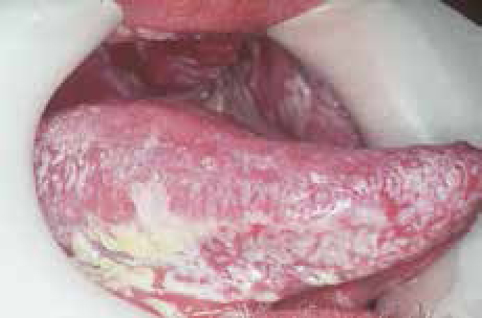
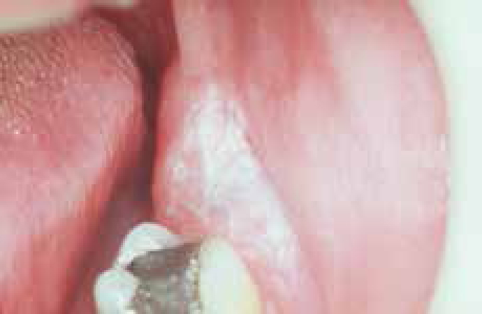
Candidosis is the state when C albicans causes lesions and these can be mainly white lesions (thrush particularly; Figure 4) or candidal leukoplakia (Figure 7) in which hyphal forms are common, or red lesions (denture-induced stomatitis, median rhomboid glossitis, erythematous candidosis), in which yeast forms predominate, and which may be symptomless though antibiotic stomatitis and angular cheilitis can cause soreness (Article 7).
Circumstances that cause susceptibility to candidosis include local factors influencing oral immunity or ecology, or systemic immune defects, or a combination of more than one factor (Table 2).
| Local factors influencing oral immunity or ecology | Systemic immune defects |
|---|---|
| Hyposalivation | Malnutrition |
| Smoking | Immunosuppressant drugs such as corticosteroids |
| Corticosteroids | T lymphocyte defects, especially HIV infection, leukaemias, lymphomas and cancers |
| Broad spectrum antimicrobials | Neutrophil leukocyte defects, such as in diabetes |
| Cytotoxic chemotherapy | Cytotoxic chemotherapy |
| Irradiation involving the mouth/salivary glands | Anaemia |
| Dental appliances |
Diagnosis
The diagnosis of candidosis is primarily clinical but a Gram-stained smear (looking for hyphae), a microbiological swab or oral rinse for culture may help to confirm the diagnosis.
Management
Possible predisposing causes should be looked for and dealt with, if possible. Topical polyene antifungals, such as nystatin or amphotericin or imidazoles, such as miconazole or fluconazole, are often indicated.
Non-infective causes
Lichen planus (LP) is a very common cause of oral white lesions. It affects up to 2% of the adult population. Accordingly most dental practitioners will have patients afflicted with LP. It is the main skin disease that can present with oral white lesions but lupus erythematosus can present similarly. Up to 44% of patients with oral lichen planus will have skin lesions and more than 70% of patients with skin lesions will have co-incident oral lesions.
Lichen planus
Lichen planus (LP) usually affects people between the ages of 30 and 65 and there is a slight female predisposition.
Aetiopathogenesis
Lichen planus is an inflammatory autoimmune-type of disease but it differs from classic autoimmune disorders in having no defined autoantibodies, and only rarely being associated with other autoimmune diseases. There is also no definitive immunogenetic basis yet established for LP and familial cases are rare.
Many patients afflicted with LP have a conscientious type of personality with obsessive-compulsive traits and suffer mild chronic anxiety, suggesting neuro-immunological mechanisms may be at play. Stress has been held to be important in LP: patients have a tendency to be anxious and depressed, but of course the chronic discomfort may partially explain some cases in which this association has been documented.
Pathologically, there is a local cell-mediated immunological response characterized by a dense T-lymphocyte inflammatory cell infiltrate in the upper lamina propria causing cell death (apoptosis) in the basal epithelium, probably caused by the production of cytokines such as tumour-necrosis factor alpha (TNFα) and interferon gamma (IFN-γ).
The antigen responsible for this immune response is unclear but lesions very similar to LP, termed lichenoid lesions, are sometimes caused by:
Clinical features
Lichen planus can affect stratified squamous epithelium of the skin, the oral mucosa and genitalia.
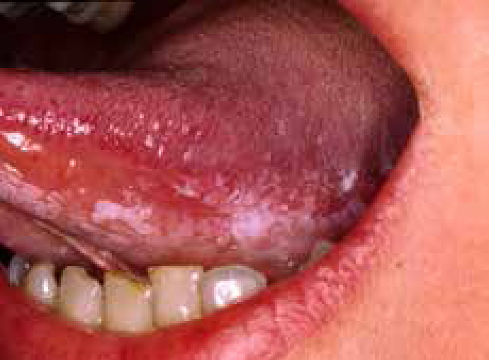
Oral LP may present a number of different clinical pictures (Figures 8–13), including:

White lesions of LP are often asymptomatic, but there may be soreness if there are atrophic areas or erosions.
Lichen planus typically results in lesions, which are usually in the posterior buccal mucosa bilaterally, but the tongue or gingivae are other sites commonly affected.
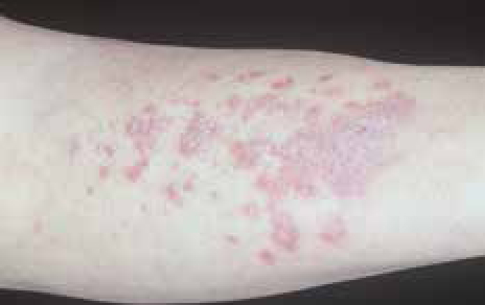
On the skin, lichen planus frequently presents as a flat-topped purple polygonal and pruritic papular rash most often seen on the front (flexor surface) of the wrists (Figure 13) in which lesions are often crossed by fine white lines (Wickham's striae; Figure 14). Nail lesions may be seen (Figure 15). Oral LP may be accompanied by vulvovaginal lesions (the vulvovaginal-gingival syndrome).

Prognosis
Often, the onset of LP is slow, taking months to reach its peak. It usually clears from the skin within 18 months but in a few people persists for many years. Oral lesions often persist. There is no sign or test to indicate which patients will develop only oral, or oral and extra-oral lesions of LP.
Non-reticular oral LP in particular has a small malignant potential, probably of the order of 1%.
Diagnosis
LP is often fairly obviously diagnosed from the clinical features but, since it can closely simulate other conditions such as:
Keypoints: lichen planus
Management
Treatment of LP is not always necessary, unless there are symptoms. Predisposing factors should be corrected:
Keypoints for patients: lichen planus
Therefore, the best management is usually to:
Websites and patient information
http://www.bcd.tamhsc.edu/outreach/lichen/index.html
http://www.aad.org/pamphlets/lichen.html
Keratoses and leukoplakias
Frictional keratosis
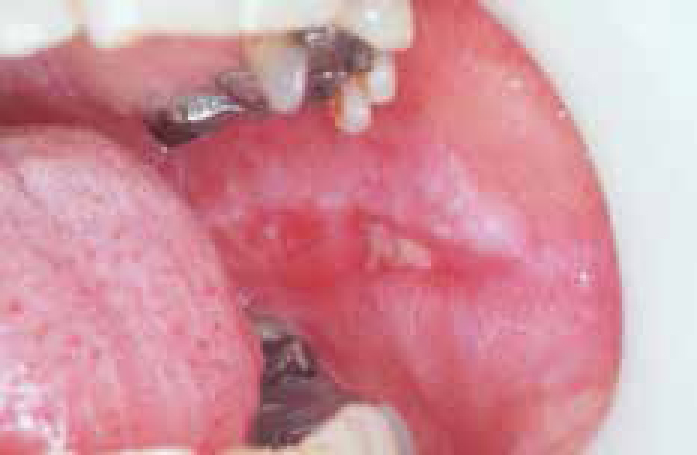
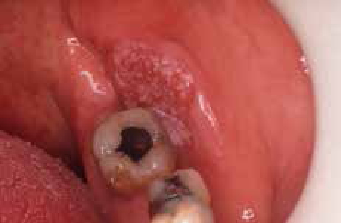
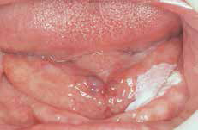
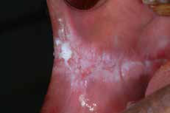
Frictional keratosis is quite common and caused particularly by friction from the teeth and seen mainly at the occlusal line in the buccal mucosae, particularly in adult females, especially in those with temporomandibular pain-dysfunction syndrome. It is also commonly seen on the lateral borders of the tongue (Figure 16). Patients with missing teeth may develop keratosis on the alveolar ridge simply related to trauma when eating (Figure 17).
Malignant change is very rare but any sharp edges of teeth or appliances should be removed and the patient counselled about the habits.
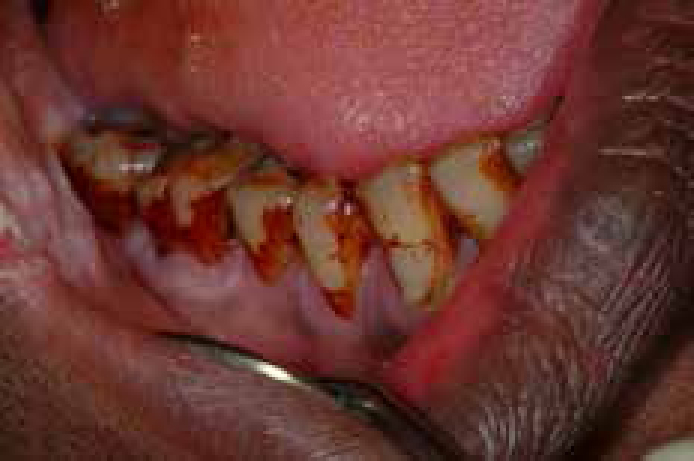
Tobacco-induced keratoses
Tobacco is a common cause of keratosis, seen especially in males, the teeth are usually nicotine-stained and there may be mucosal smoker's melanosis but malignant change is uncommon in most forms (Table 3).
| Tobacco habit | Common sites affected | Occasional sites affected | Malignant potential |
|---|---|---|---|
| Cigarette | Lip (occasionally nicotine-stained) and commissures | Palate |
Rare |
| Pipe smoking | Palate (termed smoker's keratosis or stomatitis nicotina) | Others | Rare |
| Cigar | Palate (termed smoker's keratosis or stomatitis nicotina) | Others | Rare |
| Snuff | Gingival (together with recession) | Lip | Rare |
| Reverse smoking (Bidi) cigarettes are smoked with the lit end within the mouth | Palate | Others | Common |
| Tobacco chewing | Buccal | Others | Common |
Idiopathic keratoses
Many leukoplakias are uncommon and arise in the absence of any identifiable predisposing factors and most, up to 70% in large series, are benign without any evidence of dysplasia. However, the remaining 10–30% may be, or may become, either dysplastic or invasive carcinomas. Overall, the rate of malignant transformation of all keratoses and leukoplakias is of some 3–6% over 10 years.
The lesions of greatest malignant potential are those leukoplakias which are:
In these, rates of malignant transformation up to 30% have been reported in some series.
Diagnosis
The nature of white lesions can often only be established after further investigation.
Biopsy is usually indicated, particularly where there is a high risk of malignant transformation, such as in lesions with:
Keypoints: keratosis (leukoplakia)
Therefore, the best management is usually to:
Prognosis
The finding by the pathologist of epithelial dysplasia may be predictive of malignant potential, but this is not invariable, and there can be considerable inter- and intra-examiner variation in the diagnosis of dysplasia.
Thus there has been a search for molecular markers to predict exactly which lesions are truly of malignant potential and may develop into oral squamous cell carcinoma (OSCC).
The most predictive of the molecular or cellular markers thus far assessed for OSCC development, apart from dysplasia, include chromosomal polysomy, the tumour suppressor p53 protein expression, and loss of heterozygosity (LOH) at chromosome 3p or 9p. Routine use of these is, however, hampered by their complexity and lack of facilities in many pathology laboratories.
As a surrogate for individual molecular markers, measurement of gross genomic damage (DNA ploidy) may be a realistic option, and is now available in some oral pathology laboratories.
Management
The dilemma in managing patients with potentially malignant oral lesions and field change has been deciding which mucosal lesions or areas will progress to carcinoma. At present there are no reliable markers which predict which lesions will progress. Specialist referral is indicated.
Cessation of dangerous habits such as tobacco and/or betel use (Figures 24, 25), and the removal of lesions is probably the best course of action, particularly if they are the high risk lesions or in a high risk group for carcinoma (Article 3). Perhaps surprisingly, management of leukoplakias is very controversial, since there are no randomized, controlled, double-blind studies that prove the best type of treatment. However, most specialists prefer removal of the lesion (by laser, scalpel or other means).
Keypoints for patients: keratosis (leukoplakia)
This is an uncommon condition:
Therefore, the best management is usually to:
Changes that might suggest a tumour is developing could include any of the following persisting for more than 3 weeks:
Useful websites and patient information
http://www.cochrane.org/cochrane/revabstr/ab001829.htm
http://www.emedicine.com/ent/topic731.htm
http://www.mayoclinic.com/health/leukoplakia/DS00458