
Article


Specialist referral may be indicated if the Practitioner feels:
Malignant ulcers
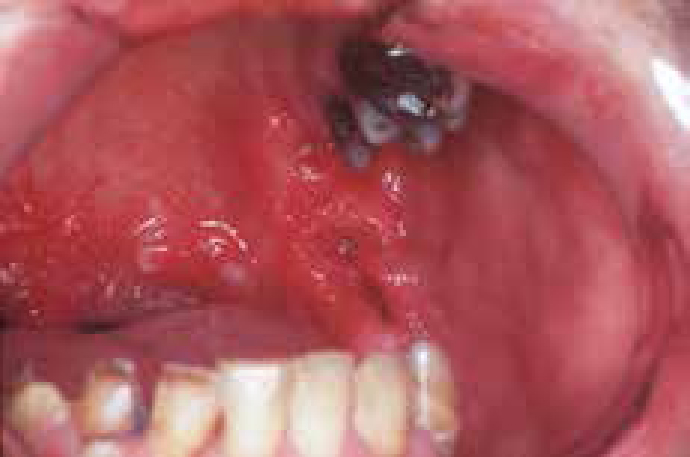
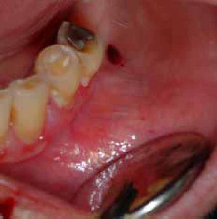
A range of neoplasms may present with ulcers: most commonly these are carcinomas (Figure 1) but Kaposi sarcoma, lymphomas and other neoplasms may be seen and are discussed in Article 3. Biopsy is required to establish a definitive diagnosis.
Systemic disease
A wide range of systemic diseases, especially, mucocutaneous diseases, blood, gut and miscellaneous uncommon disorders may cause oral lesions which, because of the moisture, trauma and infection in the mouth, tend to break down to leave ulcers or erosions. Biopsy is often required to establish the diagnosis.
Mucocutaneous disorders
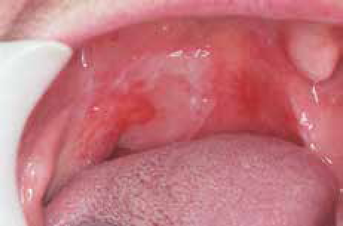
Mucocutaneous disease that may cause oral erosions or ulceration (or occasionally blisters) include particularly Behçet's syndrome, and a number of skin diseases including lichen planus (Figure 2), occasionally erythema multiforme or pemphigoid, and rarely pemphigus.
Behçet's syndrome
Behçet's syndrome (BS) is a rare condition. It is the association of recurrent aphthous stomatitis (RAS) with genital ulceration, and serious eye disease (especially iridocyclitis) but other systemic manifestations may also be seen. The disease is found worldwide, but most commonly in people from the Eastern Mediterranean countries (particularly Greeks, Turks, Arabs and Jews) and along the Silk route taken by Marco Polo across eastern Asia, China, Korea and Japan.
Aetiopathogenesis
Behçet's syndrome is a vasculitis which has not been proved to be infectious, contagious, or sexually transmitted. There are many immunological findings in BS similar to those seen in RAS, with T suppressor cell dysfunction, and increased polymorphonuclear leucocyte motility. There is a genetic predisposition. Many of the features of BS (erythema nodosum, arthralgia, uveitis) are common to established immune complex diseases.
Clinical features
Behçet's syndrome is a chronic, sometimes life-threatening disorder characterized mainly by:
Differential diagnosis
This is from a range of other syndromes that can affect the eyes, mouth and skin – such as various dermatological disorders and infections.
Diagnosis
BS can be very difficult to diagnose and there is no single diagnostic investigation. The International Study Group for Behçet's Disease (ISGBD) criteria suggest the diagnosis be made on clinical grounds alone on the basis of RAS plus two or more of:
Investigations
There is no specific diagnostic test but typing for specific human leukocyte antigens (HLA B5101) can help. Disease activity may be assessed by serum levels of various proteins raised in active BS, such as the acute phase proteins (erythrocyte sedimentation rate (ESR), C-reactive protein (CRP)) or antibodies to intermediate filaments.
Management
In the face of the difficult diagnosis and serious potential complications, patients with suspected BS should be referred early for specialist advice.
Websites and patient information
http://www.arthritisresearchuk.org/arthritis_information/arthritis_types__symptoms/behcets_syndrome.aspx
http://www.behcets.org.uk
Lichen planus
Lichen planus is discussed in Article 6.
Erythema multiforme
Erythema multiforme (EM) is an uncommon, acute, often recurrent reaction affecting mucocutaneous tissues, seen especially in younger males.
The aetiology of erythema multiforme (EM) is unclear in most patients, but it appears to be an immunological hypersensitivity reaction, leading to sub- and intra-epithelial vesiculation.
There may be a genetic predisposition with associations of recurrent EM with various HLA haplotypes.
EM is triggered by a range of usually exogenous factors, such as:
Clinical features
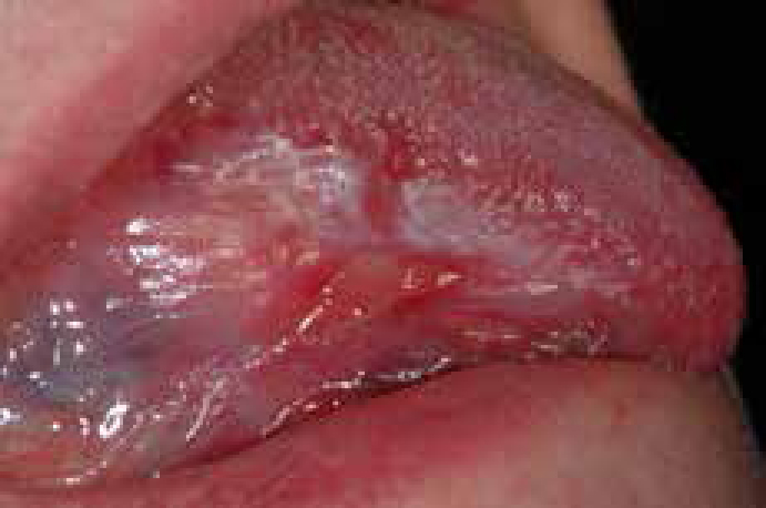
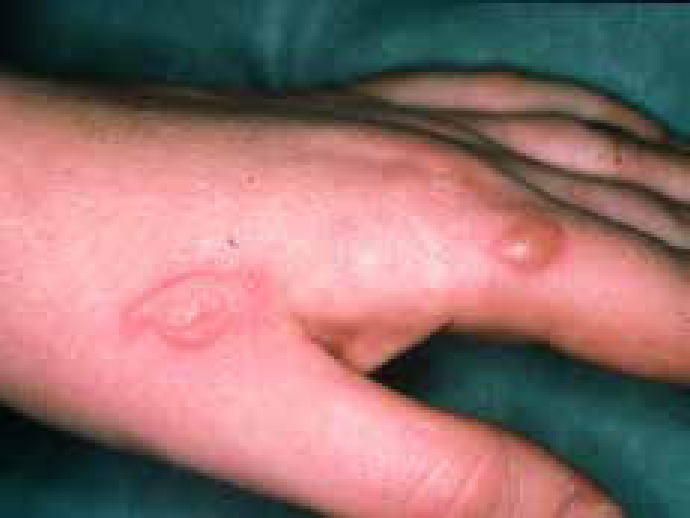
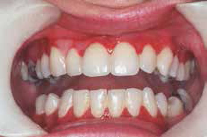
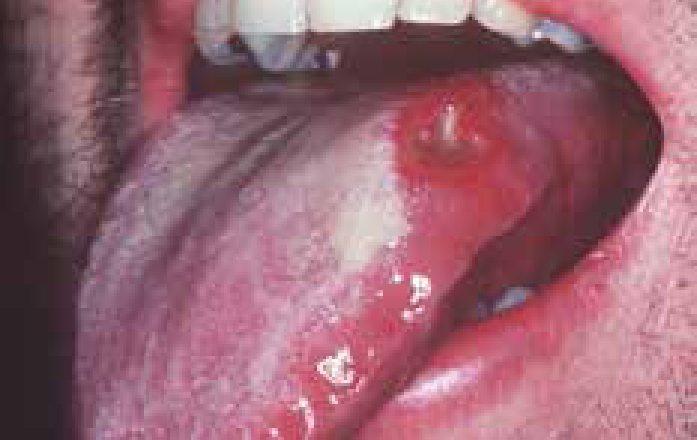
EM ranges from limited disease (Minor EM) to severe, widespread life-threatening illness (Major EM). Most patients (70%) in either form, have oral lesions, which may precede lesions on other stratified squamous epithelia (eyes, genitals, or skin), or may arise in isolation. Oral EM typically presents with macules which evolve to blisters and ulcers (Figure 3). The lips become swollen, cracked, bleeding and crusted. Skin lesions start on the hands and/or feet but subsequently spread along the limbs to involve the trunk. The lesions are initially sharply demarcated macules which evolve into target-like lesions (Figure 4).
Minor EM affects only one site and may affect mouth alone, or skin, or other mucosae. Rashes are various but typically ‘iris’ or ‘target’ lesions or bullae on extremities.
Major EM (Stevens-Johnson syndrome – SJS) almost invariably involves the oral mucosa and causes widespread lesions affecting mouth, eyes, pharynx, larynx, oesophagus, skin and genitals.
Diagnosis
There are no specific diagnostic tests for EM. Therefore, the diagnosis is mainly clinical, and it can be difficult to differentiate it from viral stomatitis, pemphigus, toxic epidermal necrolysis, and sub-epithelial immune blistering disorders. Serology for HSV or Mycoplasma pneumoniae, or other micro-organisms, and biopsy of perilesional tissue, with histological and immunostaining examination are essential if a specific diagnosis is required.
Management
Spontaneous healing can be slow – up to 2 to 3 weeks in minor EM and up to 6 weeks in major EM.
Treatment is thus indicated but controversial and thus specialist care should be sought. Supportive care is important; a liquid diet and intravenous fluid therapy may be necessary. Oral hygiene should be improved with 0.2% aqueous chlorhexidine mouthbaths.
The use of corticosteroids is controversial but minor EM may respond to topical corticosteroids. Patients with major EM, such as the Stevens-Johnson syndrome, may need to be admitted for hospital care. Major EM patients should be referred for treatment with systemic corticosteroids or other immunomodulatory drugs.
Websites and patient information
http://emedicine.medscape.com/article/1122915-overview
http://www.nlm.nih.gov/medlineplus/ency/article/000851.htm
Pemphigoid
Pemphigoid is the term given to a group of uncommon sub-epithelial immunologically-mediated vesiculobullous disorders (SEIMD) which can affect stratified squamous epithelium, characterized by damage to one of the protein constituents of the basement membrane zone (BMZ) anchoring filament components; a number of other sub-epithelial vesicullobullous disorders may produce similar clinical features (Table 1).
| Pemphigoid variants |
The main types of pemphigoid that involve the mouth are:
However, most of the literature has failed to distinguish these variants, since their distinction has only recently been recognized, and therefore the following discussion groups them together.
Mucous membrane/oral pemphigoid
Mucous membrane pemphigoid (benign mucous membrane pemphigoid) is an uncommon chronic disease, twice as common in females, and presenting usually in the fifth to sixth decades.
Mucous membrane pemphigoid is an autoimmune type of disorder with a genetic predisposition. The precipitating event is unclear in most cases but rare cases are drug-induced (eg by furosemide or penicillamine).
It is characterized immunologically by deposition of IgG and C3 antibodies directed against the epithelial basement membrane zone (BMZ). There are also circulating auto-antibodies to BMZ components, present in hemi-desmosomes or the lamina lucida.
The antibodies damage the BMZ and histologically there is a sub-basilar split. The pathogenesis probably includes complement mediated sequestration of leukocytes with resultant cytokine and leukocyte enzyme release and detachment of the basal cells from the BMZ.
Clinical features
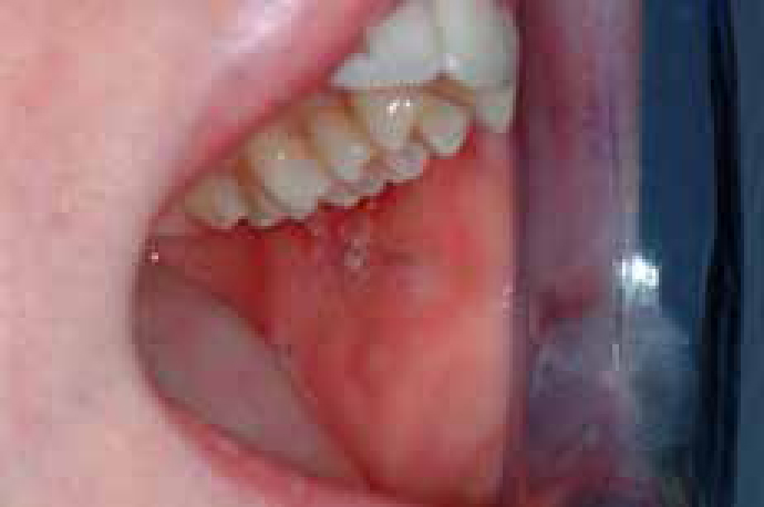
The oral lesions (Figures 5, 6 and 7) affect especially the gingivae and palate and include bullae or vesicles which are tense, may be blood-filled and remain intact for several days. Persistent irregular erosions or ulcers appear after the blisters burst and, if on the gingivae, can produce desquamative gingivitis – the most common oral finding. This is characterized by erythematous, ulcerated, tender gingivae in a patchy, rather than continuous distribution.
The majority of people with MMP have only oral lesions but genital involvement can cause great morbidity and untreated ocular involvement can lead to blindness. Nasal, laryngeal and skin blisters are rare.
Diagnosis
The oral lesions of pemphigoid may be confused clinically with pemphigus, or occasionally erosive lichen planus, erythema multiforme or sub-epithelial blistering conditions shown in Table 1.
Biopsy of perilesional tissue, with histological and immunostaining examination can therefore be essential to the diagnosis.
Management
Spontaneous remission is rare, and thus treatment is indicated. Specialist advice is usually needed.
Systemic manifestations must be given attention. For that reason, an ophthalmology consultation is essential to rule out occult ocular disease.
The majority of cases respond well to topical corticosteroids such as are used for aphthae (Article 1). Non-steroidal immunosuppressive agents, such as tacrolimus, may be needed if the response to topical corticosteroids is inadequate.
Severe pemphigoid may need to be treated with immunosuppression using azathioprine or systemic corticosteroids.
Website and patient information http://www.dent.ucla.edu/pic/members/MMP/
Pemphigus
Pemphigus is a group of, fortunately rare, potentially life-threatening chronic diseases characterized by epithelial blistering affecting cutaneous and/or mucosal surfaces. There are several variants with different auto-antibody profiles and clinical manifestations (Table 2) but the main type is Pemphigus vulgaris; this includes an uncommon variant pemphigus vegetans. Pemphigus vulgaris is seen mainly in middle-aged and elderly females of Mediterranean, Ashkenazi Jewish or South Asian descent.
| Variant | Oral lesions | Main antigens (Ags) | Localization Ags | Antibodies |
|---|---|---|---|---|
| Pemphigus vulgaris localized to mucosae (Mucosal) | Common | Dsg 3 | Desmosomes | IgG |
| Pemphigus vulgaris alsoinvolving skin/other mucosae (Mucocutaneous) | Common | Dsg 3 |
Desmosomes | IgG |
Pemphigus vulgaris is an autoimmune disorder in which there is a fairly strong genetic background. Rare cases have been triggered by medications (captopril, penicillamine, rifampicin and diclofenac are the main offenders), or other factors.
The auto-antibodies are directed against stratified squamous epithelial desmosomes, particularly the proteins desmoglein-3 (Dsg3) and desmoglein-1 (Dsg1) (Table 2). Damage to the desmosomes leads to loss of cell-cell contact (acantholysis), and thus intra-epithelial vesiculation.
Clinical features
Pemphigus vulgaris typically runs a chronic course, causing blisters, erosions and ulcers on the mucosae and blisters and scabs on the skin. Oral lesions are common, may be an early manifestation, and mimic those of pemphigoid in particular. Blisters rapidly break down to leave erosions seen mainly on the palate, buccal mucosa, lips and gingiva.
Diagnosis
To differentiate pemphigus from other vesiculobullous diseases, a careful history and physical examination are important, but biopsy of perilesional tissue, with histological and immunostaining examination, are crucial. Serum should be collected for antibody titres which provides an assessment of disease activity.
Management
Before the introduction of corticosteroids, Pemphigus vulgaris typically was fatal, mainly from dehydration or secondary systemic infections. Specialist care is mandatory. Current treatment, by systemic immunosuppression, usually with steroids, or azathioprine or mycophenolate mofetil, has significantly reduced the mortality to about 10%.
Websites and patient information http://www.pemphigus.org
Blood disorders that can cause ulcers include mainly the leukaemias, associated with cytotoxic therapy, viral, bacterial or fungal infection, or non-specific. Other oral features of leukaemia may include purpura, gingival bleeding, recurrent herpes labialis, and candidosis.
Gastrointestinal disease may produce soreness or mouth ulcers. A small proportion of patients with aphthae have intestinal disease, such as coeliac disease, causing malabsorption and deficiencies of haematinics, when they may also develop angular stomatitis or glossitis. Crohn's disease and pyostomatitis vegetans may also cause ulcers. Orofacial granulomatosis (OFG), which has many features reminiscent of Crohn's disease, may also cause ulceration. Miscellaneous uncommon diseases such as lupus erythematosus can cause ulcers.
Differential diagnosis of oral ulceration
The most important feature of ulceration is whether the ulcer is single, multiple or persistent.
Diagnosis of oral ulceration
Making a diagnosis of the cause for oral soreness or ulceration is based mainly on the history and clinical features. The number, persistence, shape, character of the edge of the ulcer and the appearance of the ulcer base should also be noted. Ulcers should always be examined for induration (firmness on palpation), which may be indicative of malignancy. The cervical lymph nodes must be examined.
Unless the cause is undoubtedly local, general physical examination is also indicated, looking especially for mucocutaneous lesions, other lymphadenopathy or fever, since it is crucial to detect systemic causes such as leukaemia, or HIV infection (Figure 8).
Biopsy
Informed consent is mandatory for biopsy, particularly noting the likelihood of post-operative discomfort, and the possibility of bleeding or bruising. Care must be taken not to produce undue anxiety; some patients equate biopsy with a diagnosis of cancer. Perhaps the most difficult and important consideration is which part of the lesion should be included in the biopsy specimen.
As a general rule, the biopsy should include lesional and surrounding tissue. In the case of ulcerated mucosal lesions, most histopathological information is gleaned from the peri-lesional tissue since, by definition, most epithelium is lost from the ulcer itself. The same usually applies for skin diseases affecting the mouth, where the epithelium in the area mainly affected will, more often than not, separate, and results will be compromised. In the case of a suspected potentially malignant or malignant lesion, any red area should ideally be included in the specimen. In some cases where no obvious site can be chosen, vital staining with ‘toluidine blue’ may first be indicated.
A biopsy punch has the advantage that the incision is controlled, an adequate specimen is obtained (typically 4mm or 6mm diameter) and suturing may not be required. However, in the skin disorders, the punch can sometimes split the epithelium or detach it from the lamina propria. When a scalpel is used, a specimen of elliptical shape is usually taken, most commonly from an edge of the lesion.
Procedure
Management of oral ulceration
| Agent | Use | Comments |
|---|---|---|
| Benzydamine hydrochloride | Rinse or spray every 1.5 to 3 hours | Effective in reducing discomfort |
| Lidocaine | Topical 4% solution may ease pain | Also reduces taste sensitivity |
| Carboxymethylcellulose | Paste or powder used after meals to protect area | Available containing triamcinolone in Canada and the Antipodes but no longer in UK or USA |
Referral of patients with oral ulceration
Patients with single ulcers persisting more than 3 weeks, indurated ulcers, or multiple persistent ulcers may benefit from a specialist opinion.
Patients with recalcitrant ulcers, or a systemic background to mouth ulcers, or needing investigation, may also benefit from a specialist referral.
Features that might suggest a systemic background to mouth ulcers include:
Investigations which may sometimes be indicated include:
Patients to refer:
Patients with conditions discussed in this section should be referred for specialist care.