
Article


Specialist referral may be indicated if the Practitioner feels:
Ulceration
Ulceration is a breach in the oral epithelium, which typically exposes nerve endings in the underlying lamina propria, resulting in pain or soreness, especially on eating spicy foods or citrus fruits. Patients vary enormously in the degree to which they suffer and complain of soreness in relation to oral ulceration. It is always important to exclude serious disorders such as oral cancer (Article 3) or other serious disease, but not all patients who complain of soreness have discernible organic disease. Even in those with detectable lesions, the level of complaint can vary enormously – some patients with large ulcers complain little; others with minimal ulceration complain bitterly of discomfort. Sometimes there is a psychogenic or cultural influence.
Terminology
Epithelial thinning or breaches may be seen in:

Causes of oral ulceration
Ulcers and erosions can also be the final common manifestation of a spectrum of conditions, ranging from epithelial damage resulting from trauma, to an immunological attack as in lichen planus, pemphigoid or pemphigus, to damage because of an immune defect, as in HIV disease and leukaemia, infections as in herpesviruses, tuberculosis and syphilis, or nutritional defects such as in vitamin deficiencies and some gastrointestinal disease (Tables 1 and 2).
| Causes | Mnemonic |
|---|---|
| Systemic diseases | So |
| Malignant disease | Many |
| Local causes | Laws |
| Aphthae | And |
| Drugs | Directives |
|
Systemic disease
|
Ulcers of local causes
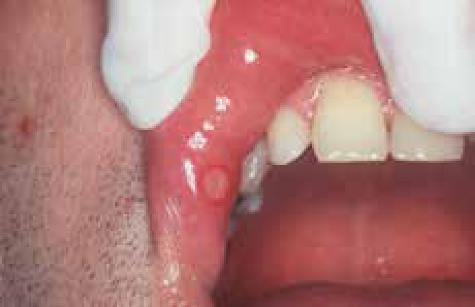
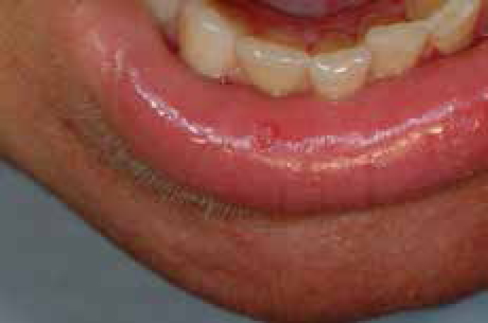
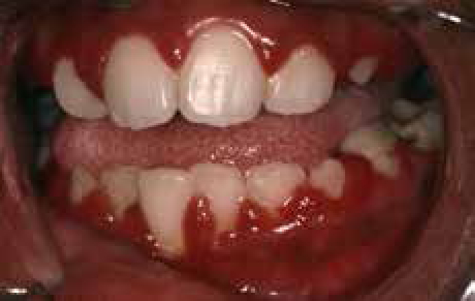
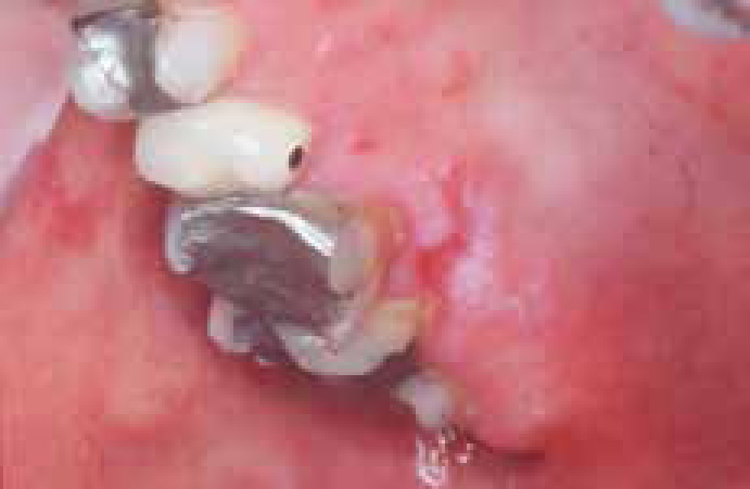
At any age, there may be burns from chemicals of various kinds, heat, cold, or ionizing radiation or factitious ulceration, especially of the maxillary gingivae (Figures 3 and 4).
Children may develop ulceration of the lower lip by accidental biting following dental local anaesthesia. Ulceration of the upper labial fraenum, especially in a child with bruised and swollen lips, or subluxed teeth or fractured jaw can represent non-accidental injury. At any age, trauma, hard foods, or appliances may also cause ulceration. The lingual fraenum may be traumatized by repeated rubbing over the lower incisor teeth in cunnilingus or in recurrent coughing as in whooping cough or in self-mutilating conditions.
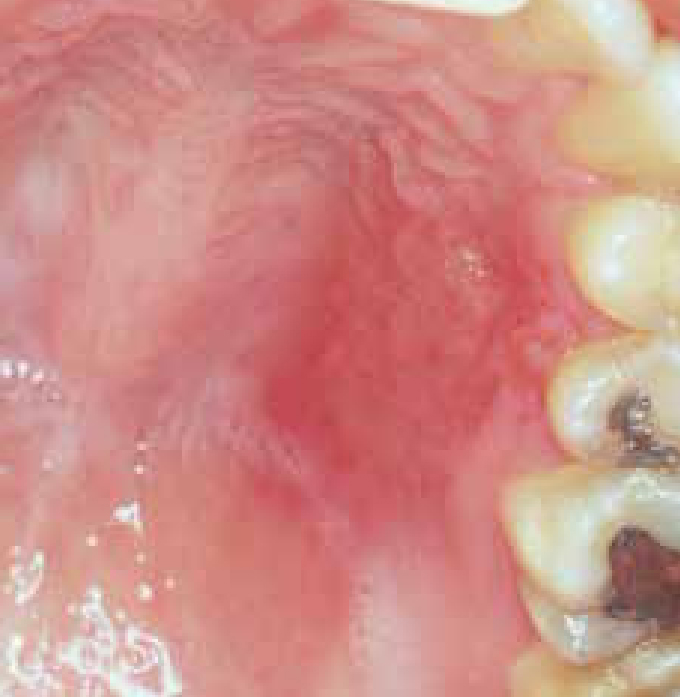
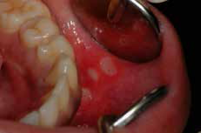
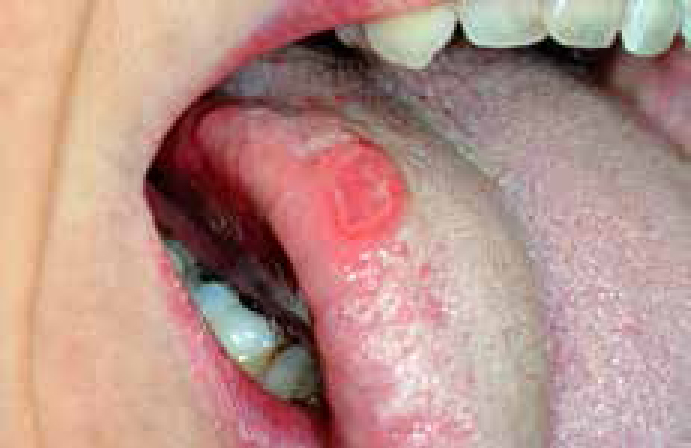
Most ulcers of local cause have an obvious aetiology, are acute, usually single ulcers, last less than 3 weeks and heal spontaneously. Chronic trauma may produce an ulcer with a keratotic margin (Figure 5).
Recurrent aphthous stomatitis (RAS; aphthae; canker sores)
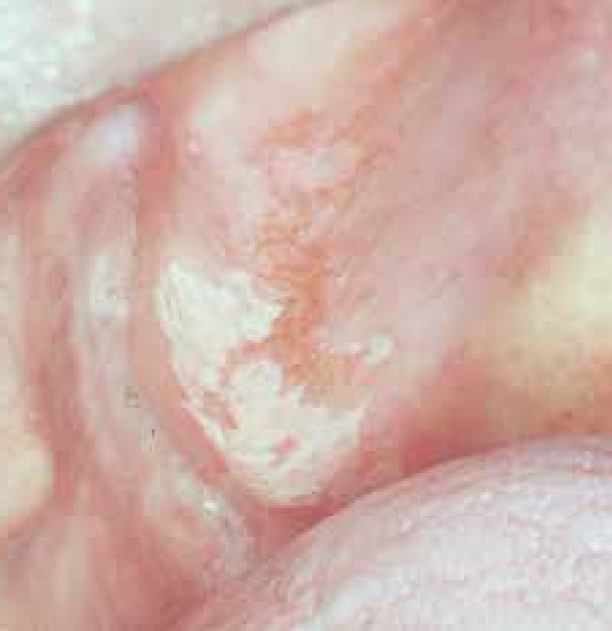
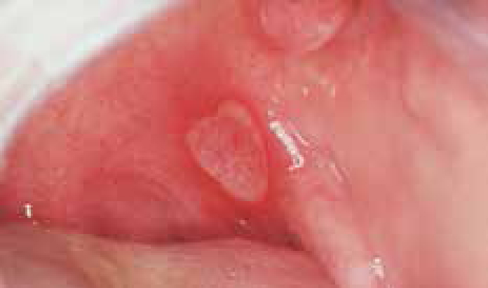
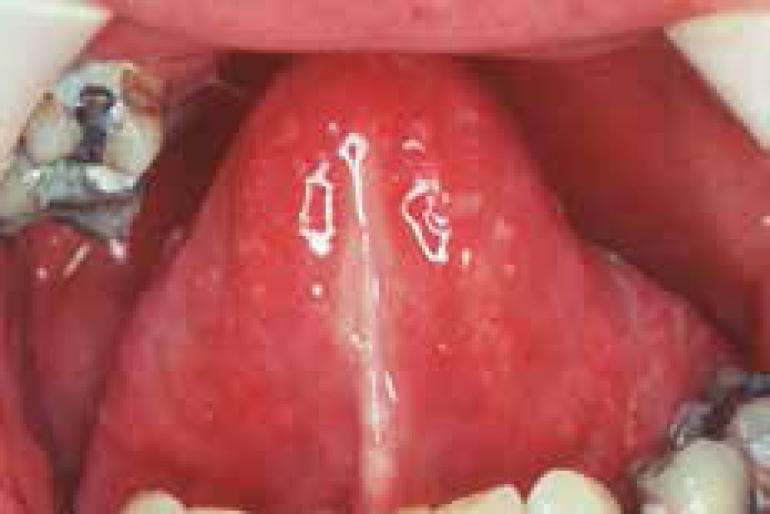
RAS is a very common condition which typically starts in childhood or adolescence and presents with multiple recurrent small, round or ovoid ulcers with circumscribed margins, erythematous haloes, and yellow or grey base (Figures 6 and 7).
RAS affects at least 20% of the population, with the highest prevalence in higher socio-economic classes. Virtually all dentists will see patients with aphthae.
Aetiopathogenesis
Immune mechanisms appear at play in a person with a genetic predisposition to oral ulceration. A genetic predisposition is present, and there is a positive family history in about one third of patients with RAS. Immunological factors are also involved, with T helper cells predominating in the RAS lesions early on, along with some natural killer (NK) cells. Cytotoxic cells then appear in the lesions and there is evidence for an antibody dependent cellular cytotoxicity (ADCC) reaction. It now seems likely therefore that a minor degree of immunological dysregulation underlies aphthae.
RAS may be a group of disorders of different pathogeneses. Cross-reacting antigens between the oral mucosa and micro-organisms may be the initiators, but attempts to implicate a variety of bacteria or viruses have failed.
Predisposing factors
Most people who suffer RAS are otherwise apparently completely well. In a few, predisposing factors may be identifiable, or suspected. These include:
Drugs (see below), Behcet syndrome, HIV, Epstein-Barr virus, auto-inflammatory states (periodic fevers) and skin diseases, such as erythema multiforme, may occasionally produce aphthous-like lesions.
Keypoints: aphthous ulcers
Clinical features
There are three main clinical types of RAS, though the significance of these distinctions is unclear and it is conceivable that they may represent three different diseases.
Diagnosis
There are no specific tests, so the diagnosis must be made on history and clinical features alone. However, to exclude the systemic disorders discussed above, it is often useful to undertake the investigations shown in Table 3.
|
|
Biopsy is rarely indicated, usually where a different diagnosis is suspected.
Management
Other similar disorders, such as Behcet syndrome, must be ruled out (Article 2). Predisposing factors should then be corrected. Fortunately, the natural history of RAS is one of eventual remission in most cases. However, few patients have spontaneous remission until after several years and, although there is no curative treatment, measures should be taken to relieve symptoms and reduce ulcer duration.
| Steroid | UK trade name | Dosage every 6 hours |
|---|---|---|
|
Medium potency
|
Betnesol | 0.5 mg; use as mouthwash |
|
High potency
|
Clenil modulite | 1 puff (200 mg) to lesions |
The major concern is of adrenal suppression with long-term and/or repeated application but there is no evidence that these cause this problem.
Topical tetracycline (eg doxycycline), or tetracycline plus nicotinamide may provide relief and reduce ulcer duration, but should be avoided in children under 12 who might ingest the tetracycline and develop tooth staining.
If RAS fails to respond to these measures, systemic immunomodulators may be required, under specialist supervision.
Keypoints for patients: aphthous ulcers
Websites and patient information
http://www.doctorsofusc.com/condition/document/11983
http://emedicine.medscape.com/article/867080-overview
http://www.cks.nhs.uk/patient_information_leaflet/mouth_ulcer
Infections
Infections that cause mouth ulcers are mainly viral, especially the herpesviruses, Coxsackie, ECHO and HIV viruses. Bacterial causes of mouth ulcers, apart from acute necrotizing ulcerative gingivitis, are less common. Syphilis and tuberculosis are uncommon but increasing, especially in people with HIV/AIDS. Fungal and protozoal causes of ulcers are also uncommon, but increasingly seen in immunocompromised persons, and travellers from the developing world.
Herpes simplex virus (HSV)
The term ‘herpes’ is often used loosely to refer to infections with herpes simplex virus (HSV), a ubiquitous virus which commonly produces lesions in the mouth and oropharynx. HSV is contracted by close contact with infected individuals from infected saliva or other body fluids after an incubation period of approximately 4–7 days.
Primary infection is often subclinical between the ages of 2–4 years. This is usually caused by HSV-1 and is commonly attributed to ‘teething’, particularly if there is a fever. However, primary infection can occur at any age and present with stomatitis (gingivostomatitis).
In teenagers or older, this may be due to HSV-2 transmitted sexually.
Generally speaking, HSV infections above the belt (oral or oropharyngeal) are caused by HSV-1 but below the belt (genital or anal) are caused by HSV-2.
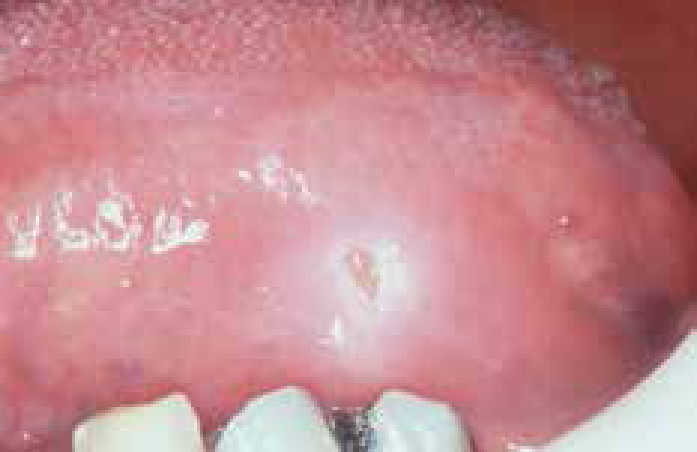
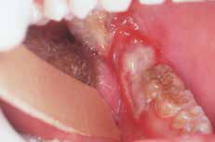
The mouth or oropharynx is sore (herpetic stomatitis or gingivostomatitis): there is a single episode of oral vesicles which may be widespread, and break down to leave oral ulcers that are initially pin-point but fuse to produce irregular painful ulcers (Figure 12). Gingival oedema, erythema and ulceration are prominent and the cervical lymph nodes may be enlarged and tender, and there is sometimes fever and/or malaise. Patients with immune defects are liable to severe and/or protracted infections.
HSV is neuroinvasive and neurotoxic and infects neurones of the dorsal root and autonomic ganglia. HSV remains latent thereafter in those ganglia, usually the trigeminal ganglion, but can be re-activated to result in clinical recrudescence (see below)
Diagnosis
Diagnosis is largely clinical. Viral studies are used occasionally and can include:
Management
Although patients have spontaneous healing within 10–14 days, treatment is indicated particularly to reduce fever and control pain. Adequate fluid intake is important, especially in children, and antipyretics/analgesics, such as paracetamol/acetaminophen elixir, help. A soft bland diet may be needed, as the mouth can be very sore. Aciclovir orally or parenterally is useful mainly in immunocompromised patients or in the otherwise apparently healthy patient, if seen early in the course of the disease, but do not reduce the frequency of subsequent recurrences.
Recurrent HSV infections
Up to 15% of the population have recurrent HSV-1 infections, typically on the lips (herpes labialis; cold sores), from re-activation of HSV latent in the trigeminal ganglion. The virus is periodically shed into saliva, and there may be clinical recrudescence. Re-activating factors include fever such as caused by upper respiratory tract infection (hence herpes labialis is often termed ‘cold’ sores), sunlight, menstruation, trauma and immunosuppression.
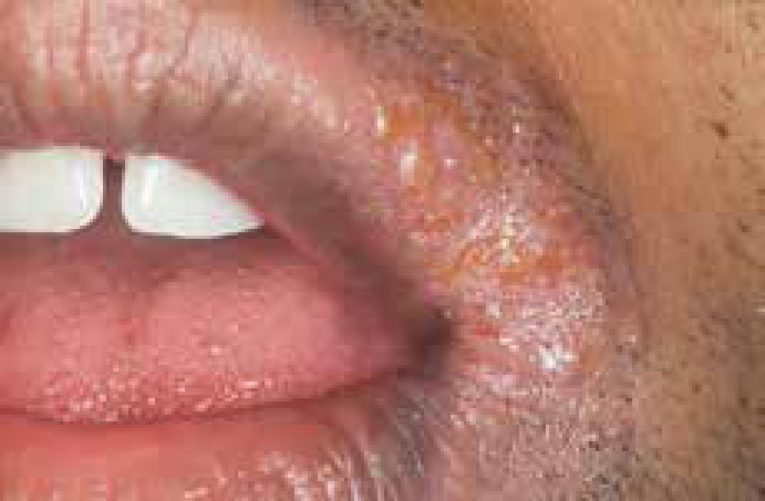
Lip lesions at the mucocutaneous junction may be preceded by pain, burning, tingling or itching. Lesions begin as macules that rapidly become papular, then vesicular for about 48 hours, then become pustular, and finally scab within 72–96 hours and heal without scarring (Figure 13).
Recurrent intra-oral herpes in apparently healthy patients tend to affect the hard palate or gingivae with a small crop of ulcers which heals within 1–2 weeks. Lesions are usually over the greater palatine foramen, following a palatal local anaesthetic injection, presumably because of the trauma.
Recurrent intra-oral herpes in immunocompromised patients may appear as chronic, often dendritic, ulcers, often on the tongue.
Diagnosis
Diagnosis is largely clinical; viral studies are used occasionally.
Management
Most patients will have spontaneous remission within one week to 10 days, but the condition is both uncomfortable and unsightly, and thus treatment is indicated. Antivirals will achieve maximum benefit only if given early in the disease, but may be indicated in patients who have severe, widespread or persistent lesions and in the immunocompromised. Lip lesions in healthy patients may be minimized with penciclovir 1% cream or aciclovir 5% cream applied in the prodrome. In severe cases where recurrences are frequent, systemic aciclovir may be indicated. Lip lesions in immunocompromised patients require systemic aciclovir or other antivirals such as valciclovir (the precursor of penciclovir).
Keypoints for patients: cold sores
Websites and patient information
http://www.cks.nhs.uk/patient_information_leaflet/cold_sore
http://www.nlm.nih.gov/medlineplus/tutorials/coldsores/htm/_no_50_no_0.htm
Drug-induced ulceration
Drugs may induce ulcers by producing a local burn, or by a variety of mechanisms, such as the induction of lichenoid lesions (Figure 14). Cytotoxic drugs (eg methotrexate) commonly produce ulcers, but non-steroidal anti-inflammatory drugs (NSAIDs), including rofecoxib, alendronate (a bisphosphonate), nicorandil (a cardiac drug) and a range of other drugs, may also cause ulcers.
A drug history is important to elicit such uncommon reactions, and then the offending drug should be avoided.