Case report: an unusual finding of a solitary bone cyst in a patient with a fractured mandible Melanie Chell Matthew Idle Jason Green Dental Update 2024 42:10, 707-709.
Authors
MelanieChell
BDS MFDS RCS(Ed)
Senior House Officer in Oral and Maxillofacial Surgery, University Hospital Birmingham, UK
Solitary bone cysts are uncommon. They have a reported incidence of 0.6% and are commonest in the mandible. The case of a 16-year-old patient who attended Accident and Emergency with a fractured mandible and the incidental finding of a solitary bone cyst is presented. Solitary bone cysts are usually asymptomatic and generally heal fully following surgical exploration.
CPD/Clinical Relevance: This case report aims to increase awareness of the general dental practitioner of solitary bone cysts as a possible finding in patients with pathological jaw fractures and radiolucencies of the jaws. It outlines the surgical management that is carried out on patients with solitary bone cysts.
Article
The World Health Organization classification of odontogenic tumours (1992) categorizes the solitary bone cyst (SBC) as a ‘non-neoplastic bone lesion’. The cyst is known by several alternative names, including simple bone cyst, traumatic bone cyst, unicameral bone cyst, progressive bone cavity, extravasation cyst, and haemorrhagic bone cyst.1,2,3 It is relatively uncommon, with a reported incidence of 0.6% (from a study of 3353 bone cysts)2 and tends to occur more frequently in males, presenting in the second and third decades.
Case report
History and examination
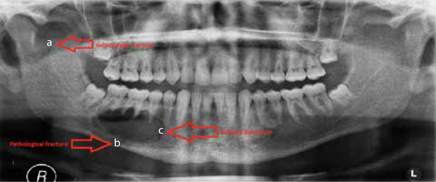
A 16-year-old male presented at the hospital A&E Department following an alleged assault. The patient was diagnosed clinically with a fractured mandible, as he presented with malocclusion and a mobile fracture site between the lower right first molar and second premolar teeth. Appropriate radiographs were taken which revealed a right subcondylar fracture of the mandible. In addition, a pathological fracture of the right body of the mandible in association with a well circumscribed, unilocular cystic area present in the lower right second premolar to lower right second molar region was identified (Figure 1). The presence of the cyst was unknown to the patient; he had been completely asymptomatic.
Figure 1. Initial orthopantomograph, showing the presence of (a) a right subcondylar fracture and (b) a pathological fracture of the right body of the mandible, associated with (c) a large cystic area.
Treatment
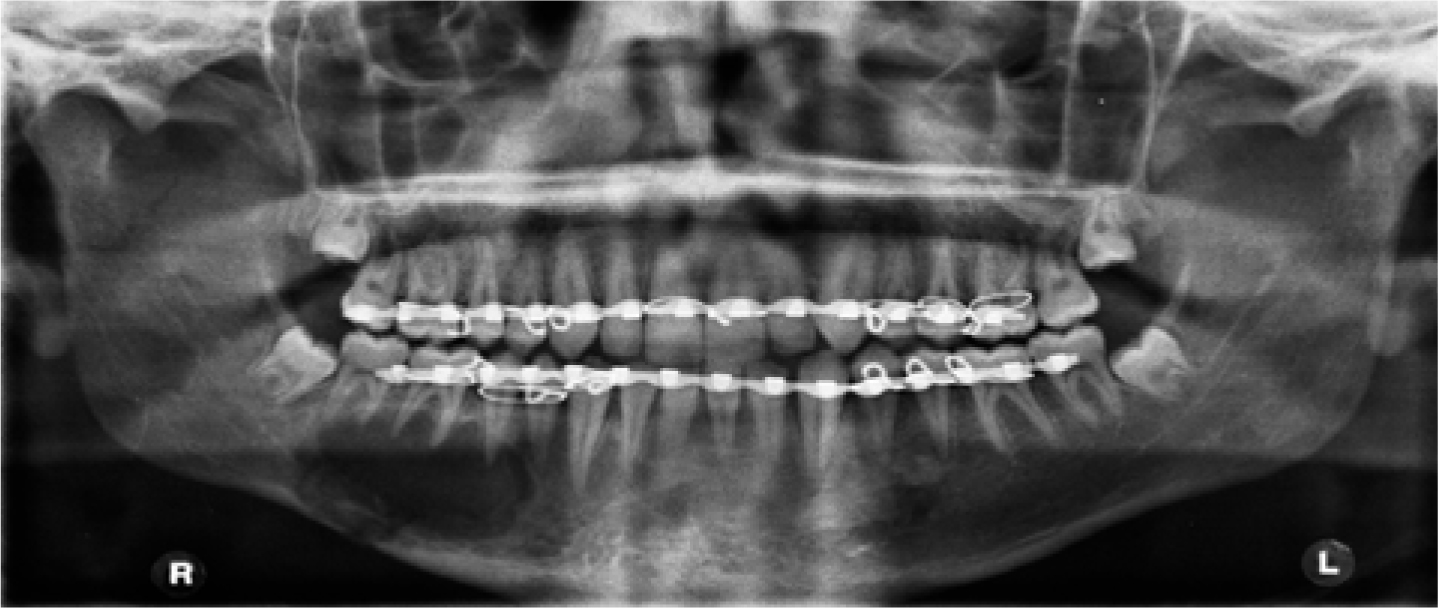
The patient was taken to theatre for repair of the fracture and exploration of the mandibular cyst. A buccal mucoperiosteal flap was raised, with access to the cyst gained through a buccal window of bone. Within the cyst there was no blood, fluid or lining detected. The fracture was minimally displaced and reduced with preformed archbars and intermaxillary elastic fixation (Figure 2). There was a small clot found in the cavity, which was sent for pathological assessment. This revealed no indication of an aneurismal bone cyst, and no cyst lining was identified in the specimen either. This led to the diagnosis of a solitary bone cyst.
Figure 2. Orthopantomograph taken immediately post-operatively showing the enucleated cyst, and the fixation of the fractures using archbars and intermaxillary fixation.
Outcome
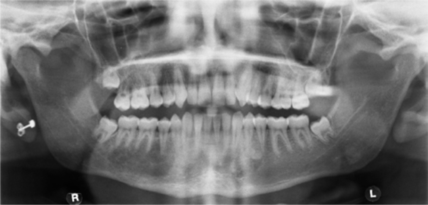
At six months post-operatively, the patient showed positive radiographic signs that the fractures were united, and the bony cavity of the cyst was filling in (Figure 3).3 The patient maintained a good functional occlusion.
Figure 3. Orthopantomograph taken six months post-operatively, following enucleation of the cyst and fixation of the fractures. The radiograph shows the solitary bone cyst cavity beginning to fill in with bone. The fractures have united.
Discussion
This patient was selected for a case report due to the uncommon finding of a solitary bone cyst in relation to his fractured mandible. At 16 years of age, the patient was just outside of the common age-range for presentation of an SBC which is quoted most commonly as presenting in the second and third decades of life, but can present later.3
In keeping with the literature, the cyst had been completely asymptomatic in presence and had almost completely healed within a few months following surgery, which is advised in order to stimulate healing as well as confirming the diagnosis.2
The cyst had a radiographic appearance typical of an SBC, well circumscribed, scalloped between the apices of the adjacent teeth, and unilocular. It met Rushton's criteria4 for pathological identification of an SBC:
Single cyst with no epithelial lining;
No signs of acute or chronic infection;
The cyst may contain fluid but not soft tissue;
The walls of the cyst should be bony but of variable thickness;
Pathological findings do not exclude the diagnosis of SBC.
As an adjunct to investigation of a potential SBC, the vitality of the teeth adjacent to the cyst could be tested pre- and post-operatively, as some cases reported found that adjacent teeth can give a non-vital response prior to surgical enucleation of an SBC, followed by a positive result post-operatively.2 This could lead to unnecessary intervention with root canal treatment. Whilst the treatment of SBCs is usually undertaken within a secondary care setting, it is important for GDPs to acknowledge the existence and management of them, in order to carry out the required investigation, make appropriate referrals and not commence unnecessary intervention themselves.