Article
I would like to share this case report of a primary diffuse large B-cell lymphoma of the oral cavity. It is essential for all general dental practitioners to be aware of this unusual presentation as early recognition, diagnosis and treatment can increase life expectancy of these patients. The prognosis is related to the disease staging.
Lymphomas are classified into Hodgkin's lymphomas and non-Hodgkin's lymphomas. Diffuse large B-cell lymphoma (DLBCLs) are non-Hodgkin's lymphomas and defined as neoplasms of large transformed B-cells with a nuclear diameter more than twice that of a normal lymphocyte. The prevalence of non-Hodgkin's lymphoma is 30–40%. Although NHLs of the oral cavity are rare (3–5%), the most frequent type of primary NHL of the oral cavity is DLBCL. DLBCL can be further classified prognostically into two subgroups, namely germinal centre B-cell like lymphomas (GCBs) and non-germinal centre B-cell like lymphoma (non-GBCs). GCB lymphomas have a better prognosis than non-GCB lymphomas.
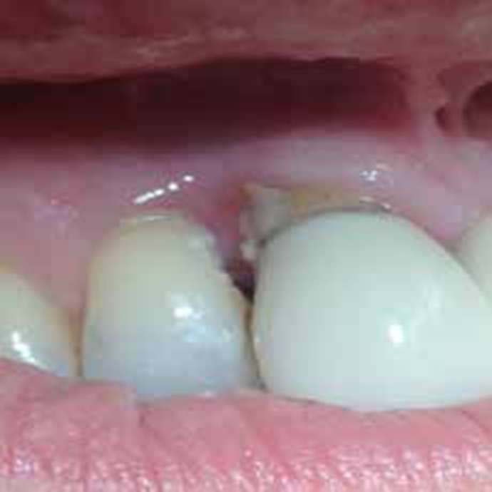
A 68-year-old female patient was initially referred by her dentist to maxillofacial surgery in Kingston Hospital. She presented with a 3 months history of rapidly enlarging growth in her UR2 and UR3 labial gingivae. The upper right incisors and upper right canine were asymptomatic. Intra oral examination revealed a soft purple-coloured sessile lump attached to the gingivae of the upper right anteriors 1cm in diameter. There wasn't any discharge associated with this lump. Upper right anteriors were not mobile (Figure 1).

A periapical X-ray did not reveal any alveolar bone resorption in this region. The growth was excised completely under local anaesthesia and was sent for histopathological analysis. This showed diffuse infiltrate beneath a thin epithelium comprising blastic-like cells with prominent nucleoli and high mitotic activity. The cells were positive with CD20, CD45 and BC12 and showed some scattered small CD3 positive T lymphocytes interspersed (Figures 2 and 3). The cells also showed a high Ki67 proliferation rate. The appearances were of high grade B-cell non-Hodgkin's lymphoma non germinal centre type. Staging with CT scan and bone marrow biopsy showed no involvement. The patient was referred to haematooncologists for treatment involving ‘R-CHOP x3’ with cyclophosphamide, doxorubicin, vincristine and prednisone followed by field radiotherapy to the actual site of the lymphoma in the gums (Figure 4).