Article


Surgery was the original treatment developed for oral cancer. It is still widely carried out, in order to achieve more than one of the following goals.
Cure
The excision of a tumour confined to one area. Surgery may then be used along with radiotherapy (RT) and/or chemotherapy (CTX), given before, during or after surgery. Transoral laser microsurgery is an innovation for complete resection of tumours with preservation of function (organ preservation).
Neck dissection to clear the neck of lymph nodes containing cancer is often also needed in addition to the excision of the primary tumour. Surgery of the neck is, however, evolving to more selective treatments. Initially radical and leading to complications such as pain and damage to the spinal accessory nerve, currently it is function-preserving, resulting in less frequent and less severe neck and shoulder pain, less depression and with less pain and better QoL. The concept of sentinel lymph node biopsy (see below) has gradually been established in the management of early oral cancer to establish whether neck dissection is required.
Reconstructive
Crucially important to restore appearance and/or the function (eg the use of tissue flaps, bone grafts, or prosthetic materials).
Debulking
Used when removing a tumour entirely would cause too much damage to an organ or vital structures.
Palliative
Used to treat complications of advanced disease, not to cure.
Surgery remains the frontline treatment for oral cancer because radical RT may be associated with substantial adverse effects such as xerostomia and the risk of osteoradionecrosis, especially in tumours close to the mandible. However, there has been a shift towards increased use of concurrent chemo-radiotherapy (CRT) as part of an organ preservation strategy. Determination of the need for other modalities and the type is dependent on careful pre-treatment evaluation.
Evaluation
Factors affecting management are related to staging of the primary disease and the presence of metastases, histopathological parameters (tumour grade, depth and pattern of invasion) and patient-related factors (eg co-morbidities and their treatment wishes). Evaluation should include:
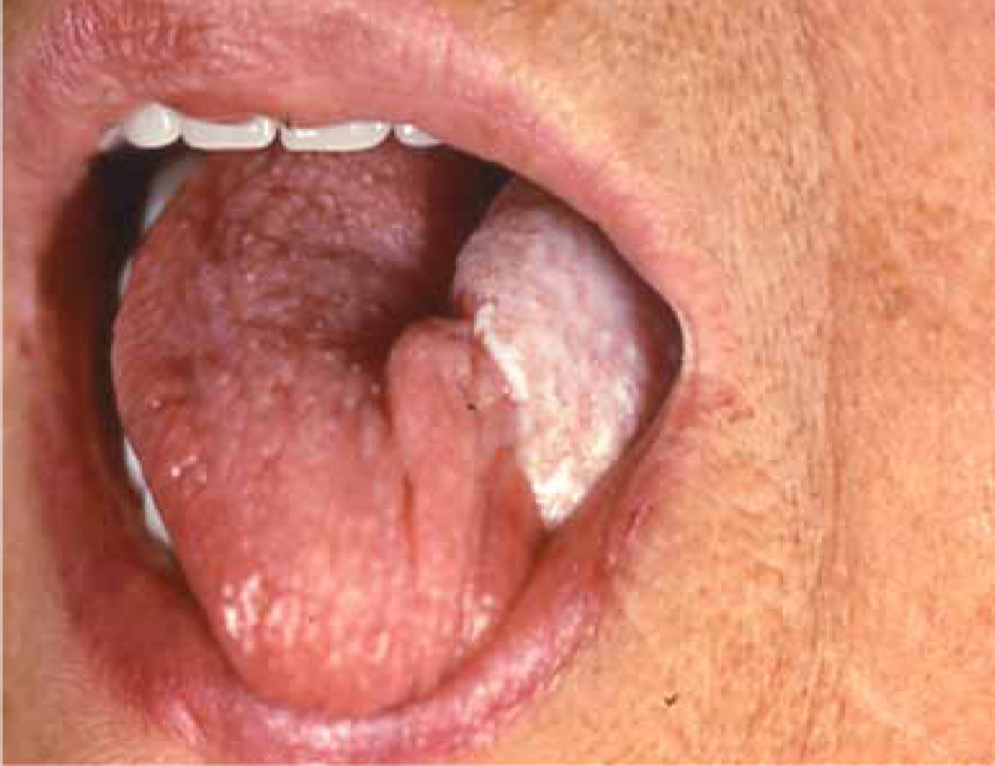
Treatment of Early Oral Cancer
T1 and T2 tumours are generally regarded as ‘early’ cancers. Surgery is the preferred modality of treatment for such lesions of the anterior tongue, buccal mucosa and floor of mouth, or when the lesion is abutting the mandible or the maxilla. Most gingival/palatal lesions are treated surgically. External beam RT alone is a valid alternative to surgery in early lesions of the retro-molar trigone
Surgical treatment includes:
T1/smaller T2 tumours may be treated effectively by single modality therapy, either surgery or RT.
Larger T2 lesions (>3 cm) may require adjuvant RT depending on whether surgical clearance has been achieved followed histological evaluation. Brachytherapy close to the submandibular salivary duct can cause scarring with need for surgical intervention to relieve obstruction.
The primary site is usually approached peri-orally and the tumour with a margin of clinically healthy surrounding tissue of at least one centimetre in all dimensions excised. Post-operative CRT is rarely indicated, unless there are adverse histopathological features, such as a discohesive pattern of invasion of the tumour front, perineural infiltration, and low lymphocytic host response – significant and independent predictors of both local recurrence and reduced overall survival. The overall prognosis for patients with such early stage oral cancer is excellent, with expected 5-year survivals of 80% and 65% for stage I and stage II disease, respectively.
Trans-oral CO2 laser resection of the primary disease without reconstruction is being increasingly advocated in some centres: survival rates compare favourably with conventional surgery and with improved functional results.
Photodynamic therapy (PDT) or Moh's micrographic surgery may help for superficial oral lesions. PDT may be an adjunct in the management of large areas of field change (condemned mucosa).
Treatment of more advanced oral cancer
Since the cancer often extends to more than one of the oral sub-sites and/or has spread to cervical lymph nodes, these patients pose a challenge. Large volume T2, T3 and T4 tumours should be treated with combination surgery/post-operative chemo-radiotherapy if the tumour can be completely excised macroscopically and repaired with satisfactory functional results and the patient is fit enough for the procedures. Post-operative CRT is dependent on surgical clearance as determined by histological evaluation, except for in T4 tumours, where it is indicated.
The principles that surgical resection are based on are:
The factors affecting the choice of surgical approach are:
Neck node involvement
Cervical lymph node metastasis is the single most adverse independent prognostic factor, with the exception of distant metastases. Prognosis is affected by the number of metastases, their level in the neck, the tumour load, extracapsular tumour spread, perineural and/or vascular invasion, previous treatment by surgery or RT and resectability. When there is a single nodal metastasis at presentation or subsequently, the survival rate halves.
Most tumours metastasize fairly predictably to particular nodal groups but some may metastasize to remote sites (eg tongue cancers to jugulo-omohyoid nodes) and the pattern of spread may also be disrupted by previous surgery or RT. The possibility of bilateral nodal disease should also be considered in tongue-based primary tumours.
However, there is no evidence that control of metastatic disease in the neck improves survival, and therefore issues of function and QoL are important in the planning.
Neck surgery once classified as ‘radical neck dissection’, which removed a large number of nodes and other structures such as the jugular vein and accessory nerve, has evolved to a less damaging ‘modified radical’ and latterly to ‘selective neck dissection’. Sentinellymph node lymphoscintigraphy (SLNL) and ultrasound-guided fine needle aspiration cytology (FNAC) biopsy has also been developed as a staging procedure in early stage I/II oral cancers, where there are no palpable lymph nodes. The theory is that the sentinel node is any node that receives lymphatic drainage directly from a tumour. This is tested by lymphoscintigraphy – the injection of a radiopaque marker such as technetium-99m-labelled nanocolloid in the tumour area and then examination of the nodes with a gamma camera to see where the radionuclide first reaches. A simpler method is to use a dye tracer. Histological scrutiny of such a node will indicate whether metastasis to cervical lymph nodes has occurred. SLN biopsy can thus help to determine the presence of lymph node metastases in patients with T1-T2, N0 oral and oropharyngeal cancers and guide as to the need for neck dissection.
Distant metastases
Patients with oral cancer may have or develop distant metastases, mainly in the lungs, liver, bone or brain. Approximately 5% of patients have metastatic disease at presentation and distant metastases may subsequently develop in one-third.
Reconstruction
The aims of reconstruction are to achieve good functional and cosmetic outcomes by:
The surgical defect, if small, is reconstructed mainly using local flaps or microvascular surgery, if necessary. Reconstruction of larger oral and oro-pharyngeal tumours may also be performed with free tissue transfer and to a lesser extent with regional pedicled flaps (Figure 1).
The flaps mainly used include:
Perforator flaps represent a safer and more reliable procedure than other flaps, offering greater freedom in choosing a donor site because flap selection is based on the quality and volume of tissue required at the recipient site. This procedure minimizes donor site morbidity (eg pain, infection, cosmetics) whilst the versatility of the flap allows easier moulding of the flap.
Bone loss is replaced either by composite flaps or bone grafts (eg from rib) or sometimes by distraction osteogenesis. Dental reconstruction is discussed in Article 18.