Moch H, Cubilla AL, Humphrey PA The 2016 WHO classification of tumours of the urinary system and male genital organs – part A: renal, penile, and testicular tumours. Eur Urol. 2016; 70:93-105 https://doi.org/10.1016/j.eururo.2016.02.029
Mancilla-Jimenez R, Stanley RJ, Blath RA. Papillary renal cell carcinoma: a clinical, radiologic, and pathologic study of 34 cases. Cancer. 1976; 38:2469-2480
Amin MB, Corless CL, Renshaw AA Papillary (chromophil) renal cell carcinoma: histomorphologic characteristics and evaluation of conventional pathologic prognostic parameters in 62 cases. Am J Surg Pathol. 1997; 21:621-635 https://doi.org/10.1097/00000478-199706000-00001
Delahunt B, Eble JN. Papillary renal cell carcinoma: a clinicopathologic and immunohistochemical study of 105 tumors. Mod Pathol. 1997; 10:537-544
Gargouri MM, Ayari Y, Ben Chehida M Clinical and pathological features of papillary renal cell carcinoma and prognostic value of its type-1 and type-2 subtypes. Afr J Urol. 2016; 22:149-152
Akhavan A, Richards M, Shnorhavorian M Renal cell carcinoma in children, adolescents and young adults: a National Cancer Database study. J Urol. 2015; 193:1336-1341 https://doi.org/10.1016/j.juro.2014.10.108
Shah A, Jahan S, Najar L Metastatic clear cell variant of renal cell carcinoma of the mandible: review and case report. Ann Maxillofac Surg. 2016; 6:144-147 https://doi.org/10.4103/2231-0746.186121
Kim JK, Kim TK, Ahn HJ Differentiation of subtypes of renal cell carcinoma on helical CT scans. AJR Am J Roentgenol. 2002; 178:1499-1506 https://doi.org/10.2214/ajr.178.6.1781499
Roy C, Sauer B, Lindner V MR Imaging of papillary renal neoplasms: potential application for characterization of small renal masses. Eur Radiol. 2007; 17:193-200 https://doi.org/10.1007/s00330-006-0271-9
NICE. Nivolumab with ipilimumab for untreated advanced renal cell carcinoma. Technology appraisal guidance (TA581). 2019. https://www.nice.org.uk/guidance/ta581 (accessed September 2021)
Yin Y, Yuan X, Gao H, Yang Q. Nanoformulations of small molecule protein tyrosine kinases inhibitors potentiate targeted cancer therapy. Int J Pharm. 2020; 573 https://doi.org/10.1016/j.ijpharm.2019.118785
Steffens S, Janssen M, Roos FC Incidence and long-term prognosis of papillary compared to clear cell renal cell carcinoma – a multicentre study. Eur J Cancer. 2012; 48:2347-2352 https://doi.org/10.1016/j.ejca.2012.05.002
Delahunt B, Eble JN. Papillary renal cell carcinoma: a clinicopathologic and immunohistochemical study of 105 tumors. Mod Pathol. 1997; 10:537-544
Zucchi A, Novara G, Costantini E Prognostic factors in a large multi-institutional series of papillary renal cell carcinoma. BJU Int. 2012; 109:1140-1146 https://doi.org/10.1111/j.1464-410X.2011.10517.x
Bianchi M, Sun M, Jeldres C Distribution of metastatic sites in renal cell carcinoma: a population-based analysis. Ann Oncol. 2012; 23:973-980 https://doi.org/10.1093/annonc/mdr362
Mir F, Alnajae H, Rupcich C 143 metastatic renal cell carcinoma to the head and neck area: a clinicopathologic study. Am J Clin Pathol. 2018; 149
Zhang R, Lee CW, Basyuni S, Santhanam V. Mandibular swelling as the initial presentation for renal cell carcinoma: a case report. Int J Surg Case Rep. 2020; 70:96-100 https://doi.org/10.1016/j.ijscr.2020.04.061
Shah A, Jahan S, Najar L Metastatic clear cell variant of renal cell carcinoma of the mandible: review and case report. Ann Maxillofac Surg. 2016; 6:144-147 https://doi.org/10.4103/2231-0746.186121
Raising awareness of acute onset of swelling and lip paraesthesia in a teenage patient Angela Boscarino Simon N Rogers Dental Update 2024 48:9, 707-709.
Authors
AngelaBoscarino
BDS, MFDS RCS (Glas)
Dental Core Trainee, Liverpool Head and Neck Centre, Liverpool University Hospital Aintree NHS Foundation Trust
Regional Maxillofacial Unit, University Hospital Aintree, Liverpool, UK and Edge Hill University, Liverpool and Evidence-Based Practice Research Centre (EPRC), Faculty of Health, Edge Hill University, St Helens Road, Ormskirk
We report a case of papillary renal cell carcinoma in a 19-year-old patient that manifested as ipsilateral numbness of the lower lip and swelling in the region of masseter insertion. Despite there being widespread metastatic disease at presentation, the diagnosis was delayed with false reassurance from a normal brain scan and the assumption that the symptoms were due to infection related to the lower wisdom tooth. Once the primary tumour site was established, the patient responded to the relatively new immunotherapy treatments for advanced renal cell carcinoma as advocated in the 2019 NICE guidance. There are no other documented case reports that discuss papillary renal carcinoma with metastatic spread to the mandible in teenagers.
CPD/Clinical Relevance: This case highlights the importance of malignancy being a differential diagnosis of lip paraesthesia irrespective of age.
Article
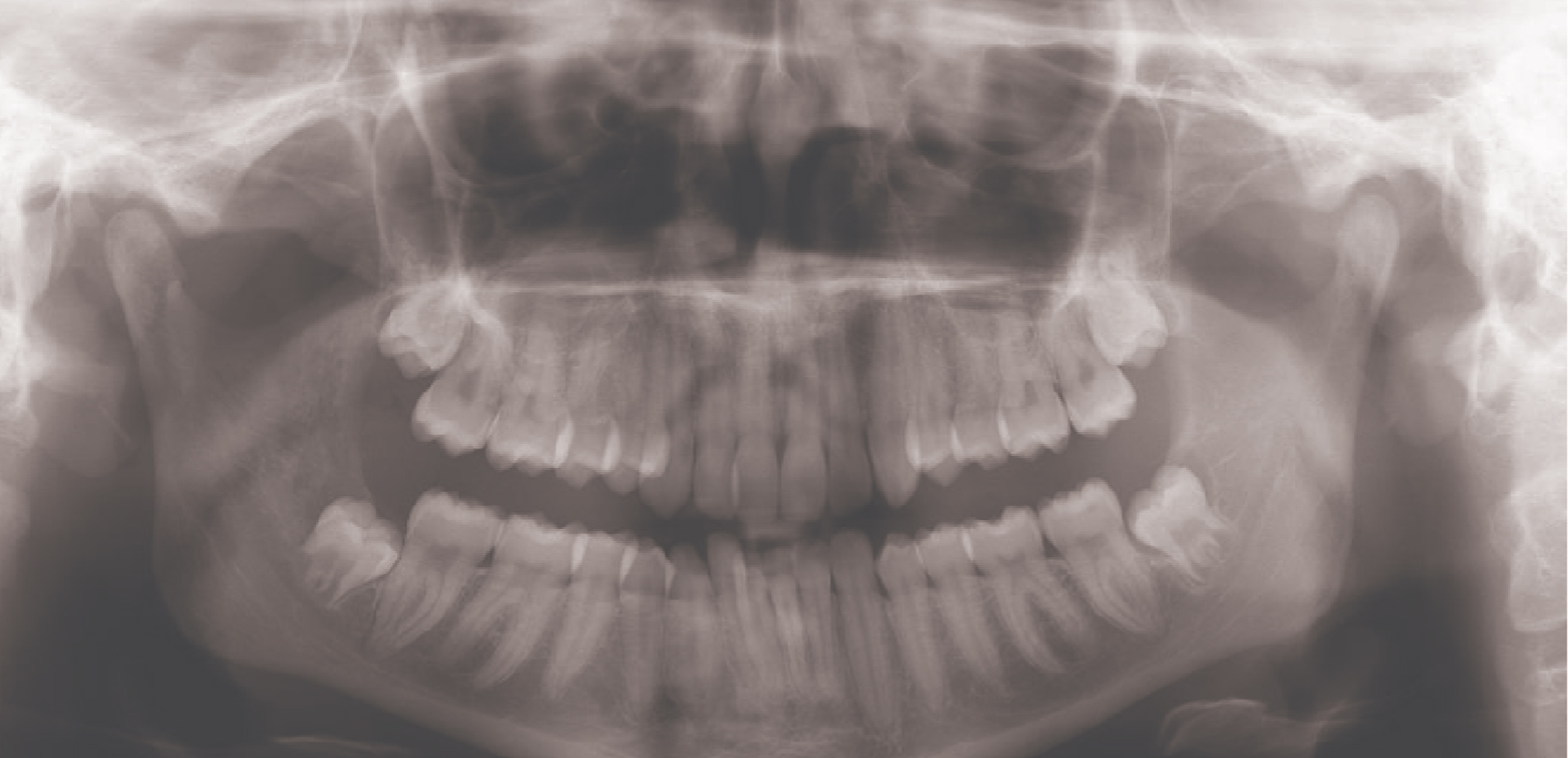
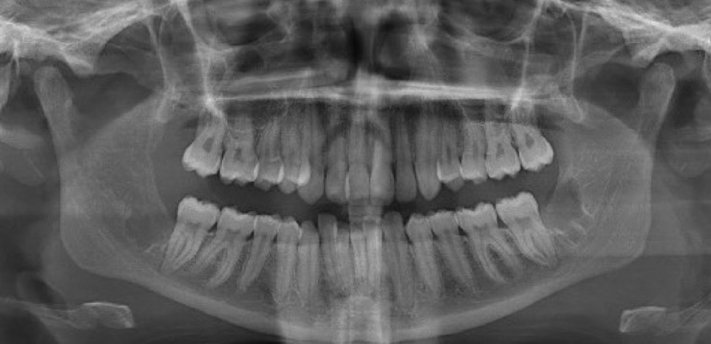
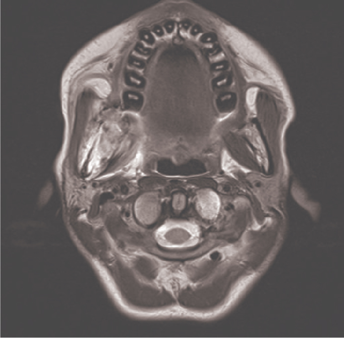
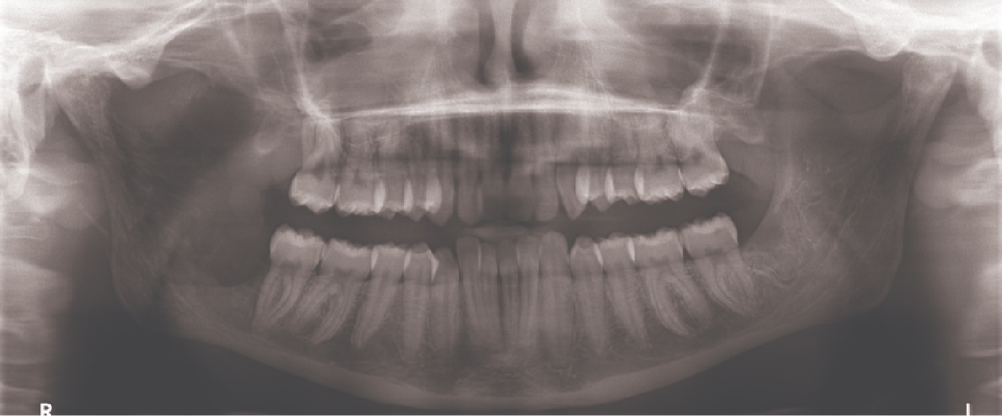
A 19-year-old female patient initially presented to her GDP with an acute onset of loss of sensation to the right side of her chin and lip. Her dentist referred her for an OPG at the local hospital, which was reported as unremarkable (Figure 1). She was also experiencing vomiting, fatigue and night sweats. Her GP requested routine bloods and a head CT, which were both reported as normal. No obvious neurological causes were noted. Three weeks after the onset of her facial numbness, she developed ipsilateral facial swelling in the right masseter region. She returned to the GDP who diagnosed pericoronitis, which was managed with irrigation of the area and antibiotics. The patient's symptoms failed to improve despite several courses of antibiotics, and they persisted during a planned trip to the USA. While in the US, she sought the attention of an oral surgeon who proceeded to extract all wisdom teeth. During the operation, he noted an unusual mass in the LR8 region, which was biopsied. The post-operative OPG revealed a radiolucency extending up to the sigmoid notch (Figure 2). The histopathology was of an adenocarcinoma that was likely to be a metastasis of a primary, which was unknown at the time. She urgently flew back to the UK and underwent further imaging that showed a 7-cm mass in the right kidney, numerous bilobar liver, lytic bone, mediastinal lymph node and bilateral adrenal metastases. A liver and supraclavicular node biopsy revealed Type 2 papillary renal cell carcinoma as the primary source. An MRI of the mandible showed a metastatic lesion surrounding the right ramus of the mandible and extending into the infratemporal fossa and to the greater wing of the sphenoid. (Figure 3).
Figure 1. OPG at initial presentation to GDP.Figure 2. OPG after the biopsy.Figure 3. Pre-treatment MRI scan of the mandible.
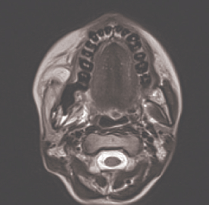
The patient received radiotherapy to her thoracic (T4–9) and lumbar (T12–L2) spine, and immunotherapy in the form of ipilimumab and nivolumab. The immunotherapy had to be discontinued owing to liver toxicity, and second-line management was with cabozantinib, a tyrosine kinase inhibitor (TKI). There was a very good response to the TKI, and necrosis of the tumour occurred. This was demonstrated in the post-treatment MRI with a high signal associated with inflammation (Figure 4), and OPG (Figure 5). The main oral symptoms of pain and trismus were slow to improve over several months and managed by spatula exercises and combination analgesia with opiates, gabapentin and NSAIDs. Five months following the start of treatment, the disease appeared to stabilize with ongoing management with cabozantinib, steroids and pain relief. Unfortunately, there was further disease progression and a subsequent MRI showed the development of new metastatic lesions in the orbital wall and occipital bone, as well as a new level 1b metastatic lymph node. The patient sadly died peacefully at home 10 months following the initial diagnosis.
Figure 4. Post-treatment MRI scan of the mandible.Figure 5. OPG after treatment.
Papillary renal cell carcinoma
In 2016, the World Health Organization updated its classification of renal tumours and expanded on the various subtypes.1 Renal cell carcinoma is the most common type of renal tumour. Papillary renal cell carcinoma (PRCC) is a histological subtype of renal cell carcinoma (RCC) and accounts for approximately 15% of all malignant renal cell carcinomas. It was first reported by Mancilla and Stanley in 1976.2 It derives from the cells of the distal convoluted tubule of the nephron and is microscopically predominantly papillary or tubulopapillary.3 In 1997, Delahunt and Eble recognized that there were two types of PRCC, Type 1 and Type 2.4 The histological pattern shows that Type 1 consists of papillae and tubular structures covered by small cuboidal cells with basophilic cytoplasm and Type 2 consists of papillae covered by large cells with eosinophilic cytoplasm. Immunohistochemical studies can also demonstrate a positive reaction to the marker cytokeratin 7.5
PRCC incidence represents approximately 2.4% of all invasive cancers and has a projected mortality rate of 1.8 per 100,000 worldwide.6 It predominantly affects males aged between 50 and 60 years. PRCC is rare in adolescents and young adults, and so there are currently no studies that give an incidence rate for this specific subtype of carcinoma in this age group. However, one national study found that of all patients under the age of 30 years who were diagnosed with RCC (all subtypes), 9.2% were aged 15–21 years.7
Risk factors for RCC include obesity, smoking, hypertension, end-stage renal failure and diabetes mellitus. Of all cases, 3–5% may have hereditary RCC syndrome, which is inherited through an autosomal dominant trait. Signs and symptoms may include backache, haematuria, unexplained weight loss, asthenia, unexplained fever and lump or mass in the side. Once the tumour metastasizes, there may also be abdominal pain, dyspnoea, neurological disturbances and a cough.8
CT and MRI are frequently used to aid diagnosis of the primary tumour, and can also assess for potential metastases. On a contrast-enhanced CT, PRCC will usually be reported as hypovascular and homogeneous in nature. On MRI, most PRCCs show hypointensity on both T1- and T2-weighted images due to either the deposition of hemosiderin and calcium, or the densely collagenous nature of PRCC.9,10
Under the 2019 NICE guidance, first-line treatment for advanced and metastatic renal cancer is with ipilimumab and nivolumab.11 These are both monoclonal antibodies that work by blocking the activity of the protein PD-1 (programmed death receptor), which prevents T cells from recognizing cancer cells and attacking them. This is turn augments the immune system's response to tumour cells. This treatment was not effective in the case described owing to liver toxicity, and the patient received second-line treatment with cabozantinib, a tyrosine kinase inhibitor (TKI). This class of drug inhibits tyrosine kinase, which is a protein that, when mutated causes unregulated cell growth. Therefore, TKI works by blocking the signalling pathways, it inhibits tumour metastases and causes an overall reduction in tumour proliferation.12
Prognosis is dependent on the PRCC subtype. Type 1 tends to have a low nuclear grade and a better prognosis, with approximately 85.1% survival rate at 5 years.13 Type 2 tends to have a higher nuclear grade, larger tumours, higher metastatic potential and with a worse prognosis.14 A multicentre review showed that approximately 10% of patients with PRCC had lymph nodes or distant metastases at initial presentation. The presence of these are independent prognostic markers for survival.15
The most common RCC metastatic sites are the lungs (45%), bone (30%) and lymph nodes (22%).16 RCC head and neck metastases are uncommon, occurring in approximately 3% of patients, with sites including the thyroid, sinuses, skull, lymph node and jaw.17 There are a few documented case reports of RCC metastases to the mandible, with symptoms including paraesthesia or anaesthesia of the chin and lower lip due to mental nerve involvement, along with swelling or loosening of teeth. However, the literature search proved inconclusive for case reports of PRCC metastases to the mandible.18,19
Mandibular metastases
In the UK, 1 in 55 males and 1 in 108 females will be diagnosed with oral cancer in their lifetime.20 Approximately 1% of all oral neoplasms will be of metastatic origin. Neoplastic metastases occur via the ‘invasion-metastasis cascade’, which allows the malignant cells to penetrate the surrounding extracellular matrix, invade the blood vessels and colonize a distant organ. Bone has a favourable micro-environment and the metastases occur via the interaction between the metastatic tumour cells.21 Metastasis to the mandible is more common than the maxilla. In particular, the premolar and molar regions have a rich vascularization and high bone marrow content and so are the most frequently involved areas.22
There is an almost equal gender distribution in mandibular metastasis. However, the nature of the primary tumour varies between men and women. The most common primary tumour site in women is the breasts, reproductive organs, thyroid gland and kidneys. In men, it is the lungs, prostate, kidneys, bones, large intestine and suprarenal glands.23
Oral metastases are usually associated with multiple organ involvement, and their presentation may be the first indication of an undiscovered primary malignancy.24 In younger patients, metastases to the jaw are more common than soft tissue metastasis. Of all jaw metastases, 81% are to the mandible, with the most frequently affected area being the molar region, followed by the premolar region.25
A patient with a mandibular tumour can present with unilateral paraesthesia along the distribution of the inferior alveolar nerve, accompanied by a rapidly progressing unilateral swelling involving the ramus and the body of the mandible. The paraesthesia is caused by either the tumour compressing the nerve or the malignant infiltration into the nerve sheath. With mandibular tumours, there may also be bony tenderness in the affected area along with unexplained tooth mobility and possible associated trismus. With progression of the disease, dysphagia along with masticatory interference can be noted.
Owing to the ease of access and low radiation dose, an OPG would be the first radiograph of choice, both in primary and secondary care, to assess osseous changes in the mandible. An OPG may show the appearance of an ill-defined lytic radiolucent lesion. It may have a ‘moth eaten’ appearance, and in some cases a pathological fracture can be present. However, an OPG may be unreliable for detecting bony invasion and a referral to secondary care for a cone beam CT or a mandibular CT may be more appropriate for a more in-depth assessment if the OPG is inconclusive.
There are NICE and NHS Scotland guidelines26,27 for identifying oral cancer signs and symptoms, which allow for a prompt referral on the urgent 2-week cancer pathway. In this case, the patient had none of these signs or symptoms, but, as demonstrated in this case, unexplained paraesthesia can be a differential diagnosis for cancer, even in a young patient. Therefore, an urgent referral to secondary care for assessment and investigations is an appropriate action.
Conclusion
This case presents a primary type 2 papillary renal cell carcinoma with mandibular and other widespread metastases (renal, adrenal, bony, liver) in a female teenage patient. This is a rare presentation, and there have been no previous case reports in the literature. It emphasizes the importance of taking a thorough, detailed history, and having a high degree of suspicion for patients who present with ipsilateral numbness and swelling, irrespective of their age. Bone metastases may not be obvious on an OPG, and so a referral to secondary care for further scans may be more conclusive. Unexplained paraesthesia is not currently part of the guidelines for the urgent 2-week cancer pathway referral, but an underlying malignancy should still be actively excluded in these cases.