
Abstract
The aim of this review is to summarize answers to common questions related to halitosis including its prevalence, the different types, aetiology, assessment, diagnosis and management in general practice.
From Volume 48, Issue 6, June 2021 | Pages 459-462
The aim of this review is to summarize answers to common questions related to halitosis including its prevalence, the different types, aetiology, assessment, diagnosis and management in general practice.

Oral healthcare professionals should be aware of the fundamentals of halitosis as they are primarily responsible for its diagnosis and management.
Halitosis (from the Latin for breath, halitus and the Greek suffix osis, meaning abnormal)1 is the presence of unpleasant or offensive breath odour independent of its origin. Halitosis can have major detrimental social implications for an individual and can significantly impact on normal social interactions.2
The true prevalence of halitosis is unknown. The majority of epidemiological studies are difficult to evaluate as they are largely based on subjective self-estimation of halitosis, which is known to be limited by inaccuracy and low sensitivity. However, the available evidence suggests that halitosis is common, and can affect individuals of all ages.
The prevalence of persistent halitosis in one of the most recent studies was reported to be 15%, was nearly three times higher in men than in women (regardless of age) and the risk was slightly more than three times greater in individuals over 20 years of age compared with those aged 20 years or under, controlling for gender.3 The methodology used was interesting because it overcame the limitations of self-reporting of halitosis, while retaining its subjective judgement; the design also facilitated the recruitment of large numbers of subjects.
Overall, the majority of studies report that approximately 30% of people have halitosis,4,5,6 but some studies do estimate that more than 50% of the population have halitosis.7
Intra-oral halitosis is also known as oral malodour and describes cases where the source of halitosis lies within the mouth. Extra-oral halitosis is where the source of halitosis lies outside the mouth. This can be further subdivided into blood-borne and non-blood-borne halitosis.8 Pseudo-halitosis and halitophobia are used to describe patients who think or persist in believing they have halitosis, even after professional assessment and a diagnosis that they do not have halitosis. Temporary, or transient halitosis, as well as morning bad breath are other forms of halitosis.
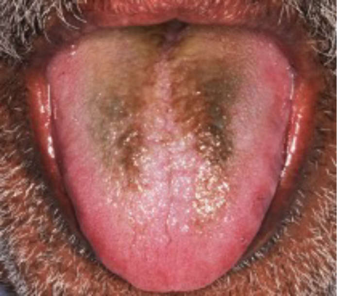
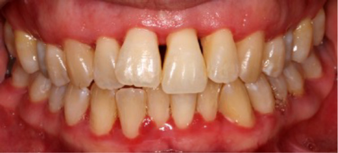
In about 85% of patients with persistent genuine halitosis, the odour originates from the mouth.9 The aetiology of intra-oral halitosis is primarily a tongue coating (Figure 1) and to a lesser extent gingivitis/periodontitis (Figure 2) or a combination of these.10 A number of other acute conditions can cause malodour including pericoronal infections, oral ulceration, acute herpetic gingivostomatitis and necrotizing gingivitis, all of which produce a characteristic strong ‘fetor oris’. The presence of xerostomia, candidal infections, medications, overhanging restorations and caries may also contribute to intra-oral halitosis.10,11,12,13
Intra-oral halitosis arises from the production of volatile malodorous compounds by the action of bacteria in breaking down components of epithelial cells, salivary and serum proteins, and food debris.
A wide range of molecular species are able to contribute to the overall production of malodour. Notably, much attention has been given to volatile sulphur compounds, including methyl mercaptan, hydrogen sulphide and dimethyl sulphide. However, a number of other compounds may also contribute to malodour (Table 1).14,15,16 The production of these compounds is mediated by the putrefaction of debris and protein substrates by a wide range of oral anaerobic bacteria (Table 2), particularly those which exhibit proteolytic activity.17,18,19,20 In the case of volatile sulphur compound generation, bacteria act on the sulphur-containing amino acids cysteine, cystine and methionine, which are available following proteolysis.
| Volatile sulphur compounds | Methyl mercaptan, hydrogen sulphide and dimethyl sulphide |
| Diamines | Putrescine, cadaverine |
| Short-chain fatty acids | |
| Butyric acid, propionic acid | |
| Phenyl compounds | Indole, skatole, pyridine |
| Porphyromonas gingivalis |
| Prevotella intermedia |
| Treponema denticola |
| Fusobacterium nucleatum |
| Tannerella forsythensis |
| Porphyromonas endodontalis |
| Eubacterium species |
The dorsum of the tongue provides a suitable environment for the growth of anaerobic organisms, as favourable redox potentials are found in the deep crypts of the tongue associated with the structure of the tongue papillae.2 Tongue coatings include desquamated epithelial cells, food debris, bacteria and salivary proteins and provide an ideal environment for the generation of volatile sulphur compounds and other compounds that contribute to malodour.2 Studies suggest that malodour may be associated with total bacterial load of Gram-negative anaerobes, both in saliva and in tongue coatings.21,22,23,24 The extent of tongue coating can vary considerably between individuals, and may be increased in patients with periodontitis and in response to systemic upset.
These anaerobic bacteria are also associated with subgingival plaque in periodontitis, and indeed, high concentrations of volatile sulphur compounds are present in periodontal pockets, and in gingival crevicular fluid of those with gingivitis or periodontitis.
Extra-oral halitosis is far less frequent than intra-oral halitosis and originates from pathological conditions outside the mouth. In non-blood-borne extra-oral halitosis, these may include nasal, paranasal and laryngeal regions, lungs or upper digestive tract. In the case of a blood-borne extra-oral halitosis, the malodour is emitted via the lungs and originates from disorders anywhere in the body, for example due to hepatic cirrhosis.10Table 3 summarizes the common extra-oral causes of halitosis.25
| Respiratory system (microbial aetiology) | Sinusitis |
| Antral malignancy | |
| Cleft palate | |
| Foreign bodies in the nose | |
| Nasal malignancy | |
| Tonsilloliths | |
| Tonsillitis | |
| Pharyngeal malignancy | |
| Lung infections | |
| Bronchitis | |
| Bronchiectasis | |
| Lung malignancy | |
| Gastrointestinal tract | Oesophageal diverticulum |
| Gastro-oesophageal reflux disease | |
| Malignancy | |
| Metabolic disorders (blood-borne) | Acetone-like smell in uncontrolled diabetes |
| Uraemic breath in renal failure | |
| Fetor hepaticus in liver disease | |
| Trimethylaminuria (fish odour syndrome) | |
| Hypermethioninaemia | |
| Cystinosis | |
| Drugs (blood-borne) | Amphetamines |
| Chloral hydrate | |
| Cytotoxic agents | |
| Dimethyl sulphoxide | |
| Disulfiram | |
| Nitrates and nitrites | |
| Phenothiazines | |
| Solvent abuse |
Transient halitosis may be caused by tobacco use, garlic and some spicy foods, and by alcohol. Morning bad breath is caused by the decrease in saliva production during the night, ie lack of the natural cleaning mechanism.10 It is common on waking in many people, but is usually short lived and typically resolves shortly after rising, especially after eating breakfast and performing morning oral hygiene procedures.2
A thorough medical history questionnaire, soft tissue examination to check for a tongue coating, periodontal examination and an organoleptic assessment will form the key steps in making a diagnosis in practice.
In terms of the organoleptic assessment of exhaled air, the clinician sniffs the air exhaled from the mouth, as well as the nose.26 Smelling both nose and mouth air is important as halitosis detectable from the nose alone (asking the patient to breathe while the mouth is closed) is likely to come from the nose or the sinuses, or from respiratory or gastrointestinal tracts. At its simplest, a clinician may use their own judgement to decide on the presence or absence of malodour in a patient. In clinical investigations, trained breath assessors may assess exhaled air from subjects, scoring their assessments on a 6-point scale from 0 to 5, according to the assessed severity of the condition.27 As a general rule in practice, it is advisable that the patient abstains from eating odoriferous foods for 48 hours before the assessment, and that both the patient and the examiner refrain from drinking coffee, tea or juice, smoking and using scented cosmetics before the assessment.28 More objective measurements of halitosis are available, but they are rarely used in routine clinical practice as they are expensive and time-consuming.
Gas chromatography can precisely measure specific gases. However, traditional forms need inert column carrier gas and require technicians or specialists with adequate training, and are thus clinically impractical.29 However, a newly developed portable gas chromatograph (OralChroma, Abi-medical, Abilit Corp, Osaka, Japan) has now been described,30 which does not use a special carrier gas (using air instead) and is highly sensitive, yet relatively low in cost compared with a standard gas chromatograph. Suphide monitoring is an easily used method, but has the limitation that important odours are not detected. An example of this is the halimeter (Interscan Corp, Chatsworth, CA, USA).
The scientific and practical value of alternative measurement methods, such as the BANA (benzoyl–arginine–naphthyl–amide) test, chemical sensors, the salivary incubation test, quantifying β-galactosidase activity, ammonia monitoring, the ninhydrin method and the polymerase chain reaction, have yet to be fully established.31,32
If no halitosis can be found during the initial examination, the assessment can be repeated on two or three different days. Thereafter, if halitosis is still not present, but the patient feels they are suffering from it, the patient can be diagnosed with pseudo-halitosis – a diagnosis that can be supported by established questionnaires.28
The management of halitosis depends largely on the cause. Once the diagnosis of intra-oral halitosis has been confirmed, the clinician should:
It is important to be aware that a limited number of patients with extra-oral halitosis and halitophobia (<10% together) will need to be referred to an appropriate medical professional.10 This may involve an oral medicine opinion, an otorhinolaryngologist to rule out the presence of chronic tonsillitis or chronic sinusitis, a physician to rule out gastric, hepatic, endocrine, pulmonary or renal disease, or a psychologist or psychiatrist.