Blum IR, Schriever A, Heidemann D Repair versus replacement of defective direct composite restorations in teaching programmes in United Kingdom and Irish Dental Schools. Eur J Prosthodont Restor Dent. 2002; 10:151-155
Blum IR, Lynch CD. Repair versus replacement of defective direct dental restoration in posterior teeth of adults. Prim Dent J. 2014; 3:62-67
Eltahlah D, Lynch CD, Chadwick B An update on the reasons for placement and replacement of direct restorations. J Dent. 2018; 72:1-7
Blum IR. The management of failing direct composite restorations: replace or repair?. In: Lynch CD, Wilson NHF, Blunton PA (eds). London: Quintessence; 2008
Deligeorgi V, Mjör IA, Wilson NHF. An overview of reasons for the placement and replacement of restorations. Prim Dent Care. 2001; 8:5-11
Wilson NHF, Lynch CD, Brunton PA Criteria for the replacement of restorations: Academy of Operative Dentistry European Section. Oper Dent. 2016; 41:(S7)S48-S57
NHS Digital. Adult dental health survery 2009. Summary report and thematic series. 2011. https://tinyurl.com/y39nbhpg (Accessed 8 October 2020)
Blum IR, Jagger DC, Wilson NHF. Defective dental restorations: to repair or not to repair? Part 1: direct composite restorations. Dent Update. 2011; 38:78-84
Blum IR, Jagger DC, Wilson NHF. Defective dental restorations: to repair or not to repair? Part 2: all-ceramics and porcelain fused to metal systems. Dent Update. 2011; 38:150-158
Kanzow P, Wiegand A, Schwendicke F. Cost-effectiveness of repairing versus replacing composite or amalgam restorations. J Dent. 2016; 54:41-47
Wilson NHF, Lynch CD. The teaching of posterior resin composites: planning for the future based on 25 years of research. J Dent. 2014; 42:503-516
Bogacki RE, Hunt RJ, del Aguila M, Smith WR. Survival analysis of posterior restorations using an insurance claims database. Oper Dent. 2002; 27:488-492
Lucarotti PSK, Holder RL, Burke FJT. Analysis of an administrative database of half a million restorations over 11 years. J Dent. 2005; 33:791-803
Burke FJT, Lucarotti PSK. How long do direct restorations placed within the general dental services in England and Wales survive?. Br Dent J. 2009; 206
Gordan VV, Riley J III, Geraldeli S The decision to repair or replace a defective restoration is affected by who placed the original restoration: findings from the National Dental PBRN. J Dent. 2012; 42:1528-1534
Cvar JF, Ryge G. Reprint of criteria for the clinical evaluation of dental restorative materials. 1971. Clin Oral Investig. 2005; 9:215-232 https://doi.org/10.1007/s00784-005-0018-z
Ryge G. Clinical criteria. Int Dent J. 1980; 30:347-358
Wilson NH, Norman RD. Five-year findings of a multiclinical trial for a posterior composite. J Dent. 1988; 19:153-159
Marquillier T, Domejean S, Le Clerc J The use of FDI criteria in clinical trials on direct dental restorations: a scoping review. J Dent. 2018; 68:1-9
Hickel R, Roulet JF, Bayne S Recommendations for conducting controlled clinical studies of direct restorative materials. Clin Oral Investig. 2007; 11:5-33
Hickel R, Roulet JF, Bayne S Recommendations for conducting controlled clinical studies of direct restorative materials. J Adhes Dent. 2007; 9:121-147
Hickel R, Roulet JF, Bayne S Recommendations for conducting controlled clinical studies of direct restorative materials. Int Dent J. 2007; 57:300-302
Hickel R, Peschke A, Tyas M FDI World Dental Federation: clinical criteria for the evaluation of direct and indirect restorations – update and clinical examples. J Adhes Dent. 2010; 12:259-272
Blum IR, Özcan M. Reparative dentistry: possibilities and limitations. Curr Oral Health Rep. 2018; 5:264-269
Mjör IA, Toffenetti F. Secondary caries: a literature review with case reports. Quintessence Int. 2000; 31:165-179
Gordan VV, Garvan CW, Blaser PK A long-term evaluation of alternative treatments to replacement of resin-based composite restorations: results of a seven-year study. J Am Dent Assoc. 2009; 140:1476-1484
Martin J, Fernandez E, Estay J Minimal invasive treatment for defective restorations: five-year results using sealants. Oper Dent. 2013; 38:125-133
Moncada G, Martin J, Fernandez E Sealing, refurbishment and repair of class I and class II defective restorations: a three-year clinical trial. J Am Dent Assoc. 2009; 140:425-432
Martins BMC, Silva EJNLD, Ferreira DMTP Longevity of defective direct restorations treated by minimally invasive techniques or complete replacement in permanent teeth: a systematic review. J Dent. 2018; 78:22-30
Özcan M. Evaluation of alternative intra-oral repair techniques for fractured ceramic-fused-to-metal restorations. J Oral Rehab. 2003; 30:194-203
dos Santos JG, Fonseca RG, Adabo GL, dos Santos Cruz CA. Shear bond strength of metal-ceramic repair systems. J Prosthet Dent. 2006; 96:165-173
Clinical Lecturer/Hon Specialist Registrar in Restorative Dentistry, University of Bristol Dental Hospital & School at Guy's, King's College and St Thomas' Hospitals, London, UK
This article provides an overview of current knowledge and understanding of existing criteria for the assessment of dental restorations and encourages dental practitioners to shift, if not already doing so, to considering minimally invasive interventions for manging deteriorating restorations. The repair of restorations in such a way extends longevity of the restoration without sacrificing intact, healthy tooth tissue, and is in the best interest of patients in terms of biological and economic costs. The replacement of a restoration should be only considered as a last resort, when there are no other viable alternatives.
CPD/Clinical Relevance: Standardised assessment of dental restorations, using established criteria for clinical judgement and decision-making, is particularly important when managing deteriorating restorations in clinical practice. Minimally invasive management of such restorations, in terms of restoration repair strategies, should be viewed as a safe, viable and effective alternative to other more invasive treatments. The reader should understand the clinical evaluation of dental restorations based on reported standardised parameters and appreciate the benefits of minimally invasive management of deteriorating, yet serviceable, dental restorations in clinical practice.
Article
Introduction
All dental restorations will ultimately suffer deterioration and degradation in clinical service.1,2,3 It has been suggested that the management of deteriorating restorations, that is restorations with localized defects (henceforth, partially defective restorations), is a common occurrence in clinical practice and, as a consequence, practitioners spend much of their chair-side time on the management of partially defective restorations.4 In fact, it has been reported that over half of all direct restorations placed by practitioners in adults in general dental practice worldwide are replacements of existing restorations rather than the treatment of new lesions of caries.3,5 Globally, the cost of restoration replacement runs to many millions of pounds sterling.6
The most recent Adult Dental Health Survey highlighted that 84% of dentate adults in the UK have at least one restoration.7 Of these adults, each had, on average, 7.2 filled teeth. Such figures are of concern when one considers that patients maintain their dentition for longer and with an increasingly ageing population, often presenting with increasingly complex medical histories, the challenge for managing deteriorating restorations is likely to increase in general dental practice. Typically, the strategy for early deterioration may involve implementing a prevention regimen and monitoring. If progression is apparent, and localized defects of the restoration are diagnosed clinically or radiographically, the application of minimal intervention approaches to treatment, ie restoration repair, is recommended as a safe, effective, tooth-conservative and cost-effective approach.8,9,10 Thus, demand for restoration repair (ie partial replacement of the restoration, allowing preservation of the portion of the restoration that presents no clinical or radiographic evidence of failure) is high and is likely to increase in primary dental care, leaving teeth with repaired restorations more able to withstand loading in function and with an enhanced prognosis.11
The decision-making process for reliably assessing a partially defective restoration remains problematic given ongoing debate and an ever-expanding evidence base on criteria for intervention.6 The decision to intervene in an existing restoration may be highly subjective on the part of the clinician. Factors, such as the age of the patient, caries risk, and the size and location of the restoration, can influence the rate at which existing restorations receive further intervention, as can changing dentist.12,13 In fact, an illustrative example of this can be seen from UK and US settings where patients who change dentists are more likely to experience restoration replacement than those who do not.12,14,15 However, there is potential for over-treatment. The risk of iatrogenic effects with over-treatment, notably the needless replacement of existing restorations, is significant, and inherently associated with an increase in the size of the cavity owing to the inevitable, unnecessary sacrifice of intact, healthy and salvageable tooth tissue distant from the site of defect, resulting in weakening of the tooth, and potentially irreversible harmful effects on the dental pulp.1,2,4,6 These detrimental effects can lead to an acceleration of the restorative downward spiral of the restored tooth.4 The effects on the dentition may be many and varied, including the need for extensive endodontic intervention or even the premature loss of the restored tooth, resulting in progressive deterioration in dental attractiveness and loss of occlusal function, possibly influencing quality of life.6
Assessment of restorations
Assessing an existing restoration can be difficult as there are many clinical factors to be considered. In an attempt to address this problem, in 1971, Cvar and Ryge16 proposed five assessment criteria for the clinical evaluation of dental restorations: colour match; cavosurface marginal discoloration; anatomical form; marginal adaptation; and caries. These criteria, generally referred to as the US Public Health Service (USPHS) criteria, had a remarkable impact on clinical dental research and provided certain criteria for the failure of, or need to replace, restorations: the so-called Charlie ratings. These criteria were extended (‘modified’) in 1980 and were called ‘modified Ryge criteria’ or ‘modified United States Public Health Service (USPHS) criteria’.17 In addition to the initial five criteria, new categories, such as occlusion, postoperative sensitivity, fracture, retention and others, were taken into account. For each category, different items allow scoring of the restoration as follows:
A (alpha): restoration which is clinically ideal;
B (bravo): restoration showing minor deviations from the ideal but nevertheless acceptable (except for retention and secondary caries);
C (charlie): restoration which should be replaced for preventive reasons to avoid the likelihood of future damage;
D (delta): restoration requiring immediate replacement.
Researchers from the UK were involved in the running of the largest, multicentre clinical trial of a restorative material ever undertaken, the clinical trial of Occlusin (ICI Dental, Macclesfield, UK), which contributed to the development of the modified USPHS criteria for the assessment of dental restorations.18 Unfortunately, researchers did not always use the same definitions to assign the scores. Moreover, the modified USPHS criteria were developed when amalgam restorations were commonly used and when adhesive materials had a limited longevity.6
To detect early deterioration and signs of failure, a more sensitive and discriminative scale than the modified Ryge (USPHS) criteria was required and, in 2007, Hickel et al. proposed a new system based on three criteria categories: aesthetic, functional and biological (Table 1).19,20,21 These new clinical criteria for the evaluation of dental restorations were approved by the FDI World Dental Federation as ‘standard criteria’ and simultaneously published in three dental journals.20,21,22 The criteria were categorized into three groups of parameters: aesthetic (four criteria), functional (six criteria) and biological (six criteria). Each criterion could be expressed by one of five scores: three for acceptable, and two for non-acceptable – one for repairable or one for replacement. Subsequently, the initial uses and resulting feedback on the use of the standard criteria led to modifications of some criteria and scores.23
FDI criteria (modified in 2010)
USPHS criteria
Categories
Subcategories
Five-step grading
Two-step grading
Categories (modified)
Grading
Aesthetic properties
Surface lustre
Surface texture
Staining
Cavo-surface marginal discolouration
Surface Margin
Colour match and translucency
Colour match
Aesthetic anatomical form
Anatomical contour
Functional properties
Fracture of material and retention
Clinically excellent/very good
Acceptable (1,2,3)
Fracture, retention
Alpha (clinically ideal)
Marginal adaptation
Clinically good
Non acceptable (4,5)
Marginal integrity
Bravo (showing minor deviations from the ideal, nevertheless acceptable)
Wear
Clinically sufficient/satisfactory (minor shortcomings with no adverse effects but not adjustable without damage to the tooth)
Occlusion
Charlie (should be replaced to avoid further damage)
Proximal anatomical form
Clinically unsatisfactory (but repairable)
Delta (requiring immediate replacement)
Patient's view
Postoperative (hypersensitivity) and tooth vitality
Postoperative sensitivity
Biological properties
Recurrence of caries, erosion, abfraction
Secondary caries
Tooth integrity (enamel cracks) Periodontal response (always compared to a reference tooth)
Adjacent mucosa
Oral and general health
Translation into clinical practice
While widely used in the evaluation of restorations in clinical research, the USPHS Charlie ratings and the FDI World Dental Federation's ‘clinically poor (replacement necessary)’ criteria have never been promoted, let alone adopted, as criteria for the replacement of restorations in the everyday clinical practice of dentistry.6 This has left practitioners making traditional, empirical decisions about the clinical acceptability of restorations in clinical service, with all the variability that this brings.6 It is suggested that most practitioners practise what they were taught in dental school, typically tempered by experience in clinical practice and acquired skills, developed largely through self-learning, in assessments of risk of failure (need for urgent treatment) before the next time they anticipate the patient returning for routine dental care.6
Minimally invasive management of partially defective direct restorations
State-of-the-art resin composites are widely accepted as the evidence-based, minimal intervention restorative material of choice for the direct restoration of teeth. This has been driven by advances in adhesive technology, patients' demand for tooth-coloured restorations and the phase down of dental amalgam owing to the Minamata convention. The partially defective direct restorations in this article are limited to resin composite restorations. The reasons most commonly cited for replacement of a resin composite restoration include:2
Clinically diagnosed secondary caries;
Marginal defects;
Marginal discoloration and staining;
Bulk discoloration;
Bulk fracture of the restoration;
Fracture of adjacent tooth tissue;
Wear of the restoration.
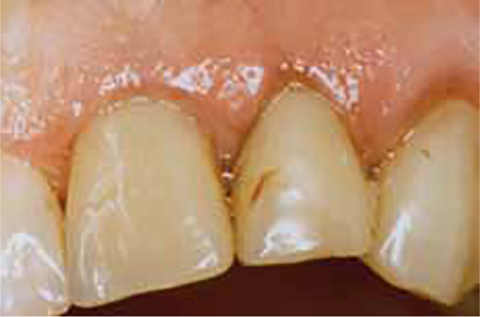
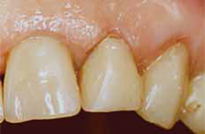
While some restorations will inevitably require replacement, by employing state-of-the-art adhesive procedures, many deteriorating yet serviceable restorations may be given extended life through repair procedures, especially if the defect is localized and accessible.24 For example, many non-carious marginal defects can be simply managed by refurbishing – a procedure that should normally pre-empt and delay repair or replacement.24 Refurbishment procedures typically involve removal of overhangs, surface recontouring, removal of discolouration and smoothening or glazing of surface, including sealing of pores and small gaps, without adding new restorative material except glaze or bonding (Figures 1 and 2).
Figure 1. Stained margins visible around composite restorations.Figure 2. Removal of stained margins by refinishing and polishing of restoration (refurbishment).
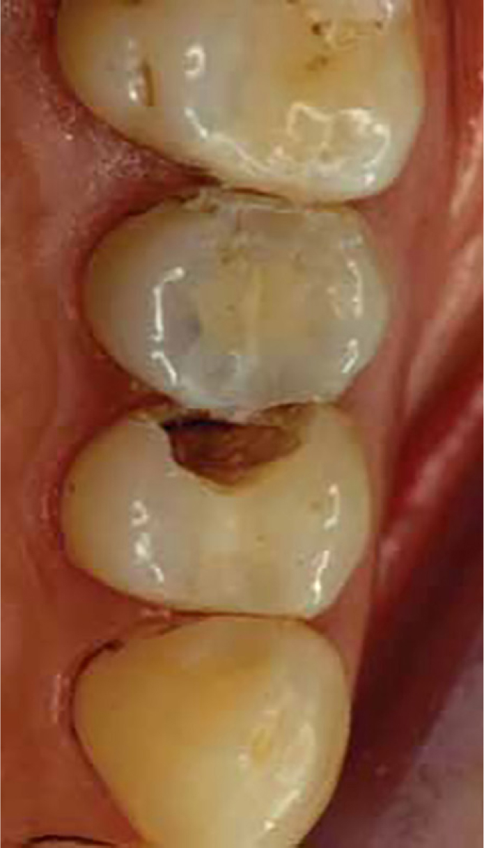
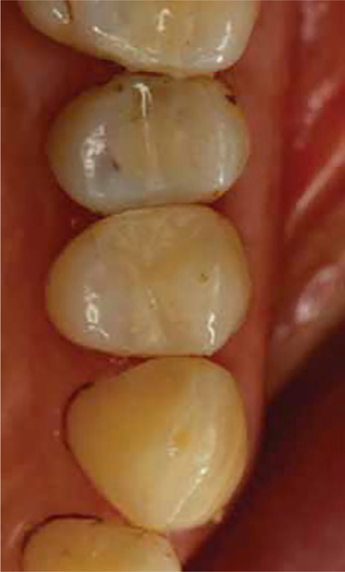
Caries adjacent to the margin of a composite restoration (secondary caries) should be treated as a new primary lesion.25 As with all patients who present with a new lesion, preventive measures should be initiated, followed by operative intervention as, and when, the lesion is shown to be active and progressing through dentine, or cavitation has occurred.8 Operative intervention should be minimally interventive, coupled with partial replacement of that portion of the adjacent composite restoration that is undermined by caries, or hinders the access required for necessary caries removal and the placement of an effective repair (Figures 3 and 4). The portion of the composite restoration that presents no clinical or radiographic evidence of failure should be left in place, unless there is good clinical indication to resort to total restoration replacement, with its various consequences. With a tendency to practise ‘defensive dentistry’ in a society that is increasingly litigious, it is regrettable that restorations affected by early forms of secondary caries, which are amenable to repair, may continue to be managed by total replacement.8
Figure 3. Clinical appearance of first premolar with removed distal portion of three surface composite restorations due to secondary caries at the distal floor.Figure 4. Restoration following repair procedure.
A recent systematic review concluded that direct restorations treated by minimally invasive alternative techniques, that is repair and refurbishment, did not present a significant difference in longevity in long-term clinical studies26,27,28 in comparison to the replacement technique in permanent teeth.29 It is clearly preferable, therefore, to perform restoration repair techniques, as an alternative to restoration replacement, wherever possible to preserve healthy tooth tissue distant to the site of defect. Thus, current evidence should help and encourage practitioners during the decision-making process to consider repair techniques as a useful alternative to replacement. The advantages of performing repair techniques have been reported2,8 as follows:
Preservation of tooth structure;
Increased longevity of the restoration;
Reduction of potentially harmful effects on the dental pulp;
Reduction in chair-side treatment time;
Reduced costs to the patient;
Good patient acceptance;
No need for local anaesthesia, provided the repair is not extensive;
Less risk of iatrogenic damage to adjacent teeth.
The clinical procedure for performing a composite restoration repair is outlined below:
Administer local analgesia, as indicated clinically;
Clean the tooth or teeth to be repaired, together with the adjacent teeth, using pumice;
Remove the defective part of the composite restoration and any adjacent lesions of secondary caries;
Ensure adequate moisture control (rubber dam isolation);
Pulp protection, if indicated, according to contemporary regimens;
Bevel the margins of the preparation, as indicated clinically, and place a long (1.0-mm wide) deep bevel on the margin of the composite resin to be repaired.
Appropriately prepared bevels increase the available surface area for bonding and facilitate a more aesthetic clinical outcome, as the composite resin to be used for the repair will blend in more effectively with the existing composite resin and remaining tooth tissues.
Acid etch the composite resin substrate together with the adjacent tooth tissue preparation margins for 15–30 seconds, wash thoroughly and dry the area using a three-in-one syringe. In addition to producing a favourable substrate surface for bonding, acid etching has a favourable cleansing effect.
An adhesive bonding system should be applied to the acid-etched composite substrate and adjacent tooth tissues and preparation margins, according to the manufacturer's directions for use.
A composite resin restorative material, compatible with the adhesive bonding system, is applied using an incremental technique to repair the defect, with each increment being fully photo-polymerized.
Again, the same type and brand of composite material should be used as the composite substrate, provided this information is known to the practitioner. The repair is then carefully contoured and finished using contemporary composite finishing systems, which allow the repair to be integrated imperceptibly into the restored tooth unit.
The occlusion is then checked to ensure that the repaired restoration will not be subjected to adverse occlusal loading.
Alternatively, a composite repair may be performed using sandblasting with silica-coated particles, a technique described elsewhere.8
Minimally invasive management of partially defective indirect restorations
Ceramic fracture and chipping have been reported to be the major cause for the replacement of both all-ceramic and ceramic fused to metal restorations.9 Blum et al9 proposed a classification of fractures occurring in metal–ceramic restorations, differentiating between simple and complex fractures. Simple fractures are those involving only the ceramic, originating from intra-ceramic defects, trauma and parafunctional habits.9,30,31 Complex fractures are associated with exposure of the metal substructure, resulting from failures at the metal–ceramic interface, an improper design, an inadequate framework support for the ceramic, fatigue of the metal substructure or internal stresses induced by incompatibilities between the coefficients of thermal expansion of ceramic and metal.9
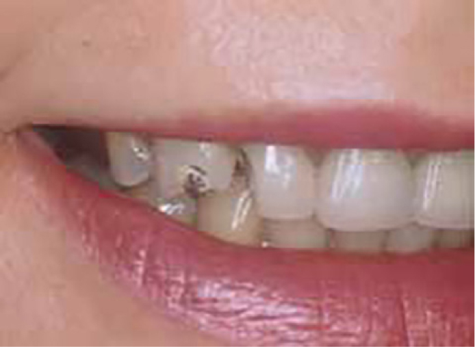
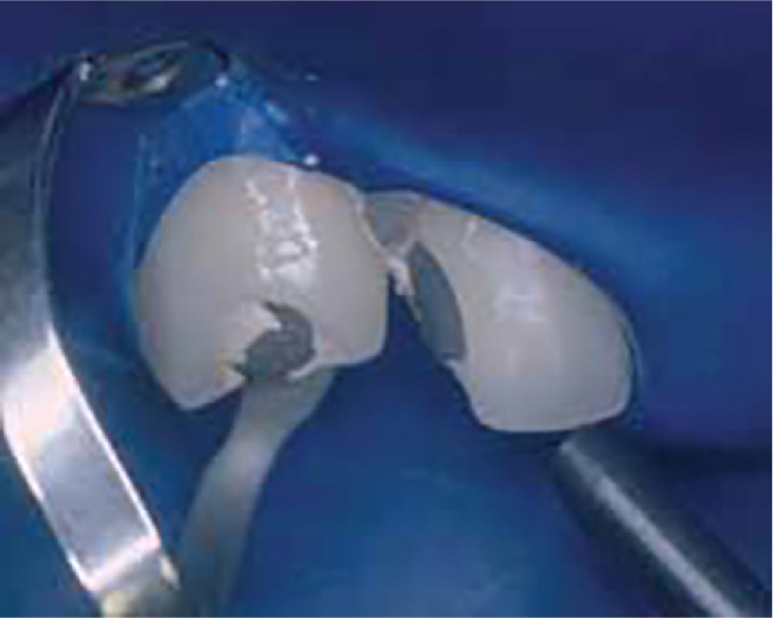
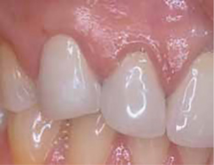
An example of a complex fracture where ceramic had fractured from the labial surfaces of the patient's maxillary right lateral incisor and canine teeth is shown in Figure 5. Clinical and radiographical examination indicated that the crown margins were sound, although some gingival recession was present. The patient stated that she was satisfied with the appearance of the crown margins as they were not visible on smiling and it was therefore decided to repair the crowns. The shade of the crowns was noted. With personal protective equipment measures for the dental team and patient in place, the defective crowns were isolated using a rubber dam. The exposed metal was sandblasted using an intra-oral sandblaster (Microetcher, Danville Engineering, USA) filled with CoJet sand (3M ESPE, Germany) at 40 psi for 15 seconds, which had resulted in a matt appearance of the exposed metal surface (Figure 6). Silane (ESPE-SIL) was then applied to the treated metal surface and allowed to dry for 30 seconds. The powder and liquid of the opaquer (Visio Gem, 3M ESPE, Germany) were mixed and applied in a thin layer to the exposed metal and light cured for 10 seconds. A layer of unfilled resin (Visio Bond, 3M ESPE) was then applied to the opaquer and light cured for 20 seconds, following which the defect was repaired using a composite resin restorative material (Figure 7).
Figure 5. The loss of ceramic from the metal-ceramic crowns following trauma is evident.Figure 6. After treatment with CoJet powder from an intra-oral sandblaster the exposed metal has a matt appearance.Figure 7. The repair was completed with the CoJet system and a composite resin restorative material (Filtek Z250; 3M ESPE).
Criteria for repair of restorations
Dentally motivated patients who attend on a regular basis, and maintain a good standard of oral health, have been deemed good candidates for restoration repair procedures.4 Patients who have complex medical histories or limited capacity to co-operate may also be viewed as suitable candidates for the repair rather than the replacement of defective restorations, in particular, if operating time needs to be kept to a minimum. In contrast, patient reluctance to accept a repair as an alternative to restoration replacement, irregular attenders, high caries risk patients or the presence of caries undermining the restoration and a history of failure of a previous repair should be viewed as contraindications for repair. In addition, repairs should not be contemplated if there is uncertainty about the procedure to be followed to ensure a satisfactory clinical outcome.
Conclusion
Proper clinical assessment and minimally invasive management of partially defective restorations increases the longevity of the remaining part of the restoration, and that of the restored tooth. Repair approaches should be carried out, wherever possible, as a viable minimally interventive treatment option. Restoration replacement should be considered as the last resort when there are no other viable alternatives.