Sham E, Leong J, Maher R, Schenberg M, Leung M, Mansour AK. Mandibular ameloblastoma: clinical 11 experience and literature review. ANZ J Surg. 2009; 79:739-744
Laborde A, Nicot R, Wojcik T, Ferri J, Raoul G. Ameloblastoma of the jaws: Management and recurrence rate. Eur Ann Otorhinolaryngol Head Neck Dis. 2017; 134:7-11
Odell EW., 9th edn. London: Elsevier; 2017
Lau SL, Samman N. Recurrence related to treatment modalities of unicystic ameloblastoma: a systematic review. Int J Oral Maxillofac Surg. 2006; 35:681-690 https://doi.org/10.1016/j.ijom.2006.02.016
Ooi A, Feng J, Tan HK, Ong YS. Primary treatment of mandibular ameloblastoma with segmental resection and free fibula reconstruction: Achieving satisfactory outcomes with low implant prosthetic rehabilitation. J Plast Reconstr Aesthet Surg. 2014; 67:498-505
Gardner DG, Pelcak AM. The treatment of ameloblastoma based on pathologic and anatomic principles. Cancer. 1980; 46:2514-2519
Pogrel MA, Montes DM. Is there a role for enucleation in the management of ameloblastoma?. Int J Oral Maxillofac Surg. 2009; 38:807-812
Sampson DE, Pogrel MA. Management of mandibular ameloblastoma: the clinical basis for a treatment algorithm. J Oral Maxillofac Surg. 1999; 57:1074-1077
Meshram M, Sagarka L, Dhuvas J, Anchlia S, Vyas S, Shah H. Conservative management of unicystic ameloblastoma in young patients: a prospective single centre trial and review of the literature. J Maxillofac Oral Surg. 2017; 16:333-341 https://doi.org/10.1007/s12663-016-0987-2
Kim H, Nam E, Yoon S. Conservative management (marsupialisation) of unicystic ameloblastoma: literature review and a case report. Maxillofac Plast Reconstr Surg. 2017; 39
Huang IY, Lai ST, Chen CH, Chen CM, Wu CW, Shen YH. Surgical management of ameloblastoma in children. Oral Surg Oral Med Oral Pathol Oral Radiol Endod. 2007; 104:478-485
Dolanmaz D, Etoz OA, Pampu A, Kalayci A, Gunhan O. Marsupialization of unicystic ameloblastoma: a conservative approach for aggressive odontogenic tumors. Indian J Dent Res. 2011; 22:709-712 https://doi.org/10.4103/0970-9290.93461
Ma ZG, XIE QY, Yang C, Xu GZ, Cai XY, Li JY. An orthodontic technique for minimally invasive extraction of impacted lower third molar. J Oral Maxillofac Surg. 2013; 71:1309-1317 https://doi.org/10.1016/J.joms.2013.03.025
Motamedi MRK, Heidarpour M, Siadet S, Motamedi AK, Bahreman AA. Orthodontic extraction of high risk impacted mandibular third molars in close proximity to the mandibular canal: a systematic review. J Oral Maxillofac Surg. 2015; 73:1672-1685
Takahashi S, Idaira Y, Sato T, Asada Y, Nakagawa Y. Unicystic ameloblastoma in a child treated with a combination of conservative surgery and orthodontic treatment: a case report. J Clin Pediatr Dent. 2019; 43:121-125 https://doi.org/10.17796/1053-4625-43.2.9
Is Less More? A conservative multidisciplinary approach to ameloblastoma Hussein Mohamedbhai Debipriya Dasgupta Charlotte Hubbett Nayeem Ali Dental Update 2024 47:6, 707-709.
This case report outlines a novel conservative surgical approach to the management of a unicystic ameloblastoma with the use of marsupialisation, enucleation, cryotherapy and orthodontic extrusion to enable successful treatment without neurological damage or deformity. It has been increasingly recognized that conservative treatment of unicystic ameloblastomas, instead of wide local excision, can reduce morbidity whilst maintaining an acceptably low recurrence rate. Several case series have also demonstrated orthodontic extrusion of impacted third molars in moving the apex of the roots away from the inferior alveolar nerve. This is possibly the first case report of the combination of these two procedures in an adult with a large unicystic ameloblastoma.
CPD/Clinical Relevance: This is not an infrequently seen neoplasia: this paper therefore has the opportunity to inform management of this condition amongst clinicians.
Article
Hussein Mohamedbhai
Case report
CH, a 22 year-old woman, presented via her general dental practitioner (GDP) with an asymptomatic swelling, which was noted on routine examination. There was no relevant medical history and no family history to note. She had never smoked and only drank alcohol occasionally. On examination, she was found to have a hard mass, along the posterior lower right buccal sulcus of the lower right second molar (LR7) extending to the alveolar ridge. This mass was tender to palpation. The LR7 was vital and the lower third molar (LR8) unerupted. Otherwise the examination was normal and there was no palpable cervical lymphadenopathy.
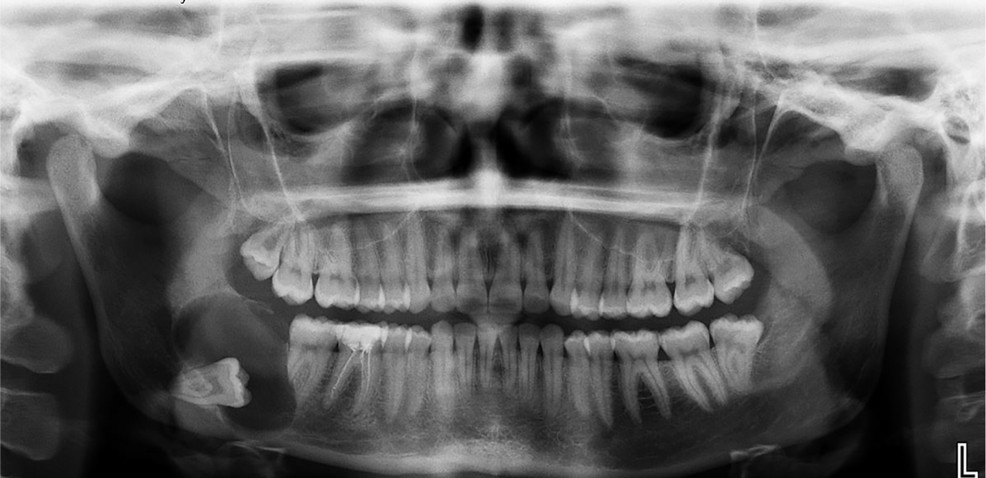
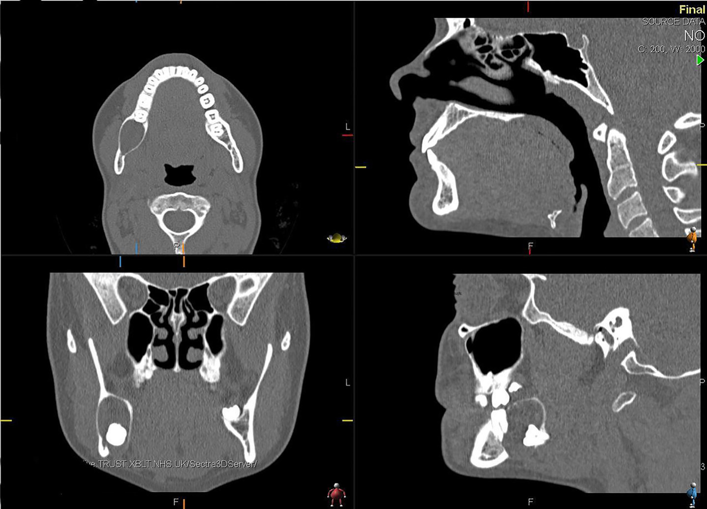
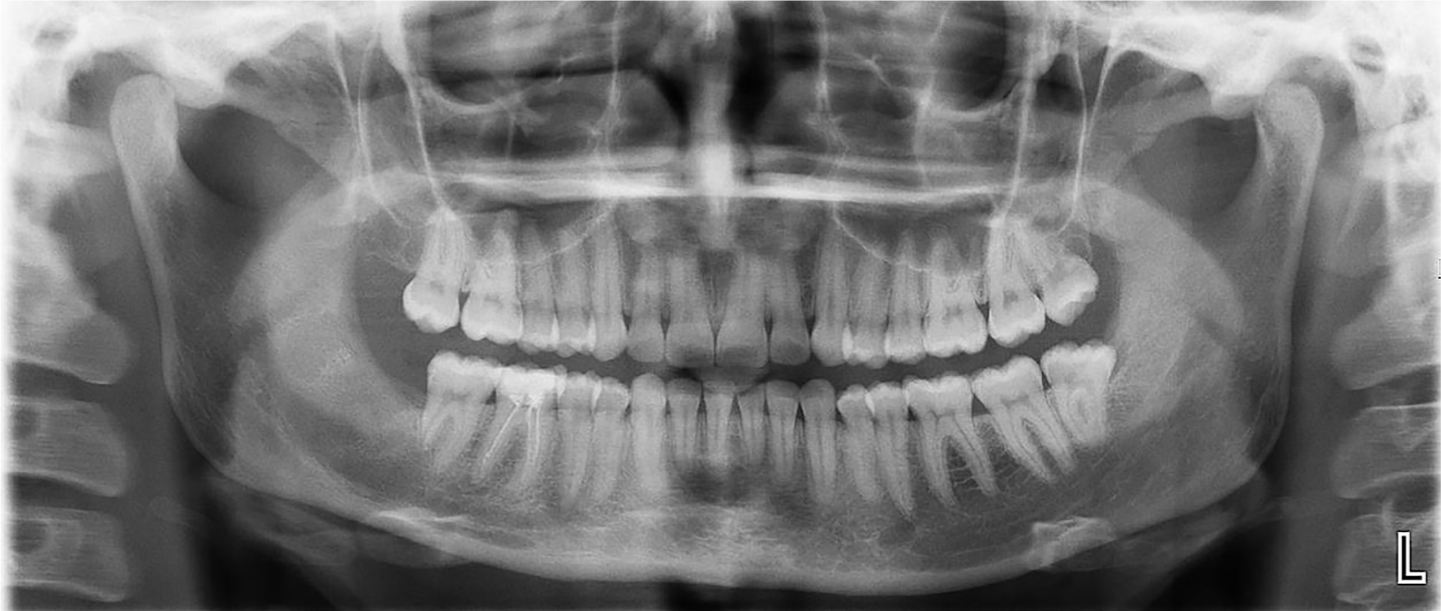
A radiographic image (Figure 1) demonstrated a unilocular, well-corticated radiolucency measuring 40 mm in diameter extending from distal of the LR7 to the angle of the mandible and 50% of the ramus. Although some characteristics were typical of a dentigerous cyst, the margins of the cyst seemed to be in contact with the cemento-enamel junction of the unerupted LR8. Other features were atypical, including the resorption of the distal root of the LR7. In addition, the cyst showed evidence of causing displacement of the LR8 distally. CT imaging (Figure 2) demonstrated that the lingual plate and mandibular cortical border were intact, as well as showing the inferior alveolar nerve was buccal to the LR8. Incisional biopsy of the abnormality and aspiration of its contents demonstrated the histological findings of a cyst lined by thin odontogenic epithelium. The stroma showed myxoid changes with nests of both active and resting odontogenic epithelium. A diagnosis of unicystic ameloblastoma was made.
Figure 1. Orthopantomogram taken on first presentation, demonstrating well defined radiolucency alongside an unerupted LR8 in the posterior mandibular region.Figure 2. CT imaging taken pre-operatively, demonstrating the intact lingual plate and mandibular cortical border, as well as showing the inferior alveolar nerve was buccal to the LR8.
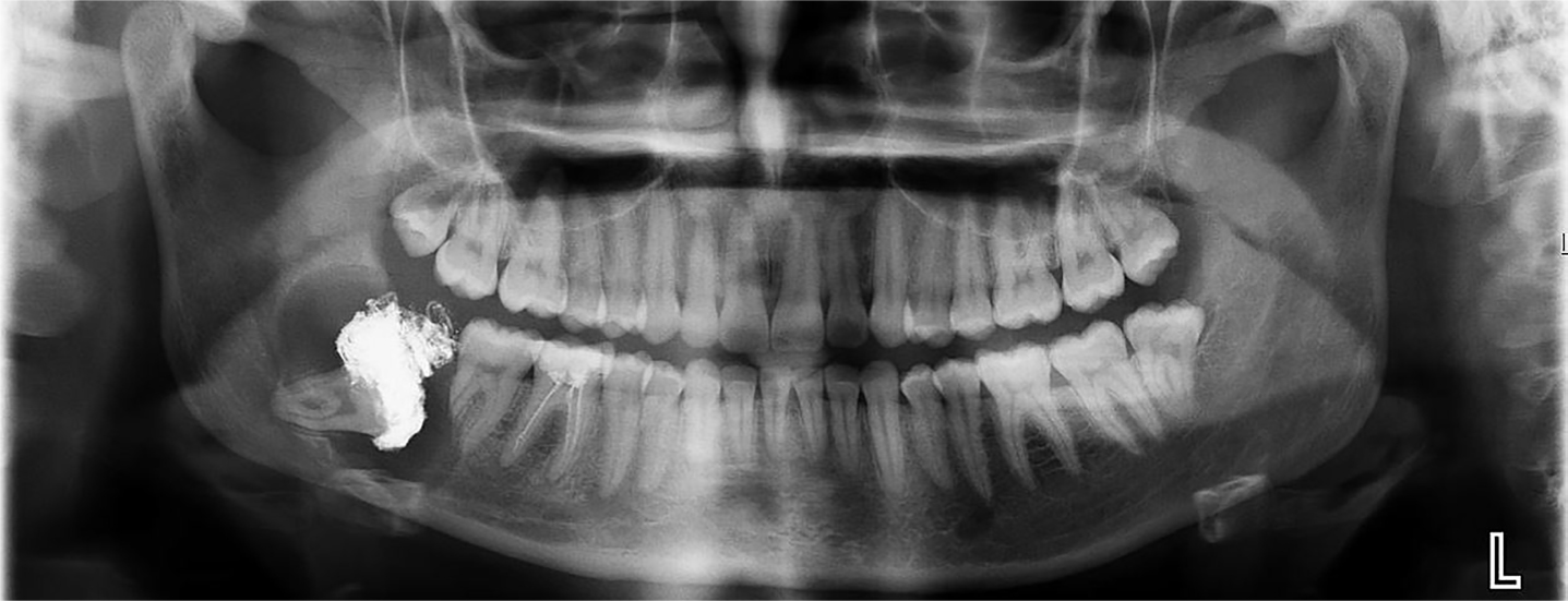
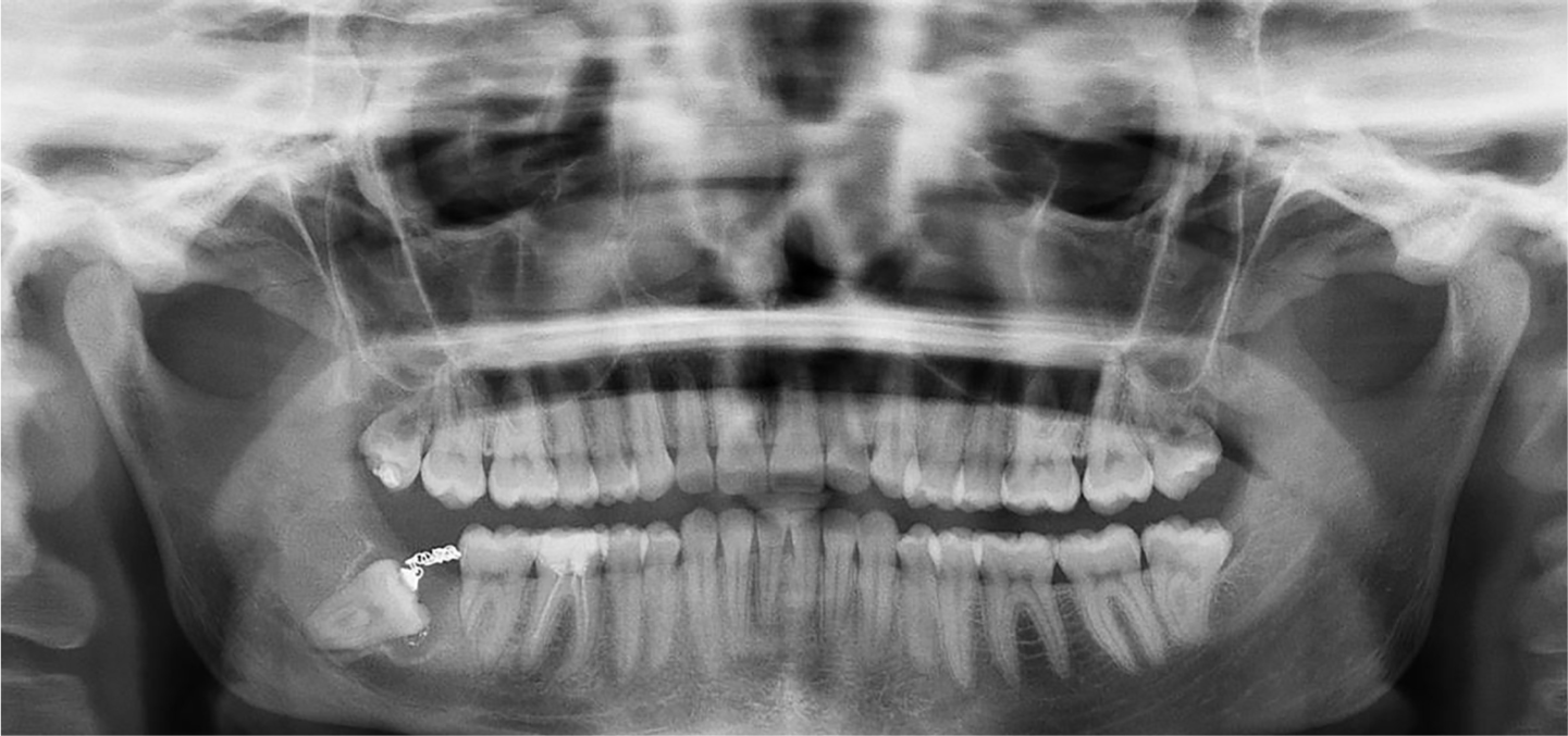
Subsequently, CH underwent marsupialisation of the cyst (and the cavity was packed with Bismuth Iodine Paraffin Paste (BIPP) impregnated ribbon gauze). The post-operative radiograph is presented in Figure 3. The cyst cavity was irrigated with normal saline and chlorhexidine solution and the pack was changed fortnightly for 6 months and until the volume of the cyst had decreased substantially. From serial radiographs, after 6 months, the cyst diameter on radiographs had reduced by 23 mm, approximately 58% in size (Figure 4), as well as also demonstrating that the wisdom tooth had also moved.
Figure 3. The post-operative radiograph following marsupialisation of the cyst and the cavity being packed with Bismuth Iodine Paraffin Paste (BIPP) impregnated ribbon gauze.Figure 4. Orthopantogram demonstrating resolution of the ameloblastoma and a gold chain attached to the crown of the LR8 to extrude the tooth orthodontically.
The aim was to reduce the size of the ameloblastoma sufficiently for planned enucleation, cryotherapy and extraction of the associated teeth. This was to minimize the risk of recurrence occurring deep in the mandible. However, a further concern was the close association of the roots of the unerupted LR8 with the mandibular canal, which would result in a high chance of nerve injury. Consequently, the LR8 was extruded orthodontically with a gold chain attached to a bracket on the UR8 (Figure 4). This was effective in moving the tooth further away from the ID nerve, but complicated by debonding of the chain. CH then underwent extraction of the LR8, with simultaneous enucleation of the ameloblastoma. Following cryotherapy, liquid nitrogen and a probe with KY jelly to form a good iceball with three freeze/thaw cycles was performed to the bony cavity, with care taken to retract and protect the surrounding soft tissues.
Six-monthly interval follow-up, for two years, has demonstrated no evidence of either clinical or radiographic recurrence. CH has full and normal inferior dental nerve sensation, no deformity (Figure 5), and no long-term complications.
Figure 5. Post-operative radiograph 2 years post treatment showing resolution of the ameloblastoma.
Patient perspective
The first words I remember my surgeon saying to me were ‘That thing is the size of a satsuma’. They were not words I had been expecting to hear, as I was absolutely convinced, until he turned the monitor towards me and showed me my x-ray picture, that everything was fine. I had had no symptoms at all, just a small swelling in the back of my mouth that I had assumed was my unerupted wisdom tooth but, on a routine dental examination, my dentist explained that she could not see the tooth on the x-ray and she referred me for a two-week wait. Admittedly, seeing the huge tumour and how I had an eggshell thickness of jawbone left scared me, and it opened up so many questions: ‘What happens next?’ ‘Will my face look the same?’ ‘What would have happened if it hadn't been found?’. I started Googling and found a young woman in the US with a near identical tumour who had had a hemimandibulectomy and reconstruction with a bone, and was worried that I, too, would be left with large scars and a different face.
My surgeon was very reassuring and, whilst I was aware that my treatment was experimental in many ways, and that there were setbacks in my treatment, such as difficulties in fitting the bracket and chain to evert the tumour, it seemed he had no doubts that it would be successful. I believe any surgery requires trust between the surgeon and the patient, but I was surprised at how big the trust was that the facial surgery required of me. Our faces are so entwined with our identities; so part of who we are. I am very lucky to have been treated by a team who earned that trust from me, and I am very grateful to have a face that is mine.
Discussion
Ameloblastoma is a benign odontogenic neoplasm of the mandible and maxilla that rarely exhibits malignant behaviour. It accounts for 1% of all tumours in the oral cavity. Generally, it occurs between the third and fifth decades, the most frequently affected site being the mandibular posterior molar region, which accounts for 60% of all cases.1 Whilst asymptomatic, an ameloblastoma usually presents as a swelling. The tumour enlarges within the jaw slowly, displacing teeth, resorbing roots and perforating through the cortical bone.2 An ameloblastoma develops from the epithelium involved in the formation of teeth, the reduced enamel epithelium and odontogenic cyst lining.3
Despite being benign, an ameloblastoma has a high recurrence rate.4 This is why, classically, an ameloblastoma has to be removed with wide local excision, involving 1–1.5 cm of normal bone around the margin. Often this necessitates reconstruction, often with free tissue transfer of a composite flap. This traditional treatment has numerous complications, such as functional and masticatory changes, facial deformities, as well as neurological sequelae from sacrificing the inferior alveolar nerve.5
An ameloblastoma can be classified into one of four types:
Conventional/multicystic;
Unicystic;
Peripheral; and
Desmoplastic.
Each subtype can present and behave differently, having implications upon treatment. Unicystic ameloblastomas typically present at a younger age than conventional ameloblastomas. Many present similarly to a dentigerous cyst with an unerupted third molar, thus histological confirmation is mandatory.3
Unicystic lesions, by definition of having a single cyst cavity, are well localized by the fibrous capsule of the cyst, and only rarely broach the peripheral tissues. This has formed the basis of why ameloblastomas may be successfully managed with conservative treatment (marsupialisation or enucleation).6 The aim of marsupialisation is to reduce the size of the tumour. The decompression of the internal contents by marsupialisation promotes remodelling of bone and osteogenesis. Once the tumour volume has been sufficiently reduced in size, it can then be treated by enucleation and/or cryotherapy. This approach has the benefits of maintaining pulp vitality, the inferior alveolar nerve and preventing jaw fracture.2
However, treatment of an ameloblastoma remains controversial. Due to debate over recurrence rates and the relative rarity of this tumour, there is clinical uncertainty over which treatment is most effective. Some case series have shown the lowest recurrence with resection,7 and that most recurrent cases occur after enucleation or excision.8 Recently, case series have shown success at conservative treatment. A series involving 15 patients, with a mean 4-year follow-up, demonstrated no recurrence with marsupialisation followed by enucleation of unicystic ameloblastoma in adolescents.9 Others have replicated this.10,11 The success of this form of treatment mostly depends on the histological nature, as well as other factors such as age, surgical technique and the size of the tumour.12
Along with the risks of treatment of the ameloblastoma, there are the risks of extraction of the unerupted LR8. The close association of the LR8 with the mandibular canal increases the risk of damage to the inferior alveolar nerve. In the present case, the additional conservative option of orthodontic extrusion of the third molar enabled an extraction which greatly reduced the risk of nerve injury. Previous studies have reported that between 3 weeks to 2 months of traction will result in radiographic evidence of the roots moving 1 mm to 3 mm from the inferior dental canal.13 Meta-analysis of 123 with a high risk of neural damage has shown success using orthodontic extrusion, with no record of permanent injury.14
It is evident that, whilst these two conservative techniques are rarely employed, they do have an evidence base with demonstrated efficacy and safety. However, the use of these two techniques in conjunction, in the case of the conservative management of a unicystic ameloblastoma, is particularly novel.
A similar report has been published by Takahashi et al.15 However, a key difference in the report, compared with the present case, is the fact that the patient was 7 years old. The majority of the published reports of conservative management of unicystic ameloblastomas relate to patients below 20 years old. Indeed, there is a paucity of evidence for this conservative management in older patients. There are different limitations of both marsupialisation and orthodontic uprighting in an adult, namely the lower rate of bone turnover and success rates. This present report adds to the limited body of literature by demonstrating efficacy of this conservative treatment in adults with unicystic ameloblastoma.
A patient perspective was included here to highlight the key features which the patient remembered and felt were important. As well as the obvious, such as maintaining aesthetics and function, it is important to highlight the trust which patients must place in this lengthy treatment course. Inspiring this trust is especially relevant when dealing with possible life-changing consequences and experiencing setbacks in treatment. As highlighted by the patient account, a key factor to building this trust is clear communication. Clearly explaining the diagnosis, treatment options, and evidence base helps to abate patient's fears. This is essential in a case like the present one, where the treatment will be complex, and time and effort needs to be taken with the patient to explain and listen.
As with all management options, there are limitations. One must consider the time frame, which can take an average of 9 months to enable full eruption.14 Also, there is the reliance upon patient compliance to tolerate, and attend, for both repeated pack changes and orthodontic treatment. Finally, in those patients with a recurrent ameloblastoma, there is a stronger argument for more aggressive management. Due to the ongoing risk of recurrence, as with any treatment of ameloblastoma, long term annual follow-up is warranted.
Summary
This case report outlines the effective conservative management of a large unicystic ameloblastoma with the added complication of an unfavourably positioned unerupted lower wisdom tooth. The report demonstrates the efficacy of the combination of conservative surgery and orthodontic treatment in management.