Ferreira O, Cardoso CL, Alvares Capelozza AL, Faria Yaedu RY, da Costa AR. Odontogenic keratocyst and multiple supernumerary teeth in a patient with Ehlers-Danlos syndrome – a case report and review of the literature. Quintessence Int. 2008; 39:251-256
Gazit Y, Nahir AM, Grahame R, Jacob G. Dysautonomia in the joint hypermobility syndrome. Am J Med. 2003; 115:33-40
Pope FM. Ehlers-Danlos syndrome. Baillieres Clin Rheumatol. 1991; 5:321-349
Letourneau Y, Perusse R, Buithieu H. Oral manifestations of Ehlers-Danlos syndrome. J Can Dent Assoc. 2001; 67:330-334
Hagberg C, Berglund B, Korpe L, Andersson-Norinder J. Ehlers-Danlos Syndrome (EDS) focusing on oral symptoms: a questionnaire study. Orthod Craniofac Res. 2004; 7:178-185
Hakim AJ, Grahame R, Norris P, Hopper C. Local anaesthetic failure in joint hypermobility syndrome. J R Soc Med. 2005; 98:84-85
Abel MD, Carrasco LR. Ehlers-Danlos syndrome: classifications, oral manifestations, and dental considerations. Oral Surg Oral Med Oral Pathol Oral Radiol Endodontol. 2006; 102:582-590
Steinmann B, Royce PM, Superti-Furga A. The Ehlers-Danlos Syndrome, 2nd edn. In: Roce PM SB (ed). New York: Wiley-Liss; 2002
Castori M. Ehlers-danlos syndrome, hypermobility type: an underdiagnosed hereditary connective tissue disorder with mucocutaneous, articular, and systemic manifestations. ISRN Dermatol. 2012; 2012:751-768
Castori M, Camerota F, Celletti C, Grammatico P, Padua L. Ehlers-Danlos Syndrome Hypermobility Type and the excess of affected females: possible mechanisms and perspectives. Am J Med Genet. 2010; 152A:2406-2408
Beighton P, De Paepe A, Steinmann B, Tsipouras P, Wenstrup RJ. Ehlers-Danlos syndromes: revised nosology, Villefranche, 1997. Am J Med Genet. 1998; 77:31-37
Malfait F, Francomano C, Byers P, Belmont J, Berglund B, Black J The 2017 International Classification of the Ehlers-Danlos Syndromes. Am J Med Genet – Seminars in Medical Genetics. 2017; 175C:8-26
De Paepe A, Malfait F. Bleeding and bruising in patients with Ehlers-Danlos syndrome and other collagen vascular disorders. Br J Haematol. 2004; 127:491-500
Hakim AJ, Grahame R. A simple questionnaire to detect hypermobility: an adjunct to the assessment of patients with diffuse musculoskeletal pain. Int J Clin Pract. 2003; 57:163-166
Arendt-Nielsen lKS, Bjerring P, Hogsaa B. Insufficient effect of local analgesics in Ehlers-Danlos type 3 patients (Connective tissue disorder). Acta Anaesthesiol Scand. 1990; 34:358-361
Mitakides J, Tinkle BT. Oral and mandibular manifestations in the Ehlers-Danlos Syndromes. Am J Med Genet – Seminars in Medical Genetics. 2017; 175C:220-225
Brady AF, Demirdas S, Fournel-Gigleux S, Ghali N, Giunta C, Kapferer-Seebacher I The Ehlers-Danlos Syndromes, rare types. Am J Med Genet – Seminars in Medical Genetics. 2017; 175C:70-115
Kapferer-Seebacher I, Pepin M, Werner R, Aitman TJ, Nordgren A, Stoiber H Periodontal Ehlers-Danlos Syndrome is caused by mutations in C1R and C1S, which encode subcomponents C1r and C1s of complement. Am J Hum Genet. 2016; 99:1005-1014
Norton LA, Assael AL. Orthodontic and temperomandibular joint considerations in the treatment of patients with Ehlers-Danlos syndrome. Am J Orthod Dentofacial Orthop. 1997; 111:75-84
Jones ML. Orthodontic treatment in Ehlers-Danlos syndrome. Br J Orthod. 1984; 11:158-162
Beighton P. Serious ophthalmological complications in the Ehlers-Danlos syndrome. Br J Ophthalmol. 1970; 54:263-268
Gorlin RJ Syndromes of the Head and Neck.New York: Oxford University Press; 1990
Klingberg G, Hagberg C, Noren JG, Nietzsche S. Aspects on dental hard tissues in primary teeth from patients with Ehlers-Danlos syndrome. Int J Paediatr Dent. 2009; 19:282-290
Carr RJ, Green DM. Multiple odontogenic keratocysts in a patient with type-ii (mitis) Ehlers-Danlos syndrome. Br J Oral Maxillofac Surg. 1988; 26:205-214
Hakim A, Grahame R. Joint hypermobility. Best Pract Res Clin Rheumatol. 2003; 17:989-1004
Prophylaxis against infective endocarditis: antimicrobial prophylaxis against infective endocarditis in adults and children undergoing interventional procedures. NICE Guideline [CG64]. 2008;
Antibiotic Prophylaxis Against Infective Endocarditis.Dundee: SDCEP; 2018
Mathias CJ, Low DA, Iodice V, Owens AP, Kirbis M, Grahame R. Postural tachycardia syndrome-current experience and concepts. Nature Rev Neurol. 2012; 8:22-34
Morgan AW, Pearson SB, Davies S, Gooi HC, Bird HA. Asthma and airways collapse in two heritable disorders of connective tissue. Ann Rheumat Dis. 2007; 66:1369-1373
Bendik EM, Tinkle BT, Al-shuik E, Levin L, Martin A, Thaler R Joint hypermobility syndrome: a common clinical disorder associated with migraine in women. Cephalalgia. 2011; 31:603-613
Castori M, Morlino S, Celletti C, Celli M, Morrone A, Colombi M Management of pain and fatigue in the joint hypermobility syndrome (a.k.a. Ehlers-Danlos syndrome, hypermobility type): principles and proposal for a multidisciplinary approach. Am J Med Genet. 2012; 158A:2055-2070
Martin VT, Neilson D. Joint hypermobility and headache: the glue that binds the two together – Part 2. Headache. 2014; 54:1403-1411
Henderson FC, Austin C, Benzel E, Bolognese P, Ellenbogen R, Francomano CA Neurological and spinal manifestations of the Ehlers-Danlos syndromes. Am J Med Genet – Seminars in Medical Genetics. 2017; 175C:195-211
Neilson D, Martin VT. Joint hypermobility and headache: understanding the glue that binds the two together – Part 1. Headache. 2014; 54:1393-1402
Hakim A, De Wandele I, O'Callaghan C, Pocinki A, Rowe P. Chronic fatigue in Ehlers-Danlos syndrome-hypermobile type. Am J Med Genet – Seminars in Medical Genetics. 2017; 175C:175-180
Hershenfeld SA, Wasim S, McNiven V, Parikh M, Majewski P, Faghfoury H Psychiatric disorders in Ehlers-Danlos syndrome are frequent, diverse and strongly associated with pain. Rheumatol Int. 2016; 36:341-348
Seneviratne SL, Maitland A, Afrin L. Mast cell disorders in Ehlers-Danlos syndrome. Am J Med Genet – Seminars in Medical Genetics. 2017; 175C:226-236
Ehlers-Danlos Syndrome (EDS) affects the metabolism of collagen which can have implications throughout the body, impacting on not only the skin, but also the joints, muscles, cardiovascular and gastrointestinal systems. The condition can have dental implications such as poor wound healing, mucosal fragility, prolonged bleeding, temporomandibular joint dislocation and, in some forms, periodontal disease. Three clinical cases of patients with EDS will be discussed and their dental management described. The patient with EDS may be seen in primary care and, if input from secondary care is required, a multidisciplinary, shared care approach will ideally be utilized.
CPD/Clinical Relevance: Ehlers-Danlos Syndrome is a condition that can be multifactorial, with medical implications as well as dental; depending on the manifestations of the condition, the patient may be seen in primary and/or secondary care with or without a shared care approach.
Article
Ehlers Danlos Syndrome (EDS) is a rare syndrome characterized by 13 types of connective tissue disorders which manifest primarily in dermatological and joint conditions. EDS affects collagen metabolism, leading to deficiency and/or disordered deposition of collagen.1 Within the literature there is an overlap between the EDS subtypes and other connective tissue diseases, including hypermobility disorders, for example Joint Hypermobility Syndrome (JHS), which is often referred to interchangeably with EDS.2
The condition was first described in 1657 when a young Spanish man was found to be able to stretch the skin overlying his right pectoral muscle over the left angle of his mandible.3 Further descriptors were added in 1901 by Ehlers, 1908 by Danlos, and then by Poumeau-Delille and Soulie in 1934 when the condition was described as Ehlers-Danlos Syndrome.4
Epidemiology and diagnosis
The prevalence of EDS is reported to be 1 in 5,000,5 however, recent clinical studies suggest that joint hypermobility syndromes are under diagnosed6 and the condition is more common than these ratios indicate. EDS is equally distributed throughout the world and theoretically affects men and women equally without racial predilection,7,8 however, in practice a greater proportion of females appear to be affected.9,10
Classification of EDS
Each phenotype of EDS has a unique collagen defect which may lead to different symptoms and signs. The most recent criteria, produced in 2017, recognizes 13 subtypes10–11,12 (Table 1). Each subtype has a set of clinical criteria that help guide diagnosis; a patient's physical signs and symptoms are matched up to the major and minor criteria to identify the subtype that is the best fit. This classification also recognizes the type of collagen involved and the genetic basis.12
Clincal EDS Subtype
Abbreviation
Inheritance Pattern
1
Classical EDS
cEDS
Autosomal Dominant (AD)
2
Classical-like EDS
cIEDS
Autosomal Recessive (AR)
3
Cardiac-valvular
cvEDS
AR
4
Vascular EDS
vEDS
AD
5
Hypermobile EDS
hEDS
AD
6
Athrochlasia EDS
aEDS
AD
7
Dermatosparaxis EDS
dEDS
AR
8
Kyphoscholiotic EDS
kEDS
AR
9
Brittle Cornea Syndrome
BCS
AR
10
Spondylodysplastic EDS
spEDS
AR
11
Musculocontractural EDS
mcEDS
AR
12
Myopathic EDS
mEDS
AD or AR
13
Periodontal EDS
pEDS
AD
Characteristics of EDS
The classic signs of EDS are:
Hypermobility of the joints; joints are often loose and unstable resulting in frequent subluxations and dislocations. Chronic musculoskeletal pain and early onset arthritis are common;7
Hyperelasticity, fragility and softness of the skin;7
Deficient wound healing with the development of scars with a ‘cigarette-paper like quality’;13
Tendency to excessive bleeding manifested by bruises, ecchymoses and haematomas.4
Oral and dental implications of Ehlers-Danlos Syndrome
Oral and facial manifestations of EDS are variable depending on the type of disease present, but in all types defects of the collagen-processing enzymes or collagen structure lead to collagen fragility throughout the body.7Table 2 outlines the main extra-oral and intra-oral manifestations of EDS, which may be present.
When treating patients with EDS there are many medical and dental factors to consider. Medical conditions and their effect on the provision of dental care are outlined in Table 3.
Associated Medical Conditions
Impact on Dental Care
Impaired MobilityMobility problems are frequent
Access to dental clinic and surgery
Wheelchair recliner or hoist may be required
May need hospital transport
Positioning in dental chair – may require pillows or need to be treated upright
Cardiac ConsiderationsAortic and mitral valve prolapses are common in the Cardiac-Valvular type of EDS27
Postural Orthostatic Tachycardia Syndrome (POTS)POTS is characterized as a significant increase in heart rate of 30 beats per minute or greater occurring within 10 minutes of standing or a heart rate when upright of greater than 120 beats per minute, but with no decline in blood pressure30
Increased risk of palpitations, dizziness and syncope30
Stress and anxiety can bring on episodes
Treat in supine position
Manage as per current resuscitation council guidelines
AsthmaDue to underlying connective tissue defects affecting the lungs27,31
Increase risk of asthma attack
Take a thorough history and ensure patients bring inhalers to appointments
MigrainesIncreased incidence of TMJ disorder, atlantoaxial and craniocervical instability, fibromyalgia, POTS, sleep disorders and anxiety are all associated with EDS and can all lead to migraines32-33,34,35
Stress or anxiety associated with dental treatment may precipitate a migraine
Patients may cancel at short notice
Chronic Pain and FatigueCausative factors include:
Lying back in the dental chair for long periods may be uncomfortable
Chronic pain may affect manual dexterity and therefore oral hygiene
Conscious sedation – strong analgesics may interact with sedative drugs
Psychiatric DisordersIncreased prevalence of depression and anxiety38
Possible associated dental anxiety
Drug-induced xerostomia as a result of anti-depressant medication
Mast Cell DisordersIncreased number and/or activity of mast cells. Signs and symptoms include:39
Skin urticaria
Flushing
Hypotension
Asthma
Diarrhoea
Abdominal bloating or cramping
Rhinitis
Anaphylaxis
Enquire regarding potential triggers
Dental treatment, drugs, stress, infection can all trigger an episode
Refer to current anaphylaxis guidelines
Oral surgery
Failure of anaesthesia has been reported in some patients with EDS, and so when treating these patients the clinician should be mindful of the risk of LA failure.14,15 This could lead to increased levels of dental anxiety. Conscious sedation or general anaesthesia may need to be considered.
Bleeding can occur with all types of EDS but is more common with the vascular and kyphoscoliotic types.7 Excessive gingival haemorrhage is commonly seen during toothbrushing and hygiene therapy.7 When undertaking extractions, care must be taken when raising mucoperiosteal flaps and sutures must be tied only under slight tension due to mucosal fragility.16 If excessive bleeding occurs following an extraction, haemostatic agents must be placed in the socket and consideration should be given to the use of a tranexamic acid mouthwash to prevent premature breakdown of the blood clot.
Some patients with EDS suffer from recurrent TMJ dislocation.7 If this is known, the clinician must be careful not to open the patient's mouth too wide as this may increase the risk of dislocation.16 Allow the patient frequent respites during lengthy treatment and the use of a small mouth prop may be helpful.
Restorative
When carrying out endodontic treatment, it may be complicated by the presence of pulp stones or calcification of the pulp chamber or abnormal root morphology.7
The new EDS classification system formally identified peridontal EDS (pEDS) as subtype 13.12 Although the exact prevalence of pEDS is unknown, the key features include extensive intra-oral periodontal destruction, the presence of pretibial plaques on the legs and generalized tissue fragility.17 Diagnosis of true pEDS can be difficult as some patients will present with an EDS diagnosis and separate coincidental periodontal disease. However, the periodontal destruction in pEDS often manifests as recession, extensive gingival inflammation with minimal plaque,17 and the absence of attached keratinized gingiva leading to greater fragility and increased risk of breakdown.18 When managing patients with pEDS, strict adherence to oral hygiene and regular non-surgical debridement is advised.
Oral medicine
As well as deficiency/ineffectiveness of collagen leading to collagen fragility in the skin, this is also reflected in the oral mucosa, with even minor trauma leading to oral ulceration or haematoma formation.7,16 Patients may have to be advised regarding oral ulceration and care should be taken to minimize trauma with dental instruments during dental treatment.
Orthodontic
Orthodontic treatment is not contra-indicated in patients with EDS but it has been reported that the teeth move more rapidly and that there is greater tooth mobility during the migratory phase, thus leading to unpredictable results.7,19 Lighter forces should be used as it is believed that stressed periodontal fibres on the tension side may tear during treatment and be slow to repair.19 This, in addition to the disorganized arrangement of collagen fibres in the periodontium, results in the need for a prolonged period of retention.19
Patients undergoing orthodontic therapy may also be at higher risk of ulceration from fixed and removable appliances.7,20
Case study 1
A 58-year-old Caucasian female patient, Patient A, was referred to the department of special care dentistry by her rheumatologist due to recurrent failed local anaesthesia (LA), and having experienced prolonged bleeding times following dental extractions in the past. The patient reported that she had developed anxiety relating to dental treatment following repeated failed LA and requested if it would be possible to be treated under sedation. The patient had been diagnosed with EDS in 1990 following repeated dislocations of her shoulders and TMJ. EDS had also manifested as irritable bowel syndrome, recurrent bruising, poor wound healing, asthma and osteoarthritis in her hips and knees; this necessitated the use of crutches so her mobility was impaired. Additionally, she reported recurrent oral ulceration and a dry mouth. She had also been recently diagnosed with mild angina. Patient A reported that there was a family history of EDS; although she was single and had no children, all of her seven nieces and nephews were affected by EDS to varying degrees.
On intra-oral examination, there was an ulcer on her soft palate approximately 5–6 mm in diameter with a surrounding ring of erythema and a further ulcer in her right buccal mucosa approximately 4 mm in diameter. Patient A gave a history of oral ulceration occurring frequently, lasting between 8 and 14 days and resolving spontaneously. Oral hygiene was good, but the patient was found to have occlusal caries on her lower left first and second molars (LL6, LL7) and a vertical root fracture on her non-vital upper left second premolar (UL5).
The initial treatment plan was as follows:
Restoration of carious teeth under LA with inhalation sedation (IHS). This was the preferred modality of anxiolysis as the patient was anxious but did not have a suitable escort for intra-venous sedation (IVS).
Extraction of UL5 under LA with IHS.
Referral to the department of oral medicine regarding oral ulceration.
Upon the first episode of treatment, IHS with nitrous oxide and oxygen, titrated up to 50%, was used for the restoration of the LL6 and LL7. Although adequate LA was achieved using an inferior dental block with 2.2 ml lidocaine hydrochloride with 1:80,000 adrenaline, Patient A was extremely anxious throughout. Following discussion with the patient, it was felt that IHS would not provide sufficient anxiolysis for the extraction of the UL5, therefore the patient found a suitable escort to have the treatment carried out under LA and IVS with midazolam to achieve greater anxiolysis.21 At the next appointment, the extraction was carried out under IVS and LA and local haemostatic measures were applied without any complications.
Patient A's oral ulceration was reviewed by the oral medicine department but, on investigation, no obvious causative factors were found. Appropriate medication was prescribed to provide symptomatic relief.
Patient A was regularly reviewed at 6-monthly intervals following her initial referral and continues to have her dental treatment carried out under LA with inhalation sedation. LA has been effective, and the patient is now far less anxious about receiving dental treatment.
Case study 2
A 26-year-old female patient, Patient B, was referred to the department of special care dentistry by her rheumatologist as LA had been previously unsuccessful during dental treatment by her general dental practitioner (GDP). The patient also reported that she frequently had short periods of loss of consciousness in the dental surgery without warning.
Her medical history included Hypermobile EDS, which was diagnosed in 2007 following multiple shoulder dislocations and the patient having an awareness of increased flexibility. Patient B reported that she has frequent flare-ups of the condition causing her to become immobile and reliant on a wheelchair for long periods. She also had Postural Orthostatic Tachycardia Syndrome (POTS) and asthma, which was well controlled with inhalers. Patient B had two children, aged 3 and 4 years, both of whom had been diagnosed with EDS.
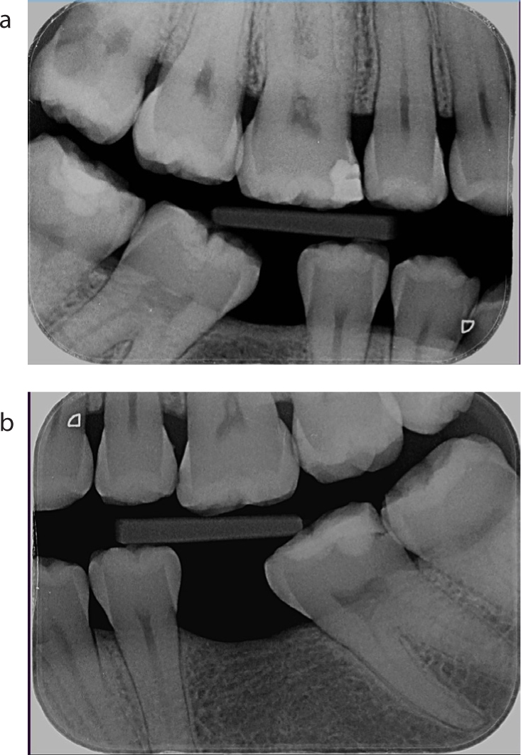
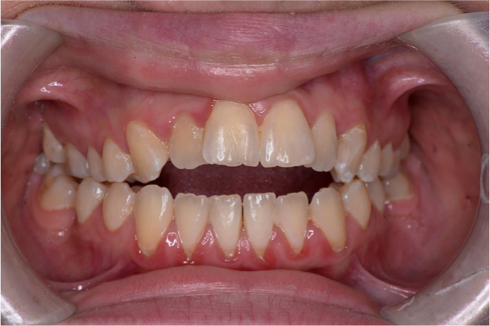
On examination, Patient B had carious lesions in several teeth, including the upper right third molar (UR8), which was unrestorable (Figures 1a and b). The patient's primary concern was an anterior open bite (Figure 2). She had previously been referred for orthodontic treatment but treatment was felt to be inappropriate due to the tissue challenges with EDS19 and her frequent episodes of loss of consciousness associated with POTS.
Figure 1.
(a) Right bitewing radiograph, patient B. (b) Left bitewing radiograph, patient B.Figure 2. Patient B in intercuspal position illustrating anterior open bite.
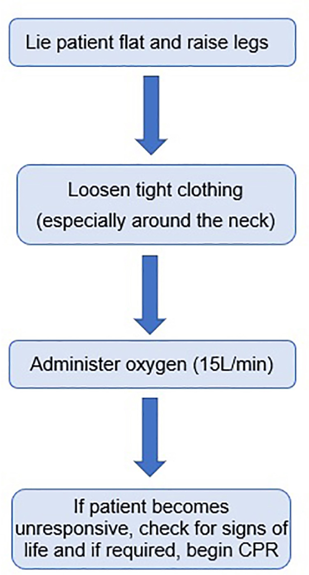
Towards the end of the initial examination, the patient suddenly lost consciousness. She was managed as per current guidelines,22 as outlined in Figure 3. Within 90 seconds she had regained consciousness and was fully alert. Following discussion with the patient, all restorations were carried out without local anaesthetic (LA). Despite suffering some minor discomfort, she was able to tolerate treatment. When extracting the UR8, 6.6 ml of lidocaine hydrochloride 2% with 1:80,000 adrenaline was administered followed by 2.2 ml articaine hydrochloride 4% with adrenaline 1:100,000. Unfortunately, the patient could still feel a degree of pain, but chose to continue with the extraction, which was then carried out without further complications. When treatment was completed, she was discharged back to her GDP for routine recall appointments.
Figure 3. Flowchart illustrating recommended management of patient collapse.22
Case study 3
A 47-year-old Caucasian female patient, Patient C, was referred to the department of special care dentistry by the department of oral medicine due to a history of fainting during dental treatment. She had a complex medical history which included paroxysmal atrial fibrillation, and supraventricular tachycardia (SVT) secondary to re-entry tachycardia, for which she underwent radioablation in 2001. This was initially thought to be the cause of the episodes of fainting. Her medical history included Hypermobile EDS, thoracic outlet syndrome, depression, gastro-oesophageal reflux disease, irritable bowel syndrome, stress incontinence, and chronic fatigue syndrome.
On examination, a cuspal fracture was noted on the lower right first molar (LR6), which was subsequently restored under local anaesthetic. Towards the end of her treatment she suffered from palpitations and loss of consciousness lasting several minutes. Vital signs were monitored and she was managed conservatively,22 following which she was transferred to the Emergency Department for further monitoring. While an inpatient, she was found to have self-terminating SVT, diagnosed on Echocardiogram (ECG), as well as postural hypotension. She was discharged with appropriate follow-up.
At a later appointment with her GDP, the patient collapsed again whilst undergoing a routine scale and polish. Due to the risk of collapse during dental treatment, a shared care protocol was agreed. It was agreed that the patient should attend the dental hospital for recalls and treatment, as required, but maintain contact with the GDP for dental emergencies.
Over the subsequent 4 years, patient C continued to attend dental recalls without any significant issues. During this period, outside of the dental setting, the patient continued to suffer from episodes of fainting and was finally diagnosed with POTS. Several months later she was also diagnosed with Mast Cell Activation Syndrome and Addison's Disease. Following an episode of Addisonian Crisis during a routine urology procedure, it was advised that planned dental treatment should be carried out in a theatre setting with a 24-hour hydrocortisone infusion post-operatively.
She attended the department of special care dentistry 6 months later with multiple carious lesions. An inpatient general anaesthetic was arranged alongside the Endocrinology and Oral and Maxillofacial Surgery (OMFS) teams, to provide comprehensive care. The treatment was carried out uneventfully, and the patient was admitted under the OMFS team, whilst the Endocrinology team managed her steroid cover.
When the patient attended several months later with a cracked tooth, the Endocrinology and Cardiology teams were contacted for advice prior to treatment. As she was on a reducing dose of steroids, no steroid cover was required. To prevent episodes of collapse associated with POTS, the patient was treated in a supine position.
The patient continues to attend for routine recalls in the department of special care dentistry, and episodes of collapse associated with POTS have been managed as before. Contact is maintained with her medical teams to ensure that she is managed appropriately when undergoing treatment.
Conclusion
As demonstrated by the clinical cases presented, EDS patients can present with a range of complications of their disease which may affect provision of their dental care. The clinician should be aware of the intra-oral manifestations of the disease as well as the concurrent medical conditions, all of which may complicate provision of care. As EDS impacts multiple aspects of care, a multidisciplinary approach is required, including good communication with medical teams. Referral to tertiary level care may be required for complex procedures, with the GDP playing an integral role in the shared dental care of the patient.