
References
Kelly JR. Evidence-based decision making: guide to reading the dental materials literature. J Prosthet Dent. 2006; 95:152-160
Kelly JR, Campbell SD, Bowen HK. Fracture surface analysis of dental ceramics. J Prosthet Dent. 1989; 62:536-541
Kelly JR, Giordano R, Pober R, Cima MJ. Fracture surface analysis of dental ceramics: clinically failed restorations. Int J Prosthodont. 1990; 3:430-440
Kelly JR. Clinically relevant approach to failure testing of all-ceramic restorations. J Prosthet Dent. 1999; 81:651-661
Scharer P. All-ceramic crown systems: clinical research versus observations in supporting claims. Signature. 1996; 1
Weinstein M, Katz S, Weinstein AB. US Patent No 3,052,982: Porcelain-covered metal-reinforced teeth. 1962;
Leempoel PJ, Van't Hof MA, Haan A. Survival studies of dental restorations: criteria, methods and analyses. J Oral Rehabil. 1989; 16:387-394
Reitemeier B, Hänsel K, Kastner C, Weber A, Walter MH. A prospective 10-year study of metal ceramic single crowns and fixed dental prosthesis retainers in private practice settings. J Prosth Dent. 2013; 109:149-155
Malament KA. Considerations in posterior glass-ceramic restorations. Int J Perio Rest Dent. 1988; 8:32-49
Spear FM. The risk of a metal-free practice. J Esthet Rest Dent. 2009; 21:71-74
Sorensen JA, Choi C, Fanuscu MI, Mito WT. IPS Empress crown system: three-year clinical trial results. J Calif Dent Assoc. 1998; 26:130-136
Fradeani M, Aquilino A. Clinical experiences with Empress crowns. Int J Prosthodont. 1997; 10:241-247
Zhao K, Pan Y, Guess PC, Zhang XP, Swain MV. Influence of veneer application on fracture behavior of lithium-disilicate-based ceramic crowns. Dent Mater. 2012; 28:653-660
Zhao K, Wei YR, Pan Y, Zhang XP, Swain MV, Guess PC. Influence of veneer and cyclic loading on failure behavior of lithium disilicate glass-ceramic molar crowns. Dent Mater. 2014; 30:164-171
Papanagiolou HP, Morgano SM, Giordano RA, Pober R. In-vitro evaluation of low-temperature aging effects and finishing procedures on the flexural strength and structural stability of Y-TZB dental ceramics. J Prosthet Dent. 2006; 96:154-164
Denry I, Kelly JR. State of the art zirconia for dental applications. Dent Mater. 2008; 24:299-307
Sailer I, Fehér A, Filser F, Gauckler LJ, Lüthy H, Hämmerle CH. Five-year clinical results of zirconia frameworks for posterior fixed partial dentures. Int J Prosthodont. 2007; 20:383-388
Christensen GJ. Porcelain-fused-to-metal versus zirconia-based ceramic restorations. J Am Dent Assoc. 2009; 140:1036-1039
Tan JP, Sederstrom D, Polansky JR, McLaren EA, White SN. The use of slow heating and slow cooling regimens to strengthen porcelain fused to zirconia. J Prosthet Dent. 2012; 107:163-169
Zhang Y, Lawn BR. Novel zirconia materials in dentistry. J Dent Res. 2018; 97:140-147
Sulaiman TA, Abdulmajeed AA, Shahramian K, Lassila L. Effect of different treatments on the flexural strength of fully versus partially stabilized monolithic zirconia. J Prosthet Dent. 2017; 118:216-220
Inokoshi M, De Munck J, Minakuchi S, Van Meerbeek B. Meta-analyasis of bonding effectiveness to zirconia ceramics. J Dent Res. 2014; 93:329-334
Heintze SD. Clinical relevance of tests on bond strength, microleakage and marginal adaptation. Dent Mater. 2013; 29:59-84
Janyavula S, Lawson N, Cakir D, Beck P, Ramp LC, Burgess JO. The wear of polished and glazed zirconia against enamel. J Prosthet Dent. 2013; 109:22-29
Kim MJ, Oh SH, Kim JH, Ju SW, Seo DG, Jun SH, Ahn JS, Ryu JJ. Wear evaluation of the human enamel opposing different Y-TZP dental ceramics and other porcelains. J Dent. 2012; 40:979-988
Mitov G, Heintze SD, Walz S, Woll K, Muecklich F, Pospiech P. Wear behavior of dental Y-TZP ceramic against natural enamel after different finishing procedures. Dent Mater. 2012; 28:909-918
Pieger S, Salman A, Bidra AS. Clinical outcomes of lithium disilicate single crowns and partial fixed dental prostheses: a systematic review. J Prosthet Dent. 2014; 112:22-30
Sulaiman TA, Delgado AJ, Donovan TE. Survival rate of lithium disilicate restorations at 4 years: a retrospective study. J Prosthet Dent. 2015; 114:364-366
Sulaiman TA, Abdulmajeed AA, Donovan TE, Cooper LF, Walter R. Fracture rate of monolithic restorations up to 5 years: a laboratory survey. J Prosthet Dent. 2016; 116:436-439
Abdulmajeed AA, Donovan TE, Cooper LF, Walter R, Sulaiman TA. Fracture of layered zirconia restorations at 5 years: a dental laboratory survey. J Prosthet Dent. 2017; 118:353-356
An evidence-based evaluation of contemporary dental ceramics
From Volume 45, Issue 6, June 2018 | Pages 541-546
Article

When placing aesthetic crown restorations, contemporary clinicians have a bewildering number of ceramic materials from which to choose. Dentists are also tasked with practising ‘evidence-based’ dentistry and, while there are numerous articles and studies published every year on ceramic materials, the quality of the ‘evidence’ is far from optimum. Almost all ceramic systems are marketed to the profession well before any clinical evidence supporting use of those systems has been published. It is clear that laboratory studies of physical and mechanical properties of ceramic materials provide little predictive evidence of clinical performance of any ceramic system.1 It is also clear that load-to-failure studies of ceramic crowns are not predictive of clinical performance.2, 3 Fatigue testing of ceramic materials under water may prove to be a viable predictive protocol, but the details of such testing have yet to be determined.4 The best method of determining clinical performance of a ceramic material is to conduct randomized, controlled clinical trials.
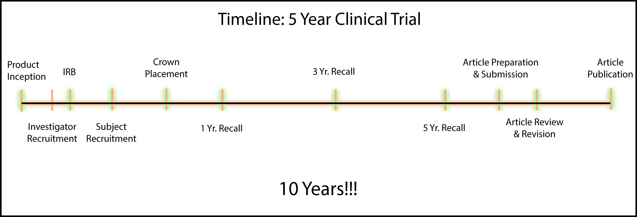
Two giants in the field of dental ceramics, Peter Sharer and John McLean, suggested that before a clinician uses a new ceramic system, he or she should be provided with evidence gleaned from 5-year independent clinical trials, and that the survival rate of restorations be at least 95%.5 While this is indeed an excellent approach, it is a fact that the time-frame from product inception until study publication for a 5-year clinical trial is close to 10 years (Figure 1). This 10-year lag in data generation renders this approach relatively impractical.
The purpose of this article is to describe an approach (laboratory survey) that provides accurate survival data on new ceramic systems to dentists in a significantly more timely manner. A second purpose is to share with readers that data generated with this laboratory survey approach may assist them in ceramic material selection to meet the specific clinical needs of their patients in an optimal manner.
History
Metal-ceramic crowns using a variety of metal alloys have been the standard aesthetic crown service offered by the majority of dentists since the patent was registered by Weinstein, Katz and Weinstein in 1962.6 For many years it was accepted that metal-ceramic crowns provided a combination of excellent clinical longevity coupled with reasonable aesthetic outcomes.7, 8 However, as patients gradually became more demanding relative to aesthetic outcomes, and dental material science attempted to meet that demand, numerous new ceramic systems were introduced to the market, with the primary driver being improved aesthetic outcomes. Table 1 is a partial list of ceramic systems introduced to the market since the early 1980s.
| Ceramic System | Manufacturer |
|---|---|
| Aluminous Core PJC | Vita Zahnfabrik |
| CaptekTM | Argen |
| Cerestore | Coors Biomedical |
| Dicor | Dentsply International and Corning Glass |
| High-Ceram | Vita Zahnfabrik |
| Willi's Glass | Dentsply |
| IPS Empress | Ivoclar Vivadent |
| Optec | Jeneric/Pentron |
| Empress II | Ivoclar Vivadent |
| IPS Eris | Ivoclar Vivadent |
| IPS e.max | Ivoclar Vivadent |
| In-Ceram (spinell, alumina, zirconia) | Vita Zahnfabrik |
| Procera® AllCeram | Nobel Biocare |
| Layered zirconia eg LavaTM | 3MTM–ESPETM (many others) |
| Monolithic zirconia eg BruxZir®, Zenostar®, KatanaTM | Glidewell, Wieland, Kuraray Noritake Dental Inc |
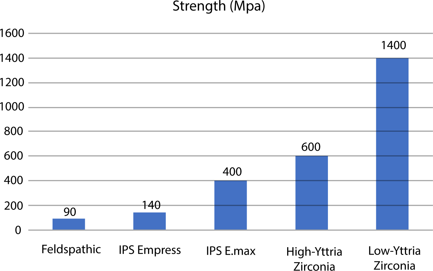
While some of the new ceramic systems did provide improved aesthetic results, it was clear that clinical longevity was significantly compromised with most of the early materials.9, 10 The first alternative to show promise in this regard was layered IPS Empress (Ivoclar), which demonstrated 95% survival at 5 years, but on anterior teeth only.11, 12 Survival rates with posterior teeth were lower at approximately 80%. IPS Empress is a leucite-reinforced glass-ceramic with a flexural strength of approximately 160 MPa. This ceramic material provides excellent aesthetic results, but is currently rarely used due to the emergence of improved materials.
Contemporary ceramic materials
While the impetus for early ceramic crown systems was the desire to obtain predictably superior aesthetic results compared to metal-ceramic crowns, the primary driver for use of contemporary ceramic materials is cost. While excellent aesthetic results are still imperative, the costs of precious metals (gold, platinum, palladium) has escalated considerably in recent years, resulting in very high laboratory costs for metal-ceramic restorations. Because of the elimination of metal costs and digital manufacturing efficiencies, the laboratory costs for contemporary ceramic restorations are substantially lower than for metal-ceramic restorations.
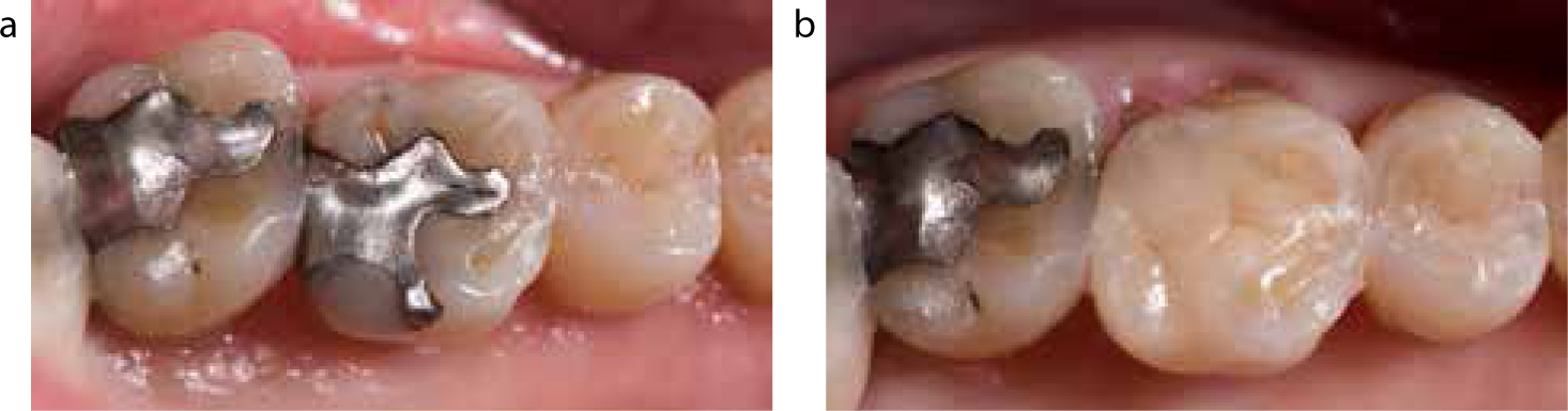
The contemporary ceramic materials that are the primary focus of this article are lithium disilicate (IPS e.MAX, Ivoclar) and zirconia (many manufacturers). Lithium disilicate and zirconia ceramics can be used both as layered or monolithic restorations. Monolithic lithium disilicate materials have flexural strengths of around 400 MPa and the layered version is somewhat weaker.13, 14 In general, monolithic restorations are stronger than layered restorations, but layered crowns provide improved aesthetic outcomes. Layered crowns are generally indicated for anterior teeth, while monolithic materials are appropriate for posterior restorations where aesthetics is not as critical. Lithium disilicate materials can be etched and bonded and can be used successfully as crowns with traditional retentive preparations and also as onlays and partial veneer restorations with contemporary conservative non-retentive preparations (Figure 2a, b).
Monolithic zirconia materials have flexural strengths as high as 1400 MPa, but lack the translucency essential for excellent aesthetic outcomes.15 Their use is primarily limited to posterior teeth. Layered zirconia restorations have improved aesthetic outcomes but early versions of layered zirconia suffered from unacceptable high levels of chipping of the veneering ceramic.16, 17 The chipping did not occur at the interface between the core and veneering ceramic, but rather presented as cohesive fractures in the body of the veneering ceramic. While such chipping did not always necessitate replacement of the restoration, chipping rates were significantly higher than chipping rates with metal-ceramic crowns.18
There are two primary potential aetiologies for the excessive chipping rates initially noted for zirconia cored restorations. The first is related to the thermal conductivity of zirconia, which is very low compared to that of metals traditionally used for ceramic bonding. As a result of this, when the zirconia-based crown is removed from the ceramic oven, the veneering ceramic cools faster than the core. After the veneering ceramic cools past its glass-transition temperature as it cools to bench temperature, the core material continues to cool and shrink very slowly. This builds up stress in the veneering ceramic which is expressed clinically sometime later in the form of chipping. This problem has been addressed by adopting modified slow cooling protocols, which has been shown to increase the strength of the veneering ceramic and, hopefully, reduce the incidence of chipping.19
The second cause of chipping has been a lack of proper support of the veneering ceramic by the core. The industry standard is that the maximum thickness of veneering ceramic should never exceed 2mm, regardless of the core material. When fabricating zirconia-cored crowns, the die is scanned and a core of uniform thickness is milled, not taking into account the thickness of the veneering ceramic. The core will exactly follow the shape of the tooth preparation which, if less than ideal, will often result in lack of support. If the laboratory technician recognized the lack of support, the shape of the core could be adjusted using computer software, but the reality is that this procedure is rarely carried out in commercial laboratories. Most manufacturers have addressed this problem and now have computer software that first completes a virtual full contour ‘wax-up’ of the restoration and then virtually cuts that back, allowing for proper support of the veneering ceramic by the core. Hopefully, the problems related to excessive chipping with layered zirconia have been addressed.
Yttria-stabilized tetragonal zirconia polycrystal restorative ceramic is the most robust ceramic material and was the basis for most of the original zirconia-based systems. However, as mentioned previously, these materials lack translucency. In an attempt to improve translucency, new generations of zirconia, using more yttria and cubic phase zirconia have been developed and have been brought to market.20 These materials have modest improvements in translucency, but have less than half the flexural strength of the original materials and have less toughness because they do not have the ability to undergo stress-induced transformation21 (Figure 3). These materials are being marketed for use in the anterior region of the oral cavity. Clinicians are advised to use these materials with caution until evidence of their clinical viability is available.
A number of protocols have been developed to attempt to bond to zirconia. All involve air-particle abrasion, use of an MDP containing primer (Clearfil SE Bond, Kuraray; Z Prime, Bisco; Scotchbond Universal, 3M-ESPE) and resin cement.22 All studies related to the effectiveness of these protocols have used shear bond testing which is notoriously limited in its ability to predict clinical performance.23All protocols achieve relatively weak bonds to zirconia and these bonds decrease significantly with any type of ageing. Air particle abrasion of zirconia is risky because it has the potential to weaken the material. In the opinion of the authors, bonding to zirconia restorations is not a predictable procedure at this time, and zirconia restorations should be used only with traditional preparations with adequate resistance and retention form.
It should be noted that zirconia materials have a strong affinity for proteins found in saliva and blood. These proteins cannot easily be removed from the intaglio surface of crowns, which are contaminated during try-in. After try-in, the internal surface of the crowns should be cleaned for 20 seconds with a sodium hydroxide solution (Ivoclean, Ivoclar) and then washed with water. Crowns should be cemented with a self-adhesive resin cement (Rely X Unicem, 3M-ESPE) or resin-modified glass ionomer cement (Rely X Luting Cement Plus, 3M-ESPE).
One of the benefits of monolithic zirconia crowns is that they require less tooth reduction than metal-ceramic or glass-ceramic crowns. Basically, 1 mm occlusal reduction with 8/10 mm axial reduction with a well-defined chamfer margin is optimal. The preparation should have adequate resistance and retention form, as mentioned previously. Crown preparations for layered zirconia and monolithic or layered lithium disilicate should have 1.5 mm reduction on the occlusal or incisal surface and 1.2 mm reduction axially.
Wear of opposing enamel or restorative material has been a concern with ceramic materials. Conventional ceramic materials will cause abrasion of enamel if they are in gliding contact with opposing natural teeth. Several studies have clearly identified that polished zirconia is extremely kind to opposing enamel.24, 25, 26, 27 It seems reasonable that the laboratory polish zirconia crowns prior to glazing so that when the glaze wears away, a polished surface results.
Survival rates
One systematic review analysed available clinical trials on lithium disilicate restorations and reported favourable results.27 The survival rate of lithium silicate single crowns was 100% at 2 years and 98% at 5 years, while the failure rate of fixed partial dentures (FPDs) was unacceptably high. Unfortunately, the studies analysed in the review were of relatively poor quality, so the validity of the findings is somewhat questionable.
An alternative approach to clinical trials has been the use of dental laboratory surveys of fractured ceramic restorations.28, 29, 30 Contemporary dental laboratories have excellent computer-based programs to track returns, and access to this data provides timely feedback on the clinical performance of their restorations. While such surveys do not replace the need for properly conducted clinical trials, they do provide relatively accurate and timely information to practitioners to help guide them in their choice of ceramic materials. The laboratory survey approach also results in evaluation of survival rates of large numbers of restorations. Successful use of these surveys requires three assumptions:
A laboratory survey of 21,337 lithium disilicate restorations in place for up to 45 months was published in 2015.28 The failure rate of monolithic lithium disilicate single crowns was very low at 0.91%, and for layered single crowns was double that of monolithic crowns at 1.83%, which is still well below Sharer's criteria. Failure rates for bonded ceramic inlays and onlays was also very low at 1.01%. Failure rate of FPDs was 4.55%.
Another laboratory survey of monolithic zirconia restorations was published in 2016, while another related to layered zirconia restorations was published in 2017.29, 30 Both surveys reported favourable results. Combining data from both studies related to single crowns, out of a total of 54,708 single crowns, the failure rate for monolithic zirconia crowns was 0.71% at 5 years. For layered zirconia single crowns, the failure rate was 3.25%, a 4–5 times higher rate of fracture, reflecting the chipping problem that occurred with the introductory layered zirconia materials. Monolithic FPDs failed at a rate of 2.62% at 5 years, which was an improvement over the fracture rate for lithium disilicate FPDs.
The data from the three referenced laboratory surveys is quite compelling (Table 2). The most commonly used ceramic materials in contemporary dental practice are lithium disilicate and zirconia. Both monolithic and layered versions of both systems are used extensively, despite the lack of convincing evidence of the efficacy of these systems. Data from these surveys validates what is happening in contemporary practices. Both systems provide aesthetic, economical alternatives to metal-ceramic restorations.
| Restoration Type | Single Crowns | Fixed Partial Dentures | Onlay Restorations | ||||||
|---|---|---|---|---|---|---|---|---|---|
| Units | Fracture | Fracture (%) | Units | Fracture | Fracture (%) | Units | Fracture | Fracture % | |
| Monolithic E.max | 11603 | 106 | 0.91 | 1494 | 68 | 4.55 | 1093 | 11 | 1.01 |
| Layered E.max | 4162 | 76 | 1.83 | - | - | - | - | - | - |
| Monolithic Zirconia | 31760 | 224 | 0.71 | 8067 | 211 | 2.62 | - | - | - |
| Layered Zirconia | 22948 | 745 | 3.25 | 8646 | 300 | 3.47 | - | - | - |
Indications
The following indications for ceramic system use are based upon the best available clinical evidence:
Conclusions
It is clear that contemporary dentists are able to utilize ceramic materials that have adequate aesthetic outcomes and improved clinical longevity. The new systems also offer considerable economic advantages over metal-ceramics. Laboratory surveys do not replace the need for properly conducted clinical trials but do provide timely information on survival rates of a large number of ceramic crowns.