
Abstract
This case report presents a case in which a patient attended because of the poor appearance of an anterior crown, but she was unaware that she had a number of teeth affected by toothwear.
From Volume 43, Issue 9, November 2016 | Pages 867-872
This case report presents a case in which a patient attended because of the poor appearance of an anterior crown, but she was unaware that she had a number of teeth affected by toothwear.

Patients may attend a dentist for a variety of reasons, some as a routine, and others because of pain, obvious disease or because of anxieties regarding their dental appearance. On occasion, attendance because of a presenting complaint may result in the patient being advised that there are other problems about which the patient is unaware. It is therefore the aim of this report to present a case in which a patient attended because of poor appearance of an anterior crown, but was unaware that she had a number of teeth affected by toothwear.
A 42-year-old female patient was referred, complaining that she was unhappy about the appearance of her anterior teeth, notably an unsightly crown on her upper left central incisor tooth.
Regarding the patient's medical history, she reported that she was a non-smoker and did not drink alcohol, but admitted to a high consumption of ‘fizzy’ drinks, namely, two litres of cola drinks daily. The patient's history also indicated a high consumption of sweets. Regarding the patient's dental history, she stated that she attended the dentist regularly, every 6 months. Her oral hygiene included brushing twice daily with a powered toothbrush.
A full intra- and extra-oral examination was undertaken. No relevant findings were made on the extra-oral examination or examination of the temporo-mandibular joint (TMJ). There was no cervical lymphadenopathy and the intra-oral mucosa was healthy. The patient had a Class I occlusion.
BPE scores were:
| 1 | 1 | 1 |
| 1 | 2 | 1 |
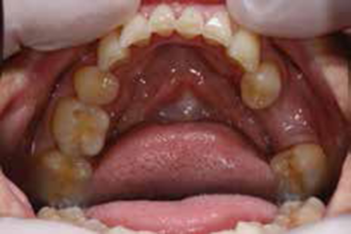
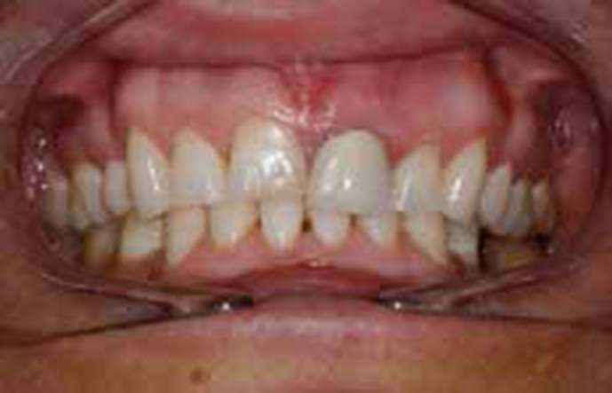
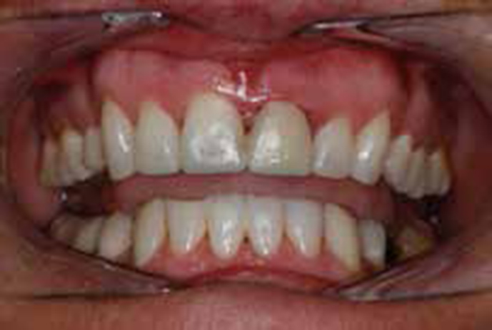
Intra-oral examination indicated a small build-up of plaque and calculus on the lingual aspect of four lower anterior teeth, with slight bleeding on probing. No caries was noted. Palatal and incisal wear was noted at UR321 and UL23 and incisally on the LL321 and LR123. A glossy appearance was noted on the occlusal surfaces of some posterior teeth (Figure 1). The UL1 metal-ceramic crown was ill-fitting, with a 2 mm marginal gap at its labial aspect (Figures 2a and b). The LL6 was missing and no restorations, other than at UL1, were present.
Bitewing radiographs and a periapical of UL1 were taken. No interproximal caries was noted and the periapical tissues at UL1 were free from pathology. There was no loss of bony support (Figure 2).
Diagnoses made were as follows:
An initial concern was the volume of the patient's consumption of carbonated drinks and sweets. She was advised to reduce both and to use a straw when drinking such beverages but, better still, to avoid drinking such beverages completely. Using intra-oral photographs, the patient was made aware of the toothwear which affected her teeth: the contributing factors were discussed. Oral hygiene instruction was given and the patient was shown how to use interdental brushes and was advised of the appropriate sizes to use. The patient was also advised to ‘spit and not rinse’ and Duraphat toothpaste 2800 ppm was recommended to reduce the chance of decay and sensitivity.1
Impressions were taken and study models made. The patient was advised that treatment was necessary to cover the worn and wearing surfaces of her upper and lower anterior teeth. She was also advised that building up these anterior teeth would effectively improve the aesthetics of these teeth, even if that was not the primary reason for treatment. The patient was given a Patient Information Leaflet derived from one previously published in Dental Update.2 She was asked to read this and discuss it at a future visit. Among the matters that this document discusses are:
Diagnostic wax-ups for the upper and lower anterior teeth were made in order to demonstrate how the teeth could look after treatment and to show the amount of tooth tissue which had been lost. The patient was also given the option of tooth whitening prior to undertaking any restorative treatment. Home and 'power whitening' were discussed.
Oral hygiene instructions:
The patient agreed to this treatment plan and understood that the composite build-ups could require occasional maintenance and that this, if necessary, might incur an additional fee. The patient was also made aware that the UL1 crown might need root-filling in future.3 The wear affecting the patient's posterior teeth was also discussed. She was appraised of the reasons for this (carbonated drinks, principally) and was advised that she should reduce the consumption of such drinks or stop them completely.
Alginate impressions were taken for the home bleaching trays and a Penta™ polyether impression (3M ESPE Impregum™, Seefeld, Germany) was taken for the study models and diagnostic waxup. The polyether impression was used owing to its high level of accuracy and dimensional stability.4 A facebow was also taken in order to have the models mounted. The laboratory was asked to use the distal aspect of the UR3 as a reference point in making the wax-ups, as this corner had not been affected by toothwear.
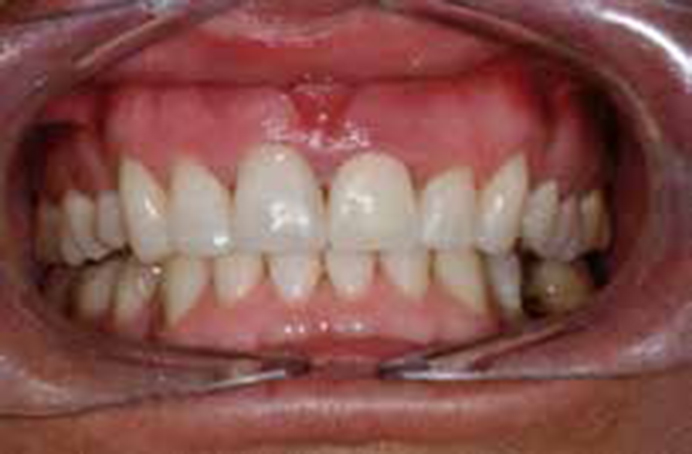
Hydrogen peroxide bleaching gels (6%) from the Pola™ range (SDI Melbourne, Australia) were given to the patient, along with a bleaching tray: she was given a demonstration on how to use this. The patient was instructed to use the bleaching gel daily for one hour using the close-fitting trays provided and to return in 2 weeks for a review and final discussion regarding the wax-ups.5 She was also advised to discontinue this treatment should her teeth become sensitive. After 2 weeks the shade of the patient's upper right central and lateral incisor teeth had shifted from shade A2 to B1 (Figure 3).
The patient was happy with the results of the bleaching and was shown the diagnostic wax-up. This was approved, with the alterations consisting mainly of increasing the length of the upper incisor teeth. (In this regard, if the patient had expressed uncertainty at the wax-up stage, a vacuum-formed stent could have been made, this then being filled with provisional crown material and seated over the teeth, in order to provide the patient with an aesthetic preview.6)
A treatment plan was finalized, consisting of:
The next appointment was a lengthy one, with the patient being booked in for a whole afternoon session. The aim was to carry out all the composite buildups and to replace the UL1 crown with a temporary acrylic crown formed at the chairside. The UL1 crown was removed first and a temporary crown made, in order to facilitate the placement of the UR1 composite.
A sandblaster using aluminium oxide powder was initially used to enhance the surface area prior to etching with 37% phosphoric acid and bonding using Scotchbond Universal™ (3M ESPE). A nano-filled resin composite (Filtek Supreme XTE™, 3M ESPE) was used as it has been considered to provide excellent aesthetics and facilitate easy polishing.7 A palatal matrix, extending just to the incisal edges of the anterior teeth, was used to aid the build-up of the incisal edges (Figure 4).
After bonding, resin composite was added to the palatal and incisal surfaces of UR321 and UL23. The two least abrasive grades of Soflex discs (3M ESPE) and diamond polishing paste were used at this stage (Figure 5). However, final polishing was not completed as it was considered that it would be necessary to complete the occlusal adjustment a week later, by which time it was anticipated that the patient would have re-established a reproducible intercuspal position and the final polishing would be carried out after any necessary occlusal adjustment. The chairside acrylic crown was not of ideal appearance. Accordingly, it was scheduled for replacement on the visit a week later.
On the review appointment after one week, the patient was content with the appearance of her anterior teeth and was coping well with the slight increase in her occlusal vertical dimension (OVD), which had caused slight dysclusion of her posterior teeth. She had felt no discomfort because of the increase in OVD and, after three days, this had not disturbed her mastication. She reported that she was again able to close her teeth in a new reproducible (intercuspal) position.
It was decided to change the UL1 acrylic crown for one constructed in a laboratory in order to improve the appearance, and also because it was anticipated that this crown would be in position for a minimum of three months, to allow for healing of the periodontal tissues. A week after fitting the new acrylic crown, the enlargement of the mesial gingiva at UL1 had also improved.
A thorough occlusal analysis was carried out in order to obtain even contacts on the new restorations in all excursions. The upper and lower composites were also finished and polished. Canine guidance was obtained on both sides, this being considered to be of importance in toothwear cases where there might be an attritional element in the toothwear.8
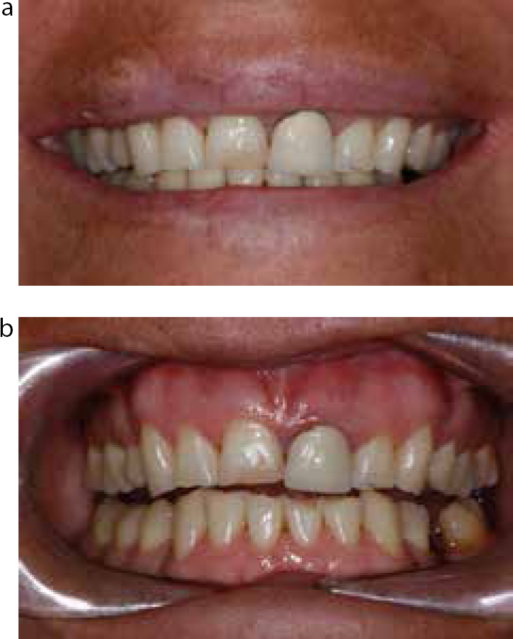
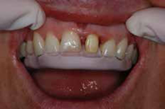
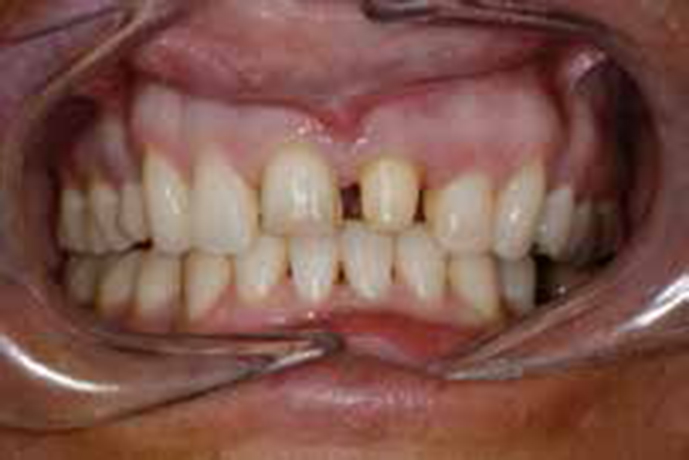
Figure 6 presents the improved appearance of the laboratory-made acrylic crown and the gingiva showing signs of improving health. At this stage, the patient was demonstrating much increased interest in the appearance of her teeth and, indeed, her oral condition in general. It was at this stage that she asked about the possibility of making the gingival margins of UL1 and UR1 equal, considering that improving the gingival height of UL12 would further enhance her dental appearance. However, on explaining that a further 3–6 months would be needed to allow for healing of the periodontal tissues following crown lengthening before a new crown would be made,9 the patient decided not to proceed with the crown lengthening. She also considered that her UR1 looked too dark in relation to the other teeth: she suggested that she should have a veneer placed on this tooth. The pros and cons of such treatment with respect to need to remove sound tooth substance were discussed, and the risk of the tooth requiring a root filling or veneer failure10 was mentioned. However, it was decided to accede to the patient's request and to proceed with the veneer. Accordingly, having previously completed the composite build-ups, it was possible to make a conservative preparation using the matrices from the wax-up (Figure 7).
The crown and veneer were made from a pressable ceramic, e-max™ (Ivoclar Vivadent, Liechtensten), with this material being utilized because of its good physical properties, the ability of its fit surface to be etched with hydrofluoric acid, thereby providing micromechanical retention, and for aesthetic reasons.11 The shade was taken at the dental laboratory. Figure 8 illustrates the crown and veneer when fitted, using Nexus™ cement (Kerr Mfg Co, Orange, CA). It was possible to obtain broad contact points, which eliminated the black triangle that was seen in Figure 7.
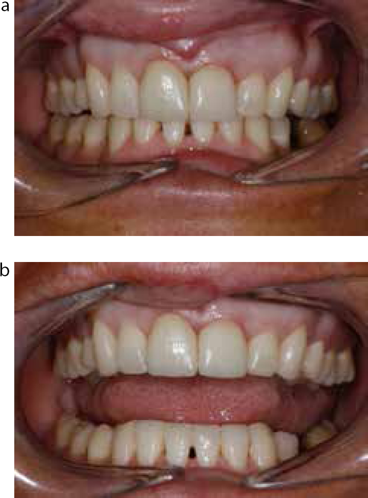
Oral hygiene instructions were once again reinforced. The patient was content and, at the six-month follow-up, it was apparent that the gingival tissues around the crown and veneer were more healthy than at the fit appointment (Figure 9).

This case demonstrates the relationship between clinical need and aesthetic treatment. The patient presented with an ill-fitting crown and worn anterior teeth, that being the clinical need. In addition, there was no canine guidance and, together with the erosive and parafunctional habits which were present, the patient's anterior teeth were likely to suffer further destruction. Restoring the patient's worn and wearing surfaces with resin composite allowed a minimally invasive approach, restored the aesthetics and function at a minimal cost to the patient, both financially and, also, just as importantly, in terms of tooth substance. However, the patient became increasingly motivated as the appearance of her teeth improved, and along with that her (dental) confidence, hence her request for the veneer at UR1. The treatment is not without risks and the patient needed to be aware of these.10 By first building up the worn teeth with composites it also allowed a more conservative approach on the veneer that was subsequently made.
The so-called Dahl concept12 with its increase in OVD has previously been considered a hazardous procedure:13 it is now an accepted treatment for worn anterior teeth, with minimal discomfort to the patient and a high degree of patient acceptance.14,15 However, it is essential that patients who receive such treatment are acquainted in advance with the potential disturbance to their occlusion and the fact that it may take several days to re-establish a reproducible intercuspal position and that, during that time, eating might be slow and/or difficult. Hence, the need for patients to be provided with a Patient Information Leaflet2 giving details of how their amended occlusion will feel, notwithstanding any change in the shape of their anterior teeth. In addition, the modest increase in the occlusal vertical dimension was best carried out prior to crowning the UL1.
Finally, there was some debate regarding the material to be used for the crown at UL1. Metal-ceramic has been the workhorse of fixed prosthodontics for decades, but the last decade has seen the appearance of zirconia with its excellent physical properties16 but a potential for less than ideal translucency, although this is a factor which recently appears to have been addressed.17 The lithium disilicate-based material e-max™ has proven excellent aesthetically and has been considered to provide sufficient strength for the forces that are applied in anterior teeth.11 In other words, zirconia may be becoming the base for crowns distal to a first premolar, and e-max™ for those anterior to that.
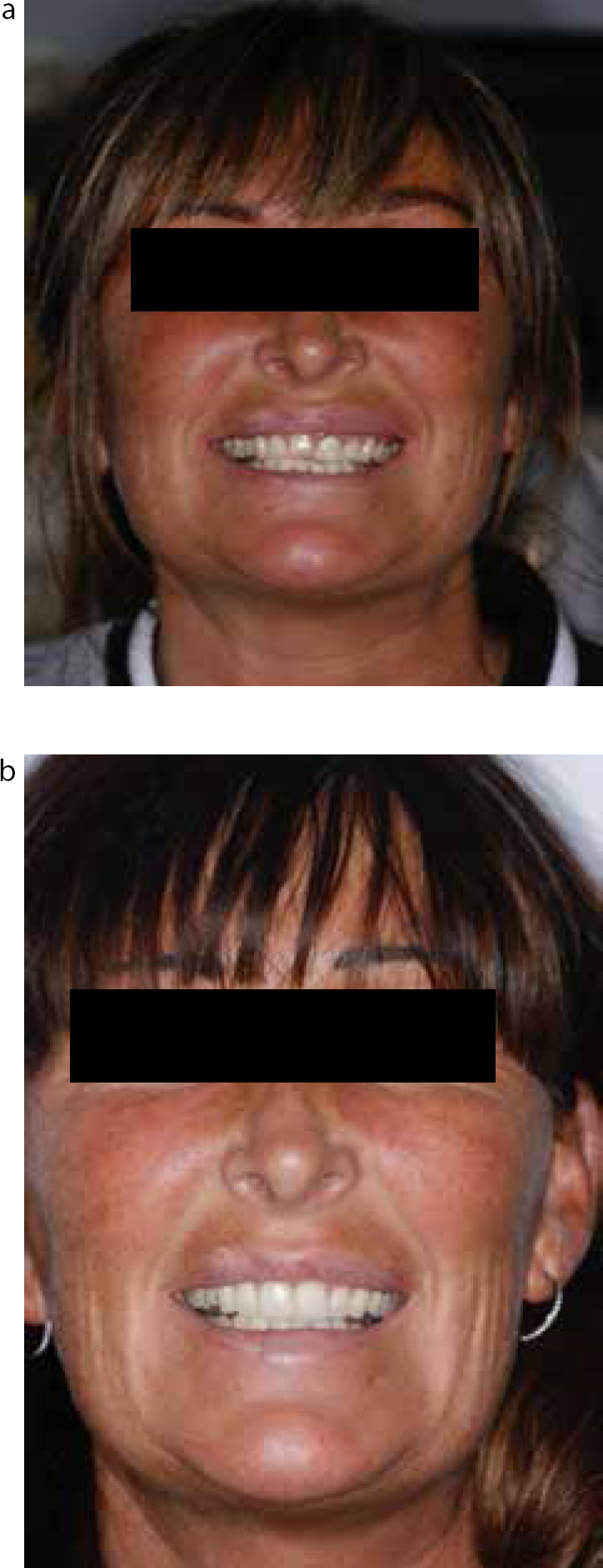
Six months from completion, the treatment has given the patient confidence and has restored her faith in regular dental attendance18 (Figure 10). The patient has cut down her consumption of carbonated drinks and sweets.
The oral hygiene is immaculate and the gingival margins around the UL1 crown have totally stabilized. The patient seems to have been really motivated by her improved dental appearance.
Relating aesthetics to clinical treatment need is an increasingly important aspect of general dentistry. A variety of minimally invasive techniques may facilitate this, but the need for minimal intervention must be in the forefront of any treatment plan.
This case was presented as a case report in part fulfillment of the requirements for the Aesthetic Dentistry module in the Masters in Advanced General Dental Practice at the University of Birmingham and was considered to be a good example of how to write a case report by the reviewer.