
Article
Recurrent multi-focal oral squamous papillomas
We saw a 73-year-old man in September 2020, who was referred by his GP for multiple lumps on his tongue and lower lip of unknown duration. His past medical history was significant for ischaemic heart disease, COPD, asthma, osteopenia and chronic kidney disease. He smoked 20 cigarettes a day (50 pack-years) and did not drink alcohol regularly.
On examination there was no obvious cervical lymphadenopathy. Multiple painless pedunculated papillomatous lumps of the lower lip, anterior mandibular ridge and ventral/dorsum of the tongue were noted (Figure 1).
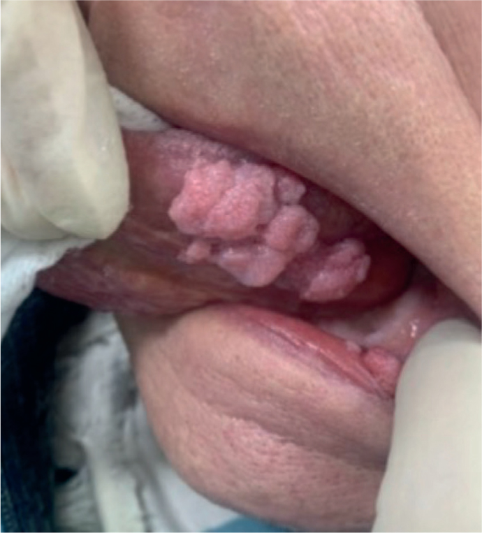
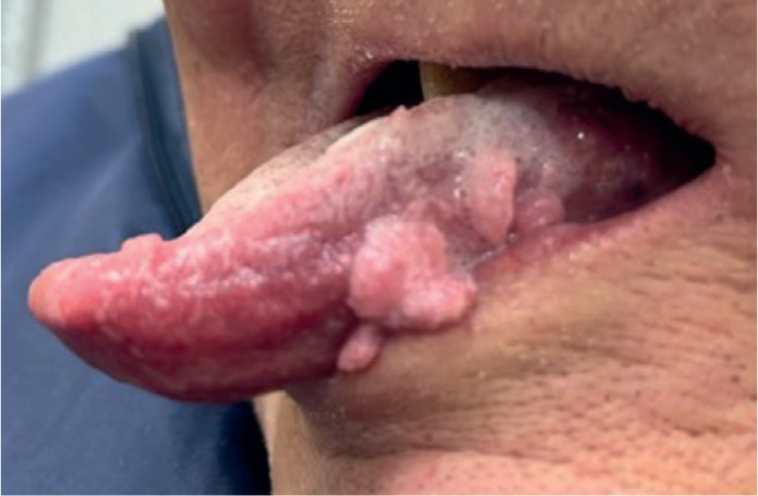
Excisional biopsies of these lesions and subsequent histopathological evaluation revealed multiple papillomatous lesions suggestive of a viral aetiology. Genotype testing confirmed human papilloma virus (HPV), X09/11 presence (low risk). Unfortunately, this patient attended again in October 2021 with recurrence of lesions and further excision of these lumps was undertaken. The results confirmed the earlier diagnosis with no sinister changes. He was seen again in November 2022 with further recurrence of lesions in the same sites noted earlier. The patient was referred to the infectious diseases team for further assessment and management of these morbid recurrent lesions.
Oral squamous papillomas are benign epithelial neoplasms arising from the stratified epithelium of the oral cavity. They are thought to be associated with HPV6 or HPV11.1 Commonly, oral squamous papillomas present as small painless exophytic projections that have a high re-occurrence rate. Risk factors include being male, having multiple sexual partners, not using protective barrier methods during oral sex, smoking and alcohol. There have been reports of malignant transformation of oral squamous papillomas; however, there has yet to be conclusive research on this.2 There are many syndromes associated with multiple oral papillomas, including dermal hypoplasia syndrome, acrodermatitis enteropatica, Cowden syndrome, nevus unius lateralis, Costello syndrome and Down syndrome.3
Management initially includes patient education and preventive advice. Preventive measures include practising safe sex, asking partners about their previous history regarding sexually transmitted infections and being regularly tested for these. It is also important for patients to note any changes in their mouths and to visit the dentist at regular intervals. The national vaccination programme in the UK recommends HPV vaccination at the age of 12 years, and it available free up to the age of 25.
Indications for removing lesions include for cosmetic or functional purposes. The most common treatment option would be surgical excision (with no margin); however, other less invasive options include laser ablation, electrosurgery and cryotherapy. Literature shows similar rates of recurrence for surgical excision and CO2 laser ablation (10% and 14%, respectively).4