A MEDLINE search early in 2015 revealed more than 250,000 papers on head and neck cancer; over 100,000 on oral cancer; and over 60,000 on mouth cancer. Not all publications contain robust evidence. We endeavour to encapsulate the most important of the latest information and advances now employed in practice, in a form comprehensible to healthcare workers, patients and their carers. This series offers the primary care dental team in particular, an overview of the aetiopathogenesis, prevention, diagnosis and multidisciplinary care of mouth cancer, the functional and psychosocial implications, and minimization of the impact on the quality of life of patient and family.
Clinical Relevance: This article offers the dental team an overview of surgery for the treatment of mouth cancer.
Article
Nicholas Kalavrezos Crispian Scully
If mouth cancer can be diagnosed at an early stage, when the lesions are small, treatment is generally less complicated and more effective. The treatment requires a multispecialty approach co-ordinated among surgeons, radiation oncologists, medical oncologists and others. Since these cancers are uncommon, patients with mouth cancer are usually treated in a specialist hospital under a multidisciplinary team (MDT) where oral squamous cell carcinoma (OSCC) should be staged according to the TNM (Tumour, Node, Metastases) classification (Article 9), since this classification relates well to overall survival rate (ie the earlier the tumour stage, the better the prognosis and the less complicated and mutilating is treatment). This article expands upon Article 9.
What are the current main treatments for mouth cancer?
Surgery and radiation are the only definitive treatment modalities for both early and locally advanced mouth cancer. Surgical resection, wherein the tumour is completely removed with uninvolved resection margins, is challenging and can involve sacrificing critical structures. Radiation, when used as definitive therapy, circumvents this difficulty, but often produces significant acute and late toxicities (Article 11). Chemotherapy alone is not a curative therapeutic modality, but may improve outcomes when used in conjunction with radiation (chemo-RT) for locally advanced disease (Article 12).
Lip cancer
In general, lip cancer is treated either surgically or with brachytherapy, a form of local radiotherapy (RT).
Intra-oral cancers
In general, intra-oral cancers are treated as follows:
T1 oral tumours are treated mainly as a local disease, mostly with surgery;
T2 to T4 tumours are treated largely by surgery and/or chemo-RT to control the primary tumour and metastases in the draining cervical lymph nodes (with a considered balance between survival benefit and the quality of life).
- T2 tumours are generally managed surgically. The potential risk of spread to the regional lymph nodes in the neck has to be considered. Surgery may involve the removal of the neck lymph nodes which are at risk en bloc if possible, with the tumour at the primary site in the mouth. Thus, for many patients, tumour excision and selective neck dissection may be the single treatment modality while, on selected cases, depending upon histopathological parameters (cancer-involved lymph nodes, clearance of oral tumour), external beam radiotherapy post-operatively is indicated. A small number of patients with tumours of the lateral margin of the tongue who may not be fit for radical surgery or a full course of radiotherapy may be treated with external beam (40 Gy) plus radioactive iridium implants (25–30 Gy).
- T3 tumours are generally treated by surgery followed by radiotherapy alone if there is no lymph node involvement. If there is extracapsular spread or multiple lymph node involvement then post-operative combined chemoradiotherapy is necessary. For many such patients, the treatment is often surgery (tumour excision with neck dissection), together with radio- or chemo-radiotherapy.
- T4 tumours may be treated with curative intent by surgery followed by chemo-radiotherapy along the lines mentioned above. Chemo-radiotherapy alone may be used in oropharyngeal tumours (organ preservation treatment) but a 2010 Cochrane review found that adding chemotherapy to surgery or radiotherapy for oropharyngeal cancer can work better than one of these treatments alone.
- Surgery may be reserved for recurrent disease after chemo-radiotherapy (‘salvage’ surgery).
- When the tumour is deemed untreatable with curative intent then a course of radiotherapy or chemotherapy alone may be used in the palliative setting.
Mouth cancer currently is thus treated largely by surgery and/or irradiation (Table 1), although few unequivocal controlled trials of any treatment modalities have been conducted. Targeted (biological) therapy is discussed in Article 12.
Treatments for mouth cancer
Beneficial
Surgery
Radiotherapy
Likely to be beneficial
Chemotherapy
Robotic surgery (for oropharyngeal tumours)
Conformal radiotherapy
Emergent treatments
Targeted therapies
What will the patient and family wish to know about surgery?
Surgery is a common treatment for mouth cancers and works very well for early cancers (Figure 1). The underlying principle is that surgical resection aims to achieve complete, microscopic clearance of the tumour with the appropriate safety margin (typically >1 cm), according to the type, site and stage of cancer.
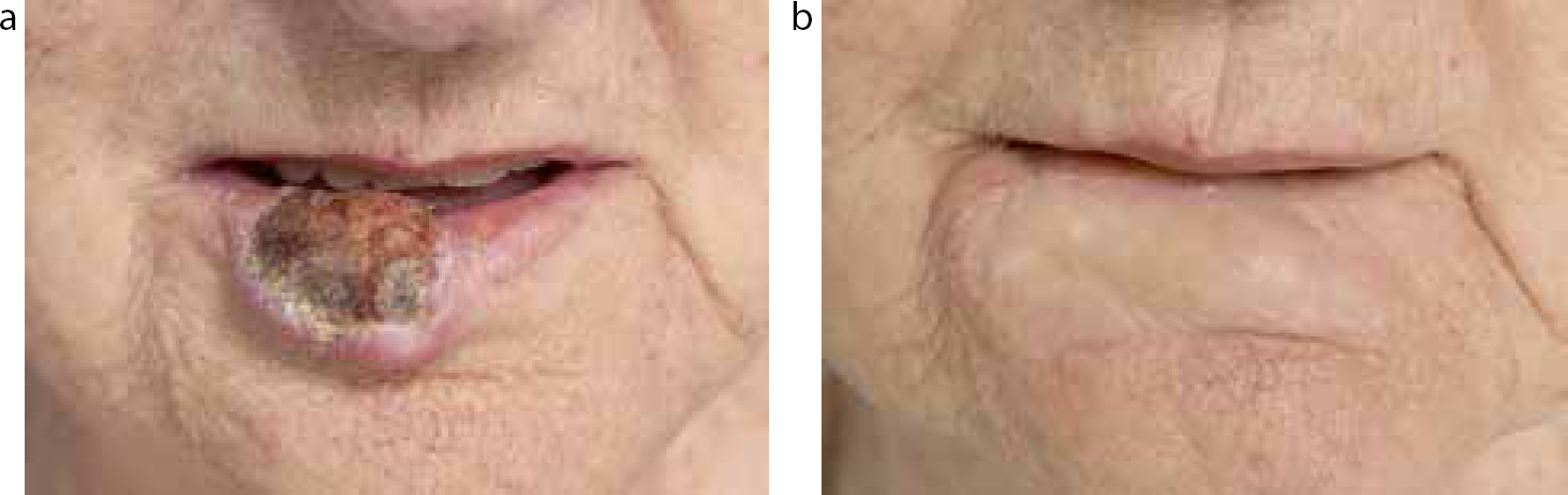
Figure 1.
(a) Lip cancer. (b) Lip cancer after surgical treatment. Lip carcinoma before (a) and after (b) excision and repair with a radial forearm skin flap.
Even larger tumours can be resected with acceptable results (Figure 2).
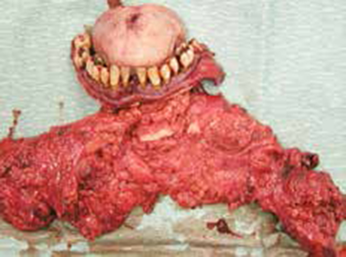
Figure 2. Advanced intra-oral tumour before (a) and after total glossectomy and reconstruction with a perforator antero-lateral thigh flap (b).
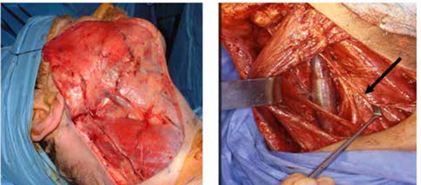
The goal of surgery then is to remove the primary tumour together with a margin of clinically normal tissue to ensure complete excision of malignant tissue and excision of any cancer-involved lymph nodes (Figures 3 and 4). A full histological examination can then be performed, for staging purposes and to help predict the prognosis and the need for adjuvant radio-or other therapy. Surgery usually provides a one-stage definitive procedure, from which the patient normally recovers within 10–14 days.
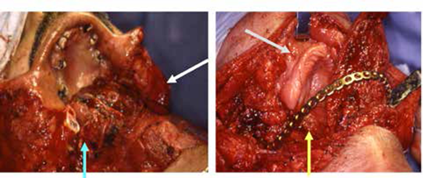
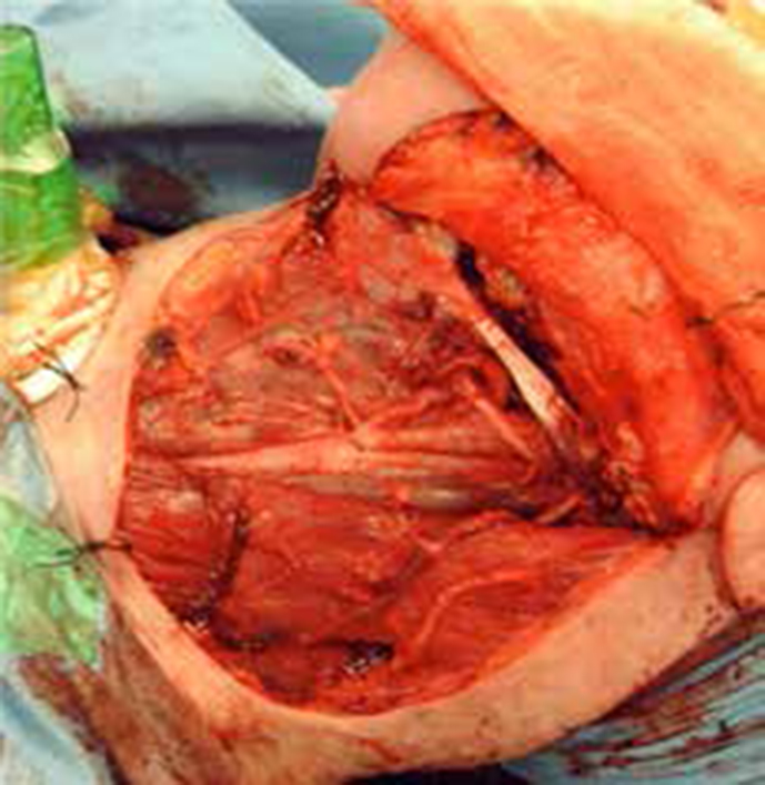
Figure 3. Defect following excision of a 'through and through' cancer of the lower third of the face (arrows) including 'angle to angle' mandibulectomy (a) and (b) titanium plate placement (arrows) prior to composite free flap reconstruction.Figure 4. Neck dissection: (a) skin flap elevated and (b) dissection of deeper neck structures. Arrow indicating the course of the accessory cranial nerve.
The size and the volume of the defect in the mouth will dictate the need for reconstruction:
Small defects of the tongue and floor of the mouth may be left to heal with no reconstruction (‘healing by second intention’);
For slightly larger or wider defects, skin grafts or flaps (grafts which maintain their own blood supply) may be used. Local flaps (eg nasolabial flaps) provide thin, reliable suitable flaps (Figure 5).
Figure 5. Skin graft to repair defect after cancer excision in lichen planus.
Large defects are usually treated with distant flaps (‘free flaps’). Defects of excised T3 and T4 tumours may require indeed sizeable tissue reconstruction. To attach the tissue the surgeon uses microsurgery to join small blood vessels in the neck to those in a new piece of body tissue. Such ‘free flaps’ are the ‘gold standard’ for these defects, taking tissues together with their vascular tree from another part of the body and bringing them into the oral cavity for reconstruction. The blood supply to the flap is then restored by joining the vessels of the flap with recipient vessels in the neck. A neck dissection allows the surgeon to connect to the neck blood vessels. If the cancer has spread to neck lymph nodes, the surgeon is likely to remove all the nodes on one or both sides of the neck, and may also remove other structures (neck dissection).
Surgery is usually under a general anaesthetic, usually with scalpel removal of tissue. Transoral laser microsurgery (TLM) is sometimes used to treat smaller cancers on the lip, mouth or throat when reconstruction is not necessary. Photodynamic therapy (PDT) uses a combination of laser light and a light-sensitive drug to destroy abnormal dysplastic cells and has occasional applications to treat large surfaces of potentially malignant disorders (PMDs) or very small, early cancers (microinvasive carcinomas).
If the cancer has spread to neck lymph nodes, the surgeon is likely to remove part or all the nodes on one or both sides of the neck, depending on the extent of the nodal spread (neck dissection). The types of neck dissection are:
Radical Neck Dissection (RND) – removal of all ipsilateral cervical lymph node groups from levels I through V, together with spinal accessory nerve (SAN), sternocleidomastoid muscle (SCM), and internal jugular vein (IJV).
Modified Radical Neck Dissection (MRND) – removal of all lymph node groups routinely removed in a RND, but with preservation of one or more nonlymphatic structures (spinal accessory nerve, sternomastoid muscle, inferior jugular vein).
Selective Neck Dissection (SND) (together with the use of parentheses to denote the levels or sublevels removed) – cervical lymphadenectomy with preservation of one or more lymph node groups that are routinely removed in a RND. Thus, for OSCC, SND (I–III) is commonly performed.
Extended Neck Dissection – removal of one or more additional lymph node groups or non-lymphatic structures, or both, not encompassed by the RND.
The concept of sentinel lymph node (SLN) biopsy has gradually been established in the management of early oral cancer to establish whether neck dissection is required.
Modern reconstructive techniques can produce very good orofacial aesthetics and function. Reconstruction is tailored to the patient's ability to cope with the long operation required (several hours) and the risk of significant morbidity.
Surgery involving the jaw, mouth, throat or tongue will postoperatively initially make eating and swallowing difficult. After such operations, patients are usually fed via a tube into the stomach for liquid feeds. This can be a nasogastric (NG) tube via the nose. If there is likely to be a lot of swelling and difficulty in tongue movement for considerable time, and if post-operative radiotherapy is anticipated, a tube put directly into the stomach as a percutaneous endoscopic gastrostomy (PEG) or, in some cases of limited mouth opening, a radiologically inserted gastrostomy (RIG) may be best.
What is needed before surgical treatment?
The patient is prepared for treatment particularly in terms of their understanding and informed consent. They are also prepared psychologically for the procedure(s) and likely outcomes, and for treatment-associated procedures such as general anaesthesia, potential blood loss and ability to metabolize drugs.
Oral health
Potential dental or oral problems should be addressed pre-operatively, to minimize or avoid later complications which may impact quality of life (Article 13). Patients are usually advised to have a complete dental/oral check-up and to have any work needed completed before treatment begins. If radiotherapy is also planned, unsaveable teeth may best be removed before treatment (Article 11).
Speech, swallowing and eating
The dietitian and/or the speech and language therapist should advise the patient before surgery or radiotherapy. The oncologist may advise a PEG (percutaneous endoscopic gastrostomy) tube or RIG (radiologically inserted gastrostomy) tube.
Alcohol
Avoiding alcohol, particularly spirits, may help make treatment more effective and reduce adverse effects.
Smoking
Stopping smoking will increase the chances of treatment being effective. Continuing to smoke increases treatment adverse effects and the risk of recurrence of the tumour. Smoking also increases the risk of developing a second primary tumour.
What procedures can be anticipated during and after the operation?
Surgery usually involves a hospital stay for days or for up to a few weeks, depending mainly on the extent of the surgery and reconstruction. Often, immediately after the operation, time in intensive care is indicated, where the patient will receive intensive nursing care, may need catheters, drips, drains and/or tubes and will be closely monitored.
Drains and dressings
Plastic drainage tubes are used to collect wound exudates for 2–7 days. Patients may need a catheter to drain urine into a collecting bag.
Drips
Post-operative swelling may well make eating and drinking uncomfortable so initially an intravenous drip is used for fluids in combination with a feeding tube.
Feeding tubes
Feeding tubes used are either:
A gastrostomy tube (PEG or RIG tube);
A nasogastric (NG) tube, passed up the nose, down the pharynx and oesophagus and into the stomach.
When the patient can eat and swallow safely, the feeding tube can be removed.
The dietitian will often prescribe a high-protein, high-calorie, liquid food to be given via the tube.
Tracheostomy tubes
If surgery is likely to embarrass the airway, the surgeon will usually create a prophylactic temporary tracheostomy or stoma. The opening, made in the lower front of the neck by removing a small portion of the front part of the windpipe, is kept patent by a small plastic tube.
Whilst with a tracheostomy, speaking with the tube in place is impossible. When swelling subsides (after about 5–7 days) and breathing is normal, the tracheostomy tube is removed, the opening then heals spontaneously and the speech is gradually restored.
What post-operative sequelae can be anticipated?
Appearance
Surgery that involves the jaw, tongue, mouth, lips and throat may well change the appearance at least temporarily. Oedema, haematomas, tissue loss or scars may be responsible.
Post-operative oedema depends largely on the extent of surgery but can be reduced by:
Minimizing the trauma, duration and extent of the operation;
Using corticosteroids systemically (eg dexamethasone);
Nursing patient in head-up position.
Facial swelling is invariable immediately post-operatively but improves with time, over a few months.
Modern surgical techniques and reconstructive surgery aim to replace lost tissue and cause little scarring. Surgeons often rebuild lost tissue using bone grafts or bone flaps from other parts. They endeavour to position scars inside facial creases. Scars are usually red or dark initially but gradually fade over time and are far less visible.
Surgery to the lips is much harder to hide. Make-up (camouflage make-up) with different colours for all skin tones can cover problem areas. There are also other ways to hide changes including wearing:
Scarves to hide any neck scars;
Hats to divert attention away from the face.
Pain
Some post-operative pain or discomfort for a few days is inevitable. Severe pain may need to be controlled by opioids. Cancer pain itself may be associated with tissue invasion, or cancer therapies, and is best managed pharmacologically with oral, rectal, transdermal, subcutaneous, or intravenous administration of NSAIDs, opioids and adjuvant drugs. Failing these, interventions that may be required include:
Peripheral nerve blocks
- maxillary
- mandibular
- glossopharyngeal;
Ganglion blocks
- sphenopalatine
- trigeminal
- stellate;
Central neuraxial techniques
- intraventricular opiates
- intrathecal pump.
Sensation
Surgery may affect the sensation in the mouth, face, neck or shoulders, and some areas may feel hypoaesthetic. It is common to have skin numbness around scars which may take several months to return to normal sensation.
Speech
Many operations can affect speech. Change in speech is most commonly associated with surgical intervention to the oral cavity or larynx (laryngectomy). For some people this is hardly noticeable, but for others speech may be temporarily or permanently altered. A speech and language therapist will be able to help the patient adapt.
Swallowing
Eating and swallowing difficulties are not uncommon early postoperatively and can be long-term, especially if there is loss of anatomical structures during surgery, reducing the range of tongue and jaw movements, diminishing pharyngeal wall or laryngeal movement, and thus increasing the risk of aspiration. Some patients have persistent longer-term effects, such as laryngeal oedema, causing dysphagia and aspiration.
Many patients require altered diet textures, but patients with advanced tumours who experience dysphagia may require gastrostomy tube-feeding. A speech and language therapist can advise about the safest and easiest types of food to have, and teach mouth and jaw exercises to improve swallowing. Patients should be weighed regularly. Dietitians should be consulted. Nutrition may need to be supplemented:
Enterally by an oro-gastric tube or NG, PEG or RIG. Continuous infusion of a liquid feed is preferred, since intermittent feeding may cause diarrhoea.
Parenterally, ie via an IV catheter (central line) in the subclavian or jugular veins (total parenteral nutrition; TPN). A central line is a silicone rubber skin-tunnelled central venous catheter (eg Hickman® or Groshong®). If this is necessary, urea, electrolytes and liver function must then be monitored.
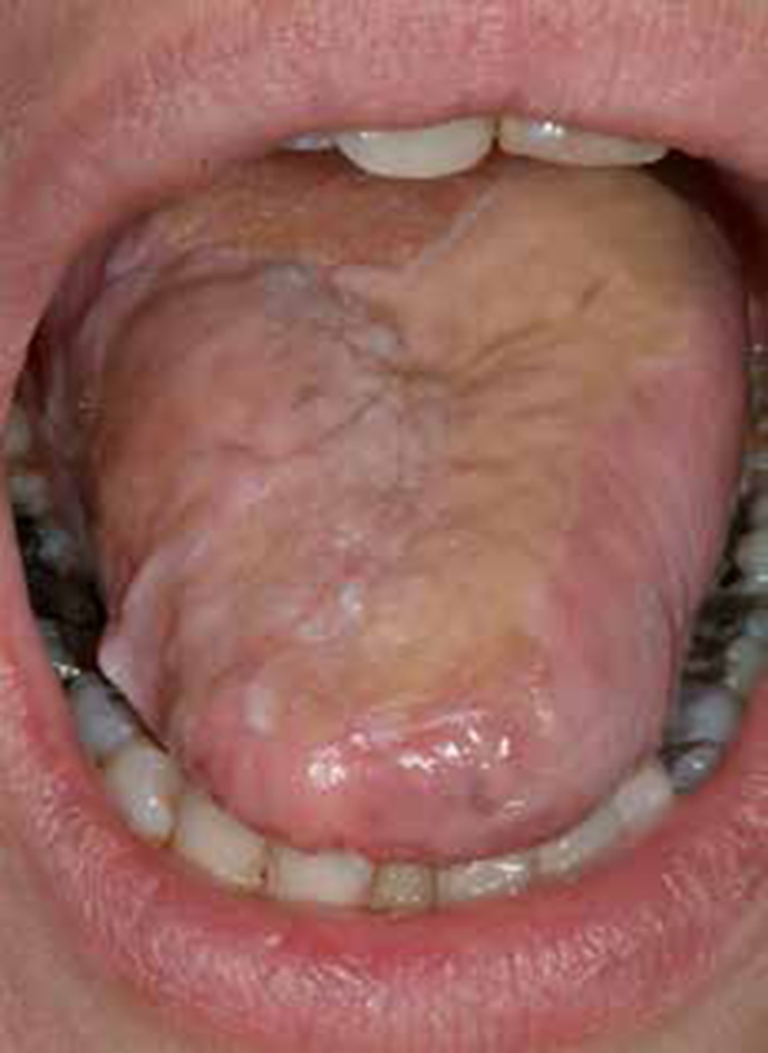
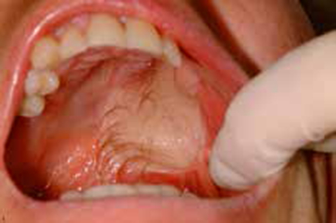
Trismus
Some operations, especially to the posterior mouth and throat, can restrict jaw opening (Figure 6). Trismus can also be reduced by minimizing trauma. Physiotherapy (eg using a Therabite) can help prevent trismus from becoming permanent.
Figure 6. Trismus after tongue cancer surgery. Note lateral tongue reconstruction with free flap.
What surgical treatments are now used for mouth cancer?
The principles of most cancer surgery are to:
Remove the whole tumour (resection): ablative surgery excises the cancer with at least a 2 cm margin of clinically normal tissue, to try and ensure that no tumour remains. Radical neck dissection, for the same reason, also removes the internal jugular vein, lymphatics and accessory nerve, as well as the cervical sensory nerves and the sternomastoid, omohyoid and often digastric muscles.
Retain as much normal anatomy and function as possible. ‘Functional’ neck dissection, preserving the above vital structures, is gaining in popularity; moderate-dose radiotherapy is, therefore, sometimes used to ‘sterilize’ such necks of residual cancer cells.
Permit normal breathing – tracheostomy may be necessary.
Support feeding also – often via a tube.
Reconstruct to achieve acceptable function and cosmetics. From both the operator's and the patient's and family's viewpoint, reconstruction is best performed at the time of initial surgery, but must be tailored to the patient's ability to cope with a long operation (up to 10 hours or more) and the risk of significant morbidity.
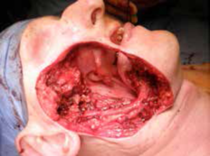
In summary, surgery provides a one-stage definitive procedure, from which the patient normally recovers within 10–14 days, and aims to remove the primary tumour together with a margin of clinically normal (‘uninvolved’) tissue and involved lymph nodes (nodes containing metastases) to ensure complete excision of all malignant tissue (Figures 7 and 8).
Figure 7. Resection.Figure 8. Bilateral neck dissection en bloc with rim resection of the mandible and sub-total glossectomy for a large floor of mouth tumour with neck metastases.
The type of surgery depends on the size and position of the cancer, and whether it has spread. A full histological examination can then be performed for staging and to help predict prognosis and any need for adjuvant radiotherapy. The surgeon will endeavour to minimize changes to functions, such as speech and swallowing, and to rebuild the area using tissue, skin or bone taken from somewhere else in the body. Modern reconstructive techniques can produce good orofacial aesthetics and function.
Surgery to the neck lymph nodes (neck dissection) is indicated if cancer has spread to neck lymph nodes and also sometimes for people with no signs of cancer in the lymph nodes, if the primary tumour is larger than 4 mm. A neck dissection will also be required if the surgeons plan to rebuild (reconstruct) part of the mouth or throat with tissue taken from another part of the body (a free flap), allowing them to reach the blood vessels in the neck (Figure 9).
Figure 9. Neck dissection.
Reconstruction is tailored to the patient's ability to cope with a long operation and the risk of significant morbidity. To attach the tissue the surgeon uses microsurgery to join blood vessels in the neck to blood vessels in the new piece of body tissue. Reconstruction of defects is usually with tissue brought into the region using split skin grafts or flaps. Intra-orally, skin flaps may bear hair (Figure 10).
Figure 10. Skin flap, with hair, over maxillary resection defect.
Local flaps (eg nasolabial flaps) provide thin, reliable flaps suitable for repairing small soft tissue defects. Distant flaps required to repair larger defects include the following:
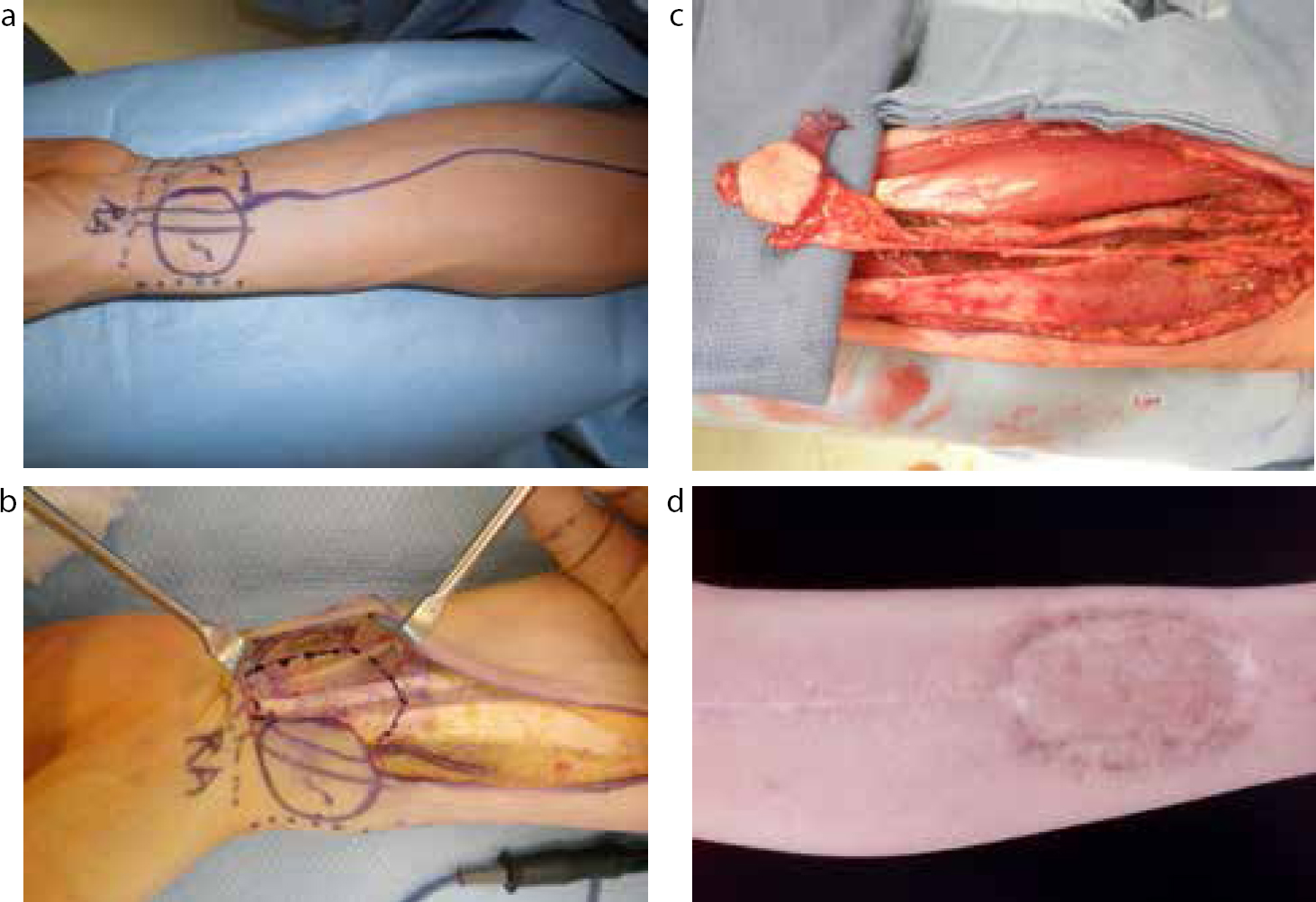
Free flaps: microvascular surgery facilitates excellent reconstruction in a single operation using, eg forearm flaps based on radial vessels (Figure 11), particularly useful to replace soft tissue, or those based on the fibula when bone is required.
Pedicle flaps: myocutaneous or osteomyocutaneous flaps based on a feeding vessel to muscle and perforators to the skin paddle (eg flaps based on the pectoralis major, latissimus dorsi, or trapezius muscle) may be used in a one-stage operation to replace skin and, because they also contain muscle, they have adequate bulk to repair defects, while in extreme cases may be used to import bone (usually rib).
Hard tissue reconstruction ideally is performed at the time of tumour resection. The ideal bone reconstruction is provided with a bone flap – the fibula bone is the most common site in use. Bone flap from the hip (deep circumflex iliac artery [DCIA] flap) is preferable in dentate patients as the depth of the hip flap corresponds well to the depth of the mandible. There are other sites as well, such as the scapula or the radial bone of the forearm, with limited use in specific cases. The more traditional non-vascularized bone grafts from the iliac crest or rib may survive poorly if contaminated or if the vascularity is impaired after irradiation and hence are not in use regularly. Modern dental rehabilitation may include dental implants that can be ideally inserted at that point to carry a prosthesis. The benefits of bone grafting for maxillary defects are less certain, and reconstruction is usually with an obturator (bung), which has the advantage that the cavity can be readily inspected.
Figure 11.
(a) Design of a radial forearm free flap. (b) Flap harvesting. Early stage of dissection. (c). Flap harvesting. Completion of dissection – flap attached to its vascular pedicle. (d) Completion of healing following skin graft repair on the forearm donor site.
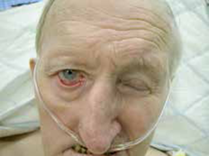
After a comprehensive ‘radical’ neck dissection, nerve control of facial and neck muscles can be damaged. Facial nerve damage can lead to ispilateral weakness and a crooked smile (Figure 12), but over a few months this may return to normal.
Figure 12. Facial nerve palsy and ectropion.
Spinal accessory nerve damage impairs shoulder movement, such as shrugging, and the shoulder misaligns; without exercise the range of shoulder movements becomes limited and painful. The trapezius muscle is responsible for the upward rotation of the shoulder during abduction and flexion and is the major stabilizer for the scapula. Patients with their spinal accessory nerve and sternocleidomastoid muscle preserved appear to have fewer difficulties with work-related, leisure-related and daily activities.
Other nerves also at risk in surgery include mainly the vagus, lingual, hypoglossal nerves and marginal mandibular branches of the facial nerves. Transection of the vagus nerve can result in intra-operative cardiac problems. Transient neuropraxia to the vagus phrenic branch often manifests with changes on plain radiography but, if a severe lung problem exists, respiration may be compromised. Bilateral phrenic nerve palsies may necessitate prolonged mechanical ventilation. The sympathetic trunk is also at risk of injury, but a subsequent Horner syndrome is rare.
‘Modified’ radical or ‘selective’ neck dissections (modified to preserve the jugular vessels, sternomastoid muscle, and accessory nerve, while ensuring complete removal of involved nodes) have thus gained popularity. Nevertheless, any type of neck dissection induces fibrosis in the neck, with some stiffness or constriction and pain, and overall loss of function. A physiotherapist can help.
Trans-oral laser surgery
Trans-oral surgery avoids external incisions and scars and is sometimes used to treat small lip, mouth or throat cancers.
Mohs surgery
Some small lip cancers may be treated with micrographic or Mohs surgery, removing the cancer in thin layers, until no cancer cells are histopathologically seen in the tissue.
Photodynamic therapy (PDT)
Photodynamic therapy uses a combination of laser light and a light-sensitive drug sometimes used to treat PMD and very small, early skin cancers (not approved/established for mucosal disease).
More serious complications of surgery
Other complications of surgery may include:
Flap failure;
Infections;
Pneumothorax;
Recurrences;
Salivary fistulae;
Seroma;
Thoracic duct leakage (chylorrhoea);
Vascular complications.
Flap failure
Free flap success rates typically exceed 95%. Micro-surgical flap transfers are, however, usually lengthy, technically demanding and therefore physiologically traumatic operations capable of potentially major complications, such as pneumonia or even myocardial infarction and stroke. Contra-indications for flap procedures therefore include significant co-morbidities. Advanced age is not necessarily a contra-indication, nor is a well-controlled, severe medical co-morbidity.
Post-operative staff must be familiar with particular care of pain, hydration, urine output, body temperature and flap vitality. Pain should be well controlled, also to prevent anxiety.
Flap vitality should be monitored by regular and frequent clinical examination for the first 48 h (flap colour, skin turgor, capillary refill, flap temperature, needle scratch test – which should result in surface oozing of bright red blood drops). In buried flaps with no skin part to monitor, vitality monitoring can be with implantable Doppler ultrasound, pulse oximetry or near-infrared spectroscopy.
Flap failure is mainly because of vascular occlusion, usually venous thrombosis. Anticoagulation (eg Dextran 40, heparin or aspirin) may be needed to avoid blood clotting in the flap for the early post-operative days. Most flap failures occur within the first 48 hours. With early recognition and intervention, salvage is often possible.
Flap vascularity, if suspected to be compromised, is managed by repositioning the patient to relieve any pedicle obstruction, and compressive dressings or tight sutures removed. Haematoma and hypotension must be excluded. If these simple manoeuvres fail, the flap must be re-explored immediately.
Infection
Wound infection can produce significant pain, swelling, restricted function and poor cosmetic results and will likely prolong hospitalization. Infection under neck flaps is particularly dangerous as the internal carotid artery may be eroded (see ‘carotid blow-out’).
Patients who smoke, have advanced cancer or require free flap reconstruction have the greatest risk of surgical wound infection. This can be minimized by:
Pre-assessment so as to enable same day admission;
Prophylactic antibiotics are indicated for clean-contaminated wounds, but not for clean wounds. Broad spectrum prophylactic antibiotics should be used on the advice of a local microbiologist. The first dose should be given at induction and continued for up to 24 hours; longer courses may increase nosocomial infection.
The diagnosis of wound infection is usually made when, at about 3–7 days after operation, the wound appears inflamed, swollen and tender. There may be pus and pyrexia. Infections may settle spontaneously within a few days, but pus or a swab should be taken to identify the organism and test sensitivity to antibiotics. If the wound is not draining but is fluctuant, one or more sutures should be removed, sinus forceps inserted and gently opened to allow pus drainage.
Pneumothorax
Pneumothorax may occur when surgery is low in the neck, particularly if the lung apex is high. Pleural tears should be closed immediately and their integrity tested by hyper-inflating the lung, placing the patient in the Trendelenburg position and irrigating the area to observe bubbles. Imaging can determine the need for open drainage.
Recurrences
When mouth cancer is fatal, it almost always is either because of failure to control the primary tumour or because of nodal metastases. Cancer centres receive many patients with advanced disease, and operations may thus fail to remove the tumour completely, resulting in a poor outcome and recurrence. Death resulting from distant metastasis, however, is unusual.
Salivary fistulae
Rarely, a salivary fistula or sialocele, due to leakage of saliva, can cause saliva to leak through the skin. Sialocele is most common over the area of the parotid following partial parotidectomy. Sialoceles usually settle spontaneously with time but may need to be managed with aspiration and the use of anticholinergic agents (eg hyoscine transdermal patch) or botulinum toxoid.
Seroma
Seroma is the accumulation of serous fluid under large flaps, and appears as a large fluctuant swelling that is neither warm nor tender (in contrast to wound infection). Seromas may have to be drained several times using large syringes and needles or cannulae. Moderate pressure dressings are then often applied.
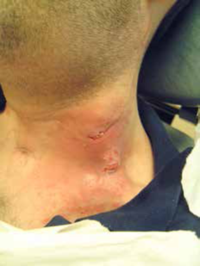
Thoracic duct leakage (chylorrhoea)
Injury to the thoracic duct during neck dissection is the most important cause of lymph (chyle) leakage (chylorrhoea)(Figure 13). Etilefrine hydrochloride, which has alpha-adrenomimetic action, is available in some countries for the management of postoperative chyle leak. Octreotide, a long-acting synthetic analogue of somatostatin, may be also effective. For persistent leaks a thoracic duct ligation or embolization may be required.
Figure 13. Chyle leakage.
Vascular complications
Vascular complications are potentially the most serious. Surgical sacrifice of the common or internal carotid arteries can produce some of the most serious complications seen. When internal carotid artery resection is planned, a balloon-test occlusion with hypotensive challenge reliably assesses the risk.
In bilateral neck dissections, where one internal jugular vein (IJV) is preserved, post-operative imaging shows thrombosis in up to 30%. If both IJVs are to be transected, then conduits in the external venous system should be preserved wherever possible. Bilateral internal jugular vein (IJV) ligation may produce raised intra-cranial pressure (ICP), along with secondary hypertension (Cushing reflex). The rise in ICP commonly requires aggressive treatment – hyperventilation, fluid restriction, corticosteroids and mannitol.
Carotid blow-out is rupture of the extra-cranial carotid arteries or their major branches, an uncommon but devastating complication, associated with over 60% morbidity and 50% mortality. Predisposing factors include prior radiation therapy, extensive surgery, wound breakdown, local infection, tumour recurrence and pharyngocutaneous fistulae. If it is anticipated that the carotid artery will be exposed, the vessels should be covered, eg dermal graft, fascia lata or levator scapulae muscle flap. If impending blow-out is suspected (eg as suggested by a sentinel bleed), stent-grafts may be preferred over emergency open carotid artery ligation (which may well be complicated by neurological sequelae, such as hemiplegia, hemi-anaesthesia, aphasia and dysarthria).
Air embolism is rare but potentially lethal and most commonly follows damage to the IVJ during surgery. Large air emboli produce sudden falls in end-tidal carbon dioxide and blood pressure and, in severe cases, air can be aspirated from the right side of the heart. A pre-cordial Doppler probe may detect a characteristic murmur. Pressure should be applied to the affected vein and the patient placed in the Trendelenburg head down position and rotated to the left. Hyperbaric oxygen is the ultimate and effective treatment.
Summary
Advantages of surgery
Advantages of surgery include the facts that the diseased tissue is removed and adverse effects of radiotherapy avoided.
Disadvantages of surgery
Disadvantages primarily are peri-operative mortality and morbidity, but modern techniques have significantly decreased these risks, as well as the aesthetic and functional defects.