Van't Spijker A, Kreulen C, Bartlett D Prevalence of tooth wear in adults. Int J Prosth. 2009; 22:35-42
O'Brien MLondon: HMSO; 1994
Mehta SB, Banerji S, Millar BJ, Saures-Fieto JM Current concepts in tooth wear managements. Part 1: Assessment, treatment planning and strategies for the prevention and passive monitoring of tooth wear. Br Dent J. 2012; 212:(1)17-27
Van't Spijker A, Kreulen C, Bartlett D Prevalence of tooth wear in adults. Int J Prosthodont. 2009; 22:35-42
Mehta SB, Banerji S, Millar BJ, Saures-Fieto JM Current concepts in tooth wear management. Part 4. An overview of the restorative techniques and materials commonly applied for the management of tooth wear. Br Dent J. 2012; 212:(4)169-177
Mehta SB, Banerji S, Millar BJ, Saures-Feito JM Current concepts in tooth wear management. Part 2 Active restorative care 1: The management of localised tooth wear. Br Dent J. 2012; 212:(2)73-82
Mehta SB, Banerji S, Millar BJ, Saures-Fieto JM Current concepts in tooth wear management. Part 3 Active restorative care 2: The management of generalised tooth wear. Br Dent J. 2012; 212:(3)121-127
Kilpatrick N, Mahoney E Dental erosion: Part 2. The management of dental erosion. NZ Dent J. 2004; 2:42-47
Hemmings K, Darbar U, Vaughan S Tooth wear treated with direct composite at an increased vertical dimension; results at 30 months. J Prosthet Dent. 2000; 83:287-293
Redman C, Hemmings K, Good J The survival and clinical performance of resin based composite restorations used to treat localised anterior tooth wear. Br Dent J. 2003; 194:566-572
Poyser N, Porter R, Briggs P, Kelleher M Demolition experts: management of the parafunctional patient: 2. Restorative management strategies. Dent Update. 2007; 34:262-268
Dahl B, Krungstad O, Karlsen K An alternative treatment of cases with advanced localised attrition. J Oral Rehab. 1975; 2:209-214
Dahl B, Krungstad O Long term observations of an increased occlusal face height obtained by a combined orthodontic/prosthetic approach. J Oral Rehab. 1985; 12:173-170
Poyser N, Porter R, Briggs P, Chana H, Kelleher M The Dahl concept: past, present and future. Br Dent J. 2005; 198:669-676
Banerji S, Mehta SB, Kamran T, Kalakonda M, Millar BJ A multi-centred clinical audit to describe the efficacy of direct supra-coronal splinting – a minimally invasive approach to the management of cracked tooth syndrome. J Dent. 2014; 42:862-887
Smales R, Berekally T Long term survival of direct and indirect restorations placed for the treatment of advanced tooth wear. Eur J Prosthodont Rest Dent. 2007; 15:2-6
Gulamali AB, Hemmings KW, Tredwin CJ, Petrie A Survival analysis of composite Dahl restorations provided to manage localised anterior tooth wear (ten year follow-up). Br Dent J. 2011; 211:(4)
Briggs P, Djemal S, Chana H, Kelleher M Young adult patients with established dental erosion – what should be done?. Dent Update. 1998; 25:166-170
Saunders W, Saunders E Prevalence of peri-radicular periodontitis associated with crowned teeth in an adult Scottish subpopulation. Br Dent J. 1998; 185:137-140
Edelhoff D, Sorenssen J Tooth structure removal associated with various preparation designs for anterior teeth. J Prosthet Dent. 2002; 87:503-509
A guided, conservative approach for the management of localized mandibular anterior tooth wear Shamir B Mehta Selar Francis Subir Banerji Dental Update 2024 43:2, 707-709.
The successful management of the worn mandibular anterior dentition may present an awkward challenge to the dental operator. The purpose of this article is to describe a case report illustrating the use of a guided, three-dimensional protocol for the ultra-conservative and predictable restoration of the worn lower anterior dentition using direct resin composite. This technique utilizes information based on established biomechanical and occlusal principles to fabricate a diagnostic wax-up, which is duplicated in dental stone. This is used to prepare a vacuum-formed modified stent, assisting the clinician to place directly bonded resin composite restorations to restore the worn lower anterior dentition. The technique, described in 2012 and referred to as ‘injection moulding’ has the potential to offer optimal form, function and an aesthetic outcome in an efficient manner.
CPD/Clinical Relevance: This article aims to describe an alternative technique to simplify the processes involved with restoration of worn lower anterior teeth.
Article
The process of tooth wear usually has a multi-factorial aetiology.1 It is often difficult to isolate a single aetiological factor when a patient presents with pathological tooth wear. Individual aetiological factors may be subdivided into erosion, attrition, abrasion and abfraction.1 Numerous epidemiological studies have reported the condition of tooth wear to be prevalent amongst the general population, with rising incidence rates.2,3,4 With an ageing population in the developed world, with patients retaining their teeth into advanced years, coupled with lifestyle and habit changes amongst younger individuals, these figures are hardly surprising.
The successful management of a patient presenting with tooth wear relies on the attainment of an accurate patient history and a meticulous examination.1 Treatment provision must aim to identify and address the possible aetiological factors so as to prevent them from inflicting further damage upon the affected dental hard tissues, as well as upon any existing or planned dental restorations.1 In order to achieve predictability, a systematic approach should be applied when managing cases of tooth wear.
Localized anterior mandibular wear can sometimes be seen to occur amongst patients who have been prescribed antagonistic porcelain occluding surfaces (on the maxillary anterior teeth), particularly where adjustments have been made to accommodate the occlusion and the surfaces have been left unpolished.5
Active restorative intervention for cases presenting with tooth wear is usually indicated where there may be aesthetic concerns, symptoms of discomfort, difficulty with adequate function, an unstable occlusal scheme or a rate of tooth wear which may be of significant concern to either the clinician or patient which, if neglected, may ultimately result in exposure of the pulp chamber or, in extreme circumstances, loss of the tooth.6 A number of techniques and materials have been described in the literature for the successful management of pathological tooth wear.5,6,7
For the restoration of the worn anterior dentition, the use of directly bonded resin composite restorations has many potential merits. Amongst these are:8
Aesthetic acceptability;
Conservative: applied with minimal or zero intervention;
Well tolerated by pulpal tissues;
Minimally abrasive to antagonistic tissues;
An inexpensive material;
May be applied in one single visit;
Ease of repair and adjustment;
May serve as a diagnostic restoration to assess tolerance and adaptability to any altered occlusal and aesthetic changes.
However, a successful outcome with this material is reliant on not only a good quality and quantity of enamel for predictable resin bonding, but also a sound working knowledge of the fundamentals of occlusion, concepts in aesthetics and adhesive dentistry (including the use of the optimal bonding agents and available composite resin-based materials) as well as operator skill.5 The limitations of resin composite as a restorative material with problems such as bulk fracture, accelerated wear (compared to alloys and ceramics), discoloration, polymerization contraction, monitoring and regular maintenance should also be carefully explained to the patient.5
A number of studies have reported promising short-term success for the use of directly bonded resin composite restorations for the treatment of anterior tooth wear (both of the maxillary and mandibular variety), including, amongst those patients where the material has been carefully applied in supra-occlusion, utilizing the concept of Relative Axial Movement.9,10,11 The latter phenomenon is commonly referred to as the Dahl concept, and is based on a combination of condylar repositioning and intrusive and extrusive movements of the dento-alveolar segments.12,13,14 Evidence also exists to support the successive application of this concept in restorative dentistry, including other conditions beyond that of the treatment of localized tooth wear. However, careful case selection is critical.15
There is also evidence, albeit of a limited variety, to support the medium-and longer-term use of this material for this purpose.16,17 However, it would appear that failures could be anticipated to occur amongst certain cases with a higher level of frequency;17 these have been listed in Table 1.
The presence of antagonistic ceramic restorations
Lack of adequate posterior support
Lack of an evenly shared anterior guidance
Presence of Class II div 2 malocclusions (possibly due to increased shear strain and tensile forces)
Reliance on dentine bonding due to lack of good quality and quantity of tooth enamel
Use of micro-filled based resin materials (increased risk of bulk fracture)
Inadequate amounts of resin composite applied to worn surfaces
Placement of material beyond the physiological and functional adaptive envelope of the patient
Poor placement technique
Amongst patients who display parafunctional habits and a tendency towards bruxism
Resin composite may be applied in a ‘preventive manner’ used simply for the protection of vulnerable (or at risk) surfaces, as ‘intermediate composite restorations’ (ICR) until it is perhaps more feasible to plan the placement of more robust and/or complex definitive restorations.18 However, it is evident that direct resin composite materials must be placed at a minimal increment thickness of 1.5 to 2.0 mm in all areas of functional loading to ensure appropriate longevity, as mentioned by Poyser et al.11
Although the required level of inter-occlusal clearance may exist in the inter-cuspal position (ICP) or where there may be a marked discrepancy between the inter-cuspal position and the retruded contact position (RCP), for many cases space may need to be created by either:
Placing the material in supra-occlusion;
The process of subtractive tooth preparation;
By planning an overall increase in the patient's occlusal vertical dimension (OVD).7
Space management is crucial in cases where the wear is limited to the anterior segment. The ultimate goal of treating a patient with pathological tooth wear is to provide the space that is required for successful restorative intervention without a need for further removal of the already compromised tooth structures, which has obvious biological implications.19,20
Methods of direct composite resin application
A variety of techniques have been described in the contemporary literature, as a means for applying resin composite for the direct restoration of anterior tooth wear.5,6 These can be broadly divided into those advocating a ‘free-hand approach’, the use of a guiding ‘silicone key’ or the use of a ‘custom, thermoplastic template’. The latter two are reliant on the taking of study casts, mounted on a semi-adjustable articulator, followed by the fabrication of a diagnostic wax-up based on a carefully prescribed occlusal scheme and aesthetic prescription.
The use of a free-hand technique offers a cost-effective approach. However, it is highly reliant on the operator having a good knowledge of the concepts of functional occlusion, aesthetics and skill with resin placement. A failure to appreciate the dynamic occlusal scheme may culminate in premature restorative failure or possible consequences elsewhere in the dentition.7
The use of a silicone key derived from the wax-up provides the operator with a template to duplicate the occlusal form established extra-orally. Resin layering becomes a controlled process permitting the anatomical development of the desired morphology and colour by using a variety of shades which may be light cured in suitable increments. The silicone key also offers an important default, should there be a need to revise restorations in the future. The use of a translucent polyvinyl siloxane material (such as Memosil 2, Heraeus Kulzer, Hanau, Germany) also offers the potential to cure transversely through the matrix. However, difficulties with matrix placement, unwanted flexion of the matrix and the management of the inter-proximal areas may be factors to consider. In the lower arch, additional difficulties may be encountered with the use of a silicone key, such as the inability to attain adequate moisture control in the presence of a bulky, lingually placed key, interference with the tongue, access in particular where teeth may be retroclined or where there may be copious loss of dental hard tissues.
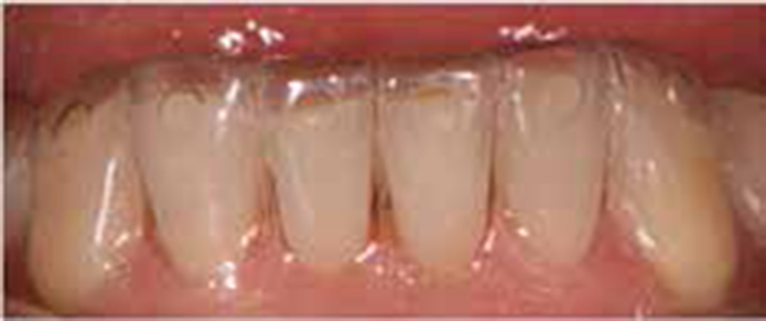
Figures 1 and 2 depict an example where a transparent silicone key has been used to restore worn lower anterior teeth using resin composite.
Figure 1. Use of a silicone index to plan resin additions to worn lower anterior teeth.Figure 2. Post-operative view, following the addition of direct resin composite, G-aenial Direct (GC Corp, Japan).
Use of an unmodified thermoplastic matrix has been described which involves the insertion of resin composite into the matrix, and then seating this following the preparation of the enamel and dentine for bonding and light curing in situ. The management of unwanted material excess (particularly in the inter-proximal areas), the inability to apply the material incrementally, the high polymerization exotherm and contraction, coupled with the tendency towards air entrapment within the material with potential subsequent voids, are clear disadvantages with this technique.5
However, the use of a ‘modified template’ to permit the ‘injection moulding of pre-warmed resin composite’ offers considerable advantages, and overcomes many of the concerns associated with the use of an unmodified template.
This technique has been described below as part of a case report.
Case presentation
A 46-year-old male patient complained of thermal sensitivity from his lower anterior teeth upon consumption of cold food and beverages and a lack of ability to chew his food properly, which had been noticed over the past year. The patient's medical history was unremarkable. However, his past dental history revealed the undertaking of numerous occlusal adjustments to the antagonistic dentition following the cementation of multiple unit maxillary anterior teeth restorations of an indirect, ceramic variety placed 15 years previously.
A careful extra-oral examination revealed that both the masseter and temporalis muscles displayed signs of muscular hypertrophy and tenderness on palpation. However, a normal range of mandibular motion was observed with the absence of any clicking, displacement or deviation of the mandible during opening and closing movements of the temporomandibular joint(s).
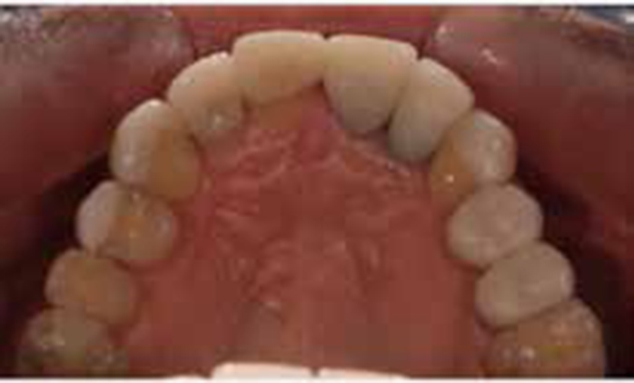
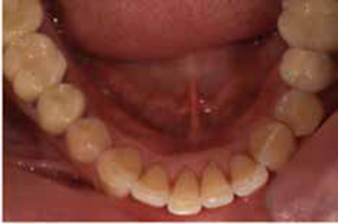
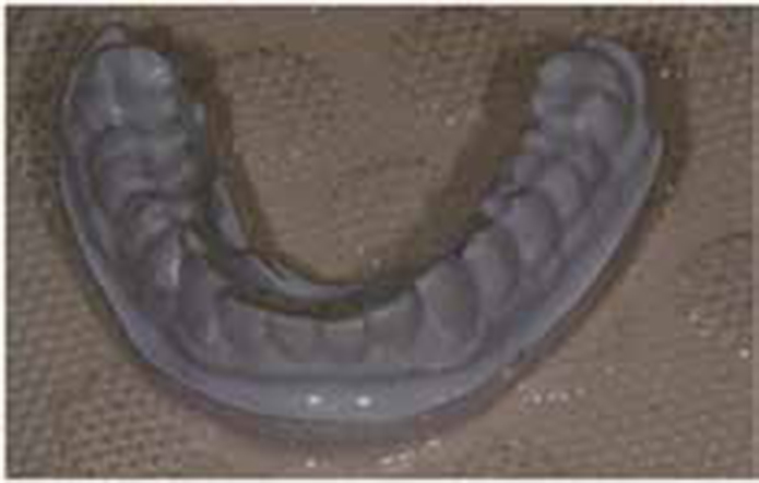
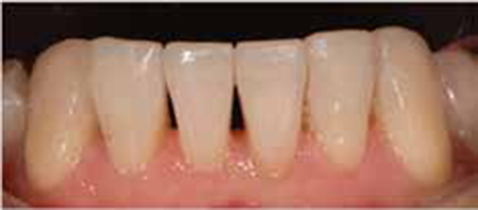
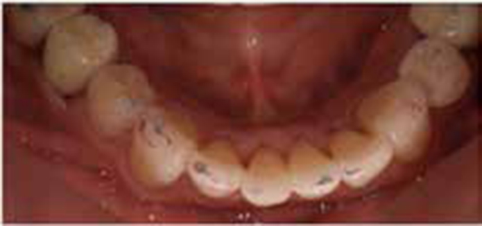
Intra-orally, the patient displayed the presence of a heavily restored dentition, with signs of localized moderate pathological tooth wear of the lower anterior teeth with the presence of exposed dentine on the incisal edges (Figures 3–5). A detailed occlusal assessment was undertaken and revealed a slide from CR to ICP with a larger horizontal component and wear of the canine teeth resulting in the loss of canine guidance, with subsequent balancing side interferences on the second molars on both sides.
Figure 3. Pre-operative facial view.Figure 4. Pre-operative maxillary occlusal view.Figure 5. Pre-operative mandibular occlusal view to show tooth wear present.
The patient also presented with a low lip line and was satisfied with the appearance of his maxillary teeth. The gingival deficiency around an implant-retained crown at UR2 was not visible. The discoloration and morphology of the incisal edges of lower anterior teeth that were visible during speech were, however, a concern for this patient.
A preventive programme was initiated comprising simple scaling, polishing, diet assessment, and oral hygiene instruction.
Following an evaluation period of 6 months, to ensure stability and compliance with the preventive regimen, a direct composite mock-up was first carried out to predetermine the aesthetic and functional outcome. This involved drying the lower affected teeth (canine to canine) and an appropriate shade of resin composite was placed onto the worn surfaces without any prior preparation for adhesive bonding. An alginate impression (Palgat™ Plus, 3M ESPE, MN, USA) of this mock-up guides the dental technician whilst fabricating the wax-up to the aesthetic and functional prescription.
Composite was then removed and accurate pre-operative upper and lower impressions were taken in Express™ PVS (polyvinylsiloxane) (3M ESPE, MN, USA) using metal rim-lock trays. A facebow record and a centric relation record was taken using an anterior deprogrammer with the aid of Dawson's bimanual manipulation technique, along with a comprehensive selection of clinical photographs.
Impressions were cast in Fujirock EP (GC Corp, Tokyo, Japan). The casts were mounted on a semi-adjustable articulator Artex CP (Amman Girrbach, Pforzheim, Germany), using the CR and facebow records.
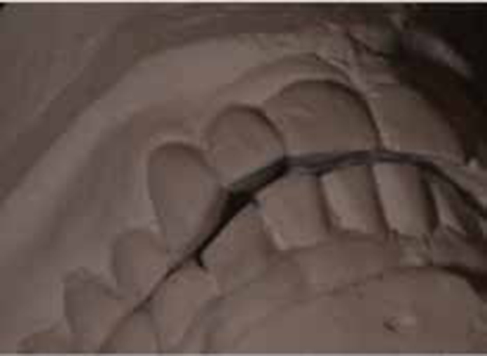
An analysis of the mounted casts in centric relation (CR) revealed the presence of a sizeable discrepancy with a dominant horizontal component between RCP (retruded contact position) and ICP (intercuspation position). In the RCP, only bilateral posterior tooth contacts were observed, thus leaving an inter-occlusal space of 1.5–2.0 mm, which may be used to restore the lower anterior teeth using direct resin composite in a predictable manner, without the concomitant need for any further tooth reduction or, indeed, an increase in the occlusal vertical dimension, thereby leaving the patient with fewer changes to which to adapt (Figure 6).
Figure 6. Casts in centric relation to show the presence of an inter-occlusal clearance, which may be used to place restorative material to restore the worn lower teeth.
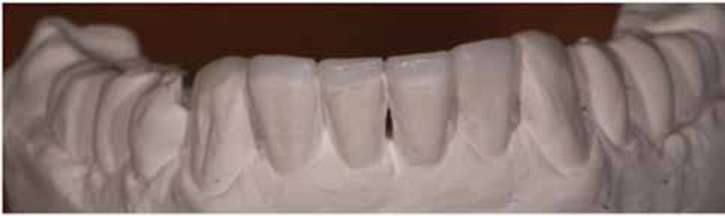
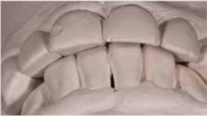
The lower six anterior teeth were waxed-up considering all elements of functional smile design and the impression from the mock-up to provide even centric contact in CR (Figure 7), with immediate disclusion of all the posterior teeth in eccentric movements.21Figure 8 provides a view of the waxed-up lower cast in relation to the upper cast, mounted in CR.
Figure 7. Diagnostic wax-up.Figure 8. Wax mock-up, with casts in intercuspation.
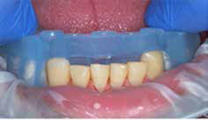
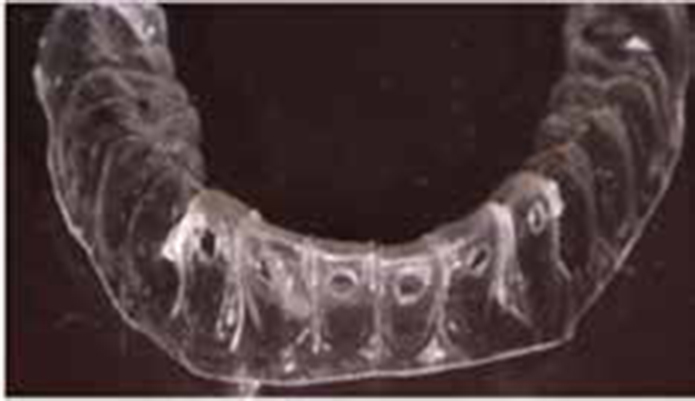
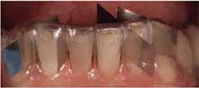
Using a stone duplicate model of the diagnostic wax-up, a 0.5 mm PVC thermoplastic template was formed by the vacuum processing of a template sheet (Acorn Plastics Ltd, Burford, Oxon) over the lower cast, including all but the distal-most teeth. The template was extended 3mm beyond the gingival margin to ensure rigidity post customization and finished with a flat marginal outline.
The template was then tried in to ascertain the desired fit, and an intra-oral mock-up performed in order to gain patient consent. The teeth to receive restorative care were lightly coated with petroleum jelly, and a suitable quantity of bis-acryl resin Luxatemp Automix Plus (DMG, Hamburg, Germany) placed into the template, which was then carefully seated in situ. Upon setting, the template was carefully removed and flash excess trimmed away. This enabled the patient to visualize the finished result.
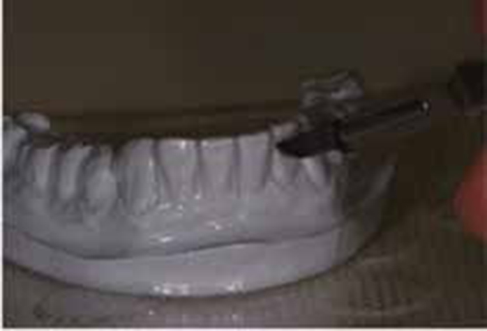
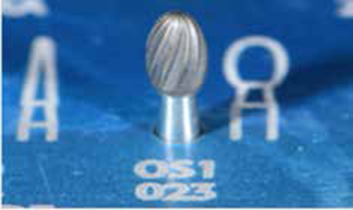
A preheated blade # 15 (Swann-Morton, Sheffield, UK) was used to section the template interdentally in the area of the lower anterior teeth, and loading holes were created on the buccal surfaces of those teeth using a large football bur OS1 023 (Brasseler, GA, USA) (Figures 9–12). Slits were extended 3–4 mm beyond the desired inter-proximal contact area, and the loading holes positioned approximately midway between the existing occlusal surface and the desired incisal edge position. Loading holes were also carefully assessed pre-operatively to ensure that the tip of the desired resin compule was able to fit passively. A second template was prepared in an analogous manner, to serve in a contingency purpose.
Figure 9. PVC stent on the dental cast.Figure 10. Customization of the PVC stent.Figure 11. Modified PVC stent.Figure 12. Bur used to prepare loading holes.
The fit of the stent was verified intra-orally, making sure that the desired metal matrices Flutrec (Pulpodent, MA, USA) could be positioned inter-proximally (as shown below) placed in a relatively passive manner. Please note, the placement of the slits extending a few millimetres beyond the contact area (as discussed above) should allow the metal matrix to extend into this area. In some cases, however, in order to attain this, a small amount of inter-proximal reduction may be required, which can be achieved using a suitable diamond-polishing strip. However, this process was not required in this case.
The lower anterior teeth were subsequently cleaned using an air abrasion device (50 μm) (Microetcher llA, Danville, CA, USA) so as to remove stains and increase the bonding strength without sacrificing any tooth structures.22
Isolation was achieved using cotton wool rolls and copious suction. Teeth were total etched with 32% orthophosphoric acid Uni-etch (Bisco Inc, IL, USA) for 15 sec. The etchant was rinsed and teeth dried (avoiding over desiccation); caution was also taken not to dehydrate the exposed dentine. Opti Bond Solo (Kerr Corp, CA, USA) was applied to both the enamel and dentine using a micro brush, dried with an air gun to permit solvent evaporation and cured for 30 seconds on each surface per tooth. G-aenial composite (GC Corp, Tokyo, Japan) was chosen for this case. Enamel composite from the selected Patient's shade (AE) was pre-heated to 39°C using a composite heater ENA Heat (Micerium Spa, GE, Italy) to improve the physical and handling characteristics of the composite.23 However, some clinicians may choose to negate the use of a resin warmer, where there may be a preference for a resin composite material that is less viscous and more readily sculptable.
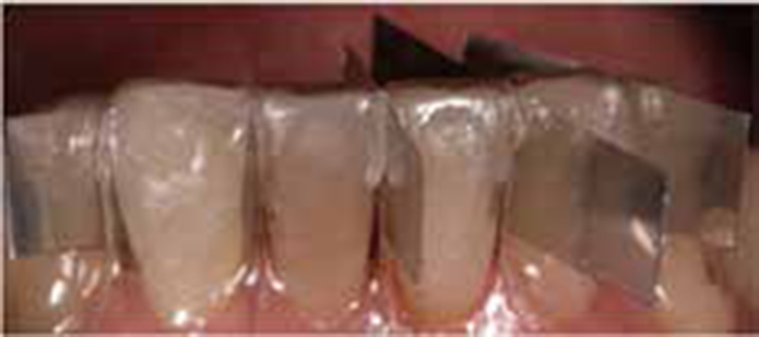
The template was placed in the patient's mouth. Thin metal matrixes were inserted interdentally between template sectioning. The preheated composite was then injected through the labial holes on the template (Figures 13, 14). During this process, digital pressure was applied to the lingual aspect of the template to avoid undesired overflow. Resin was also inserted in a careful manner, using a back-flow technique so as to avoid air entrapment. Once it was evident that an adequate quantity of resin had been inserted, the template was gently squeezed to ensure material adaptation using a flat plastic instrument. Excess resin in the sprue region was wiped away using the flat plastic instrument prior to light curing, which could otherwise render the removal of the template challenging (Figure 15).
Figure 13. PVC stent in situ.Figure 14. Use of separating matrices.Figure 15. Resin placement completed. Stent in situ.
In this case, there was only a need to replace the lost enamel tissues. However, in cases of more severe wear, it is possible to restore lost dentine using a layered approach. The latter involves the placement of ‘dentine cones’, using an appropriate shade of dentine placed in a manner to replace lost dentine. This is carried out prior to the injection moulding process. The above technique may then be used to replace the lost enamel tissue.
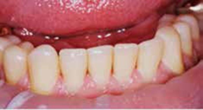
The restorations were then light cured with Demi™ Ultra LED (Kerr Corp, CA, USA). After curing of the composite material for 60 seconds per surface, the matrices were removed and template lifted off (Figure 16).
Figure 16. Restorations pre-finishing.
A 20 minute interval was then applied prior to the commencement of the finishing and polishing protocol (Figure 17), so as to permit time for undisturbed delayed polymerization, commonly referred to as dark polymerization.24
Figure 17. View after gross finishing, prior to fine polishing.
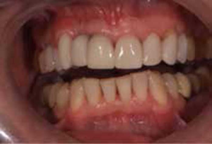
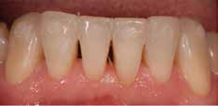
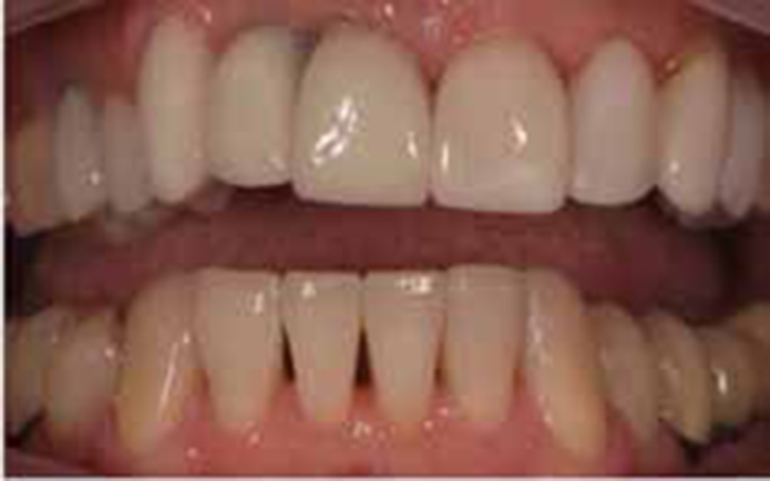
Finishing was initially performed using a fine diamond bur followed by fine carbide burs (Brasseler, GA, USA) so as to remove gross excess. Final polishing was carried out using Mini Flex disks. At this stage the occlusal scheme was evaluated. Final lustre was then achieved with Astrobrush (Ivoclar Vivadent AG, Schaan, Lichtenstein). The goal was to attain an effective anterior guidance that would provide immediate disclusion of all the posterior teeth in eccentric movement (Figure 18), and at the same time avoid interference with the envelope of function. Achieving a balanced mutually protected occlusion is desirable. Figure 19 provides a final post-operative view.
Figure 18. Post-operative view, with occlusal contacts marked using articulating paper.Figure 19. Final post-operative view.
Conclusions
The restoration of worn lower anterior teeth can pose the clinician with some difficult challenges. This technique, using direct resin composite and a modified template, simplifies finishing by allowing the information gathered using a diagnostic wax-up to be translated into the clinical scenario. It also permits the use of interdental matrices.