
Article
Comment: A new look at the diagnosis of pulpitis (Dent Update 2018; 45: 589)
I read your interesting Editorial in the recent issue of Dental Update, and your enthusiastic support of the authors' evidence from new research on vital pulp therapy facilitating, as claimed, a new biologically driven treatment protocol.
Both referenced papers are interesting but I would contest the newness of the approach. My DDS thesis (The University of Edinburgh 1981) and subsequent papers were virtually advocating an identical approach. My paper (‘Assessment of the pulpotomy technique in human first permanent mandibular molars’ Br Dent J 1983; 155: 151, and several others, by myself, in the 80s) gives details of my clinical diagnostic approach, the difficulty of making such a clinical diagnosis, as histology is unavailable, and the surgical approach using coronal pulpotomy in appropriate clinical circumstances. Long-term results are also given, indicating that pulpotomy may be successful in the treatment of the ‘painful’ deep carious lesion in adults up to the age of at least 35 years. The approach was predicated on the many papers (several cited by the present authors) also published in the 70s and 80s, which relate to the correlation between clinical and histological pulp diagnoses.
Your question ‘…is the only person who could carry out the treatment above successfully a specialist endodontist with an operating microscope? I am advised that this is not the case. In other words, has an admirable new concept been developed to a stage where general dentists (worldwide) will be able to revise their treatment strategies on an everyday basis?’
This is answered in my papers. I applaud the authors on their research. It is surprising that they did not source and cite my many papers on a virtually identical topic.
Case report: fractured instrument
Periotomes are used to aid traumatic tooth extraction by severing the fibres of the periodontal ligament to loosen the tooth and make it more easily removed from the socket. They have a thin sharp blade which is moved around the circumference of the tooth in stages to progress the blade further towards the root apex.
A 28-year-old patient was sent to the emergency department by his dentist after a periotome was fractured during the extraction of an UR6. Unfortunately, although the tooth came out, the fractured periotome segment did not (Figure 1).
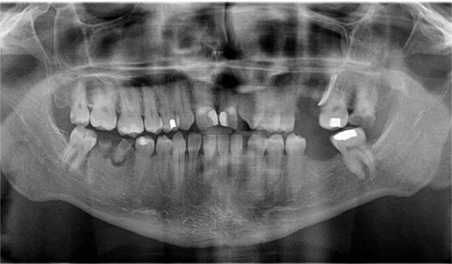
After viewing the OPG ordered by A&E, a cone beam CT of the area was performed and the patient underwent a minor surgical procedure to remove the fragment under local anaesthetic. This was done by raising a flap and removing the surrounding bone.
It is important to use periotomes, as with all instruments, in the manner in which they were designed. It is fair to assume that, in this case, a lot of pressure will have been put on the periotome for it to fracture. A periotome should be used with light apical pressure and not as an elevator. It was considered that, in this case, the periotome was not sharp enough to use as intended. Therefore, the only way to extract the tooth with the instrument was to elevate it. Periotomes must be sharpened regularly for them to be used with any success.