References

Win LC, Sands P, Bonsor SJ, Burke FJT. Ceramics in dentistry: which material is appropriate for the anterior or posterior dentition? Part 1: materials science. Dent Update. 2021; 48:680-688
Heintze SD, Rousson V, Mahn E. Bond strength tests of dental adhesive systems and their correlation with clinical results-a meta-analysis. Dent Mater. 2015; 31:423-434
Bayne SC. Dental restorations for oral rehabilitation – testing of laboratory properties versus clinical performance for clinical decision making. J Oral Rehabil. 2007; 34:921-923
Sailer I, Balmer M, Husler J 10-year randomized trial of zirconia-ceramic and metal–ceramic fixed dental prostheses. J Dent. 2018; 76:32-39
Monaco C, Llukacej A, Baldissara P Zirconia-based versus metal-based single crowns veneered with overpressing ceramic for posterior endodontically treated teeth: 5-year results of a randomized controlled clinical study. J Dent. 2017; 65:56-63
Nejatidanesh F, Abbassi M, Savabi G Five year clinical outcomes of metal–ceramic and zirconia-based implant-supported dental prostheses: a retrospective study. J Dent. 2020; 100
Pjeturrson BE, Sailer I, Makarov NA All-ceramic or metal–ceramic tooth-supported fixed dental prostheses (FPDs)? A systematic review of the survival and complication rates. Part II: multiple-unit FPDs. Dent Mater. 2015; 31:624-639
Zarone F, Di Mauro MI, Spagnuolo G Fourteen-year evaluation of posterior zirconia-based three-unit fixed dental prostheses. A prospective clinical study of all ceramic prostheses. J Dent. 2020; 101
Chaar MS, Kern M. Five-year clinical outcome of posterior zirconia ceramic inlay-retained FPDs with a modified design. J Dent. 2015; 43:1411-1415
Naenni N, Bindl A, Sax C A randomized controlled clinical trial of 3-unit posterior zirconia-ceramic fixed dental prostheses with layered or pressed veneering ceramics: 3-year results. J Dent. 2015; 1365-1370
Kern M, Passia N, Sasse M, Yazigi C. Ten-year outcome of zirconia ceramic cantilever resin-bonded fixed dental prostheses and the influence of the reasons for missing incisors. J Dent. 2017; 65:51-55
Ioannidis A, Bindl A. Clinical prospective evaluation of zirconia-based three-unit posterior fixed dental prostheses: up to ten year results. J Dent. 2016; 47:80-85
Tanner J, Niemi H, Ojala E Zirconia single crowns and multiple-unit FPDs – an up to 8-year retrospective study. J Dent. 2018; 79:96-101
Hansen TL, Schriwer C, Olio M, Gjengedal H. Monolithic zirconia crowns in the aesthetic zone in heavy grinders with severe tooth wear – an observational case-series. J Dent. 2018; 72:14-20
Esquivel-Upshaw JF, Kim MJ, Hsu SM Randomised clinical study of wear of enamel antagonists against polished monolithic zirconia crowns. J Dent. 2018; 68:19-27
Jum'ah AA, Brunton PA, Li KC, Waddell JN. Simulated clinical adjustment and intro-oral polishing of two translucent monolithic zirconia dental ceramics: an in vitro investigation of surface roughness. J Dent. 2020; 101
Becker M, Chaar MS, Garling A, Kern M. Fifteen-year outcome of posterior all-ceramic inlay-retained fixed partial prostheses. J Dent. 2019; 89
Garling A, Sasse M, Becker MEE, Kern M. Fifteen-year outcome of three-unit fixed dental prostheses made from monolithic lithium disilicate ceramic. J Dent. 2019; 89
Malament KA, Natto ZS, Thompson V Ten-year survival of pressed, acid-etched e.max lithium disilicate monolithic and bilayered complete-coverage restorations: performance and outcomes as a function of tooth position and age. J Prosthet Dent. 2019; 121:782-790
Pieger S, Salman A, Bidra AS. Clinical outcomes of lithium disilicate single crowns and partial fixed dental prostheses: a systematic review. J Prosthet Dent. 2014; 112:22-30
Coe JM. Critical summaries. J Am Dent Assoc. 2017; 148:345-346
Brandt S, Winter A, Lauer H-C IPS e.max for all-ceramic restorations: clinical survival and success rates of full-coverage crowns and fixed partial dentures. Materials (Basel). 2019; 12
Teichmann M, Gockler F, Weber V Ten-year survival and complication rates of lithium disilicate (Empress 2) tooth-supported crowns, implant-supported crowns and fixed partial dentures. J Dent. 2017; 56:65-77
Heintze SD, Zimmerli B. Relevance of in vitro tests of adhesive and composite dental materials. Part 1: Approval requirements and standardized testing of composite materials according to ISO specifications. Schweiz Monatsschr Zahnmed. 2011; 121:810-816
Zarone F, DiMauro MI, Ausiello P Current status on lithium disilicate and zirconia: a narrative review. BMC Oral Health. 2019; 19

Ceramics in dentistry: which material is appropriate for the anterior or posterior dentition? Part 2: recent clinical research

From Volume 48, Issue 8, September 2021 | Pages 690-696

Authors

Loo Chien Win

BDS (AIMST), MSc Restorative (Birmingham)

MSc Restorative (Birmingham), Quay Dental Penang, Penang Island, Malaysia

Articles by Loo Chien Win

Email Loo Chien Win

Peter Sands

MSc, BDS, LDS, MCGD

Didcot, Oxfordshire

Articles by Peter Sands

Stephen J Bonsor

BDS(Hons) MSc FHEA FDS RCPS(Glasg) FDFTEd FCGDent GDP

The Dental Practice, 21 Rubislaw Terrace, Aberdeen; Hon Senior Clinical Lecturer, Institute of Dentistry, University of Aberdeen; Online Tutor/Clinical Lecturer, University of Edinburgh, UK.

Articles by Stephen J Bonsor

FJ Trevor Burke

DDS, MSc, MDS, MGDS, FDS (RCS Edin), FDS RCS (Eng), FCG Dent, FADM,

Articles by FJ Trevor Burke

Abstract

The large choice of ceramic materials for an indirect restoration has given clinicians a dilemma when choosing a suitable ceramic material for restorations in anterior or posterior teeth. Part 1 compared the physical properties and aesthetics of lithium disilicate and zirconia materials. This article explores recent clinical research on these materials.

CPD/Clinical Relevance: Awareness of which ceramic material performs optimally on anterior and posterior teeth is clinically important.

Article

The first article in this short series1 principally described the materials science aspect of two frequently-used ceramic materials: lithium disilicate and zirconia. It is the aim of Part 2 to review recent clinical research in these materials and discuss whether the results of this narrative review reflect those in Part 1 by the present authors,1 despite reservations having been expressed in the past2,3 that there is dubious correlation between laboratory-based research and clinical results. This article, therefore, will describe recently published research on zirconia and lithium disilicate crowns and bridges, and also will include publications comparing zirconia-based and metal–ceramic restorations, given that the latter has been a clinical ‘gold standard’ for recent decades when viable alternatives were few and far between.

Zirconia-based versus metal–ceramic restorations

The authors of the present article consider that this comparison is of great relevance, given that metal–ceramic crowns and bridges have been the ‘gold standard’ for tooth-coloured indirect restorations, despite the limitations apparent in Figure 1a. Table 1 presents the recent research that has been identified, while Figure 1 presents failed, unaesthetic metal–ceramic crowns at UL1 and UR1.

Figure 1. Defective metal–ceramic crowns at UL1 and UR1, with suboptimal aesthetics

Reference, location Study details Results Conclusion
Sailer et al4 Switzerland 58 patients who had received 76 3-to 5-unit posterior fixed partial dentures (FPDs/bridges), randomly assigned to zirconia-based and metal-based prostheses 44 patients with 53 bridges were available for the 10-year review, with 17 FPDs lost to follow-up because patients dropped out. Six were considered catastrophic failures, five in the zirconia group (two framework fractures, three due to loss of an abutment tooth) and one in the metal–ceramic group (debonded and unable to re-cement). Of the 59 remaining FPDs, minor chipping of the veneering ceramic and occlusal wear were similar for both the zirconia and metal framework groups. Fracture of a bridge framework was considered by the authors to be a rare complication, only having occurred at two zirconia-based FPDs at the 10-year follow-up It is therefore apparent that, in this work, zirconia-based FPDs exhibited a higher rate of framework fracture, de-bonding, and major fractures of the veneering ceramic. Nevertheless, the authors concluded that there were similar survival rates of zirconia-ceramic and metal-ceramic FPDs at 10 years
Monaco et al5 Bologna, Italy 5-year randomized controlled clinical trial comparing the survival of 90 single crowns on posterior teeth (72 patients), constructed with either a metal or zirconia framework and pressable veneering ceramic, with the crowns being luted with a resin cement Patient recall was 94.5% at 5 years, with two crowns demonstrating complete failure – one being a zirconia crown that failed due to core fracture at 24 months and the second, a metal–ceramic crown, which was a biological failure because of tooth root fracture. Chipping of the veneering ceramic was noted in three zirconia crowns and two metal–ceramic crowns, with all patients being satisfied with the aesthetics of their restoration(s) The authors concluded that ‘veneered zirconia crowns exhibited sufficient strength and could represent an alternative for single crown restorations’
Nejatidanesh et al6 5-year retrospective study of 144 posterior zirconia-based or metal-based FPDs in 144 patients The success rate was 81.6% and 81.0% for the zirconia-based and metal-based FPDs, respectively, with fracture of the veneering ceramic occurring in 6.4% and 2.7% of the zirconia-based and metal-based restorations, respectively The authors concluded that, at 5-years, ‘zirconia-based and metal–ceramic FPDs showed similar promising clinical performance’, adding that ‘zirconia-based implant supported FPDs might be a successful restorative method with acceptable survival and patient satisfaction’
Pjetursson et al7 Systematic review of metal–ceramic or all-ceramic FPDs 40 studies include that reported a total of 1796 metal–ceramic and 1110 all-ceramic tooth-supported FPDs. The meta-analysis indicated an estimated 5-year survival of metal–ceramic FPDs to be 94.4%, for reinforced glass ceramics 89.1%, glass-infiltrated alumina FPDs 86.2% and densely sintered zirconia 90.4%. Although the survival rate of all-ceramic FPDs was lower than that of metal–ceramic FPDs, the differences did not reach statistical significance. Regarding framework fractures, significantly more were reported for reinforced glass ceramic and glass-infiltrated alumina FPDs (12.9%) compared with metal–ceramic FPDs (0.6%) and densely sintered zirconia FPDs (1.9%) The authors concluded that metal–ceramic FPDs had lower failure rates than all-ceramic FPDs after a mean observation time of 3 years, adding that ‘ceramic fractures and chipping of ceramics were frequent, but that, in the future, refinements in the production of all-ceramic reconstructions are indicated’

The conclusion to be gleaned from this section is that, from the results of recent research presented in Table 1, while metal–ceramic remains the ‘gold standard’, all-ceramic crowns and fixed partial dentures (FPDs) formed with a zirconia framework hold promise, with results approaching those of metal–ceramic in a majority of the included studies. This may be considered relevant, as the proportion of patients requesting metal-free restorations increases.

Zirconia-based crowns and bridges

Table 2 presents the recent research on zirconia-based crowns and bridges. It may be concluded that, from the results of recent research identified in Table 2, monolithic and veneered zirconia crowns and FPDs are capable of providing good clinical service. It may also be of interest to note that Esquivel-Upshaw and colleagues15 from Gainesville, USA, examined the in vivo wear of enamel when opposed by 16 polished monolithic zirconia crowns and 14 metal–ceramic crowns, with the results indicating no difference. The results from Jum'ah et al16 regarding in vitro polishing of zirconia may also be of interest, given that their results indicated that the use of zirconia burs may result in deep surface flaws, that a four-step polishing protocol that they proposed (using the DiaShine Dentist Zirconia Adjusting and Polishing Kit, VH Technologies Ltd, USA) was associated with the finest finish.


Reference, location Study details Results Conclusion
Zarone et al8 Prospective 14-year evaluation of 48 zirconia-based three-unit fixed partial dentures (FPDs) placed in 37 patients Cumulative survival rates indicated 91% and 99% survival for patients with one and two FPDs, respectively The authors concluded that zirconia was a clinical option to fabricate short-span posterior FPDs
Chaar and Kern9 The clinical outcome of posterior zirconia-based inlay-retained FPDs in 30 patients at 5 years was evaluated. They used a modified design with a 0.2–0.5-mm retainer wing placed in enamel beyond the lingual and buccal margins of the inlay box Six patients were lost to follow up. Of the remaining FPDs, one was considered failed due to eight repeated debondings, this patient being a bruxer with a deep overbite and without a canine-protected occlusion. The cumulative rate of chipping was 10.5%, but this did not affect the clinical function of the affected FPDs and 25 RPDs were free of complication The authors concluded that the performance of zirconia-based inlay-retained FPDs ‘was encouraging’
Naenni et al10 36 patients with posterior zirconia-based FPDs, 18 with pressed veneering ceramic and 18 with layered veneering ceramic were evaluated At 3 years, survival rate was 100%, but there was a trend towards more chipping in the pressed veneered ceramic group The authors concluded that posterior restorations with zirconia frameworks are a viable treatment option
Kern et al11 The outcome at 10 years (mean observation time = 92 months ± 33 months) of 108 zirconia cantilever resin-bonded bridges provided tor 87 patients was evaluated, these replacing 75 maxillary incisors and 33 mandibular incisors Six debondings and one loss of restoration were recorded, the lost restoration having been removed at the patient's request after a small chip occurred at the incisal edge, and with three of the six debondings as a result of trauma. All debonded bridges were rebonded successfully, yielding a 10 year survival rate of 98.2% The authors concluded that ‘anterior zirconia cantilever resin-bonded bridges provided excellent longevity’
Ioannidis and Bindl12 Zirconia-based three-unit posterior FPDs in 55 patients who received 59 FPDs were evaluated At 6.3 ± 1.9 years, 57 FPDs were available for examination. Biological complications, such as loss of tooth vitality, abutment tooth root fracture, periodontal complications and secondary caries, affected 17.5% of FPDs and technical complications (16 chippings) were noted in 28% of FPDs. However, it was possible to repair or polish these: no severe technical complication, such as framework fracture, occurred The authors concluded that three-unit FPDs formed in zirconia offer a possible treatment option, albeit with a high rate of chipping, but adding that manufacturing processes ‘nowadays’ are modified to avoid this
Tanner et al13 Retrospective clinical study that evaluated the survival of 40 zirconia-based restorations (17 single crowns, 23 FPDs) after a mean time of 5.7 years, placed in the student clinic at Turku University, Finland The survival rate of the crowns was 94.2% and the FPDs 95.7%, with a complication rate of 26% for FPDs crowns and 5.8% for crowns, with veneering ceramic fractures only noted in the FPDs. 85% of patients scored ‘A’ for satisfaction The authors concluded ‘zirconia is a suitable material for single crowns and FPDs, with a high survival rate, but that technical and biological complications can be expected and that attention must be paid to the shape of the framework, especially on connector areas, in order to maintain gingival health’
Hansen et al14 Bergen, Norway In a prospective observational case-series study, monolithic zirconia crowns in the aesthetic zone in ‘heavy grinders’ with severe tooth wear were assessed. A total of 84 crowns in 13 patients (all male) were assessed at times between 1 and 3 years One crown had a total fracture after 16 months and was replaced by another crown. Chipping was noted in 5.3% of crowns and all patients expressed satisfaction with their crowns The authors concluded that ‘monolithic zirconia crowns may provide a valid treatment modality of severe tooth wear in the aesthetic zone where minimally invasive treatment fails’

Lithium disilicate crowns and FPDs

Table 3 presents the recent research on lithium disilicate-based crowns and bridges. It may be concluded from the results of the identified, that crowns formed in lithium disilicate provide good clinical survival in the anterior and premolar areas of dentitions in which optimum aesthetics is an overriding factor (Figure 2). However, conventionally supported FPDs and inlay-retained FPDs formed in this material are not generally to be recommended.


Reference, location Study details Results Conclusion
Becker et al17 Kiel, Germany Long-term clinical outcome of 45 inlay-retained FPDs (in 42 patients) made from lithium disilicate glass-ceramic (IPS e.max Press, Ivoclar Vivadent) were evaluated. These replaced eight premolars and 37 first molars. Clinical treatment was carried out by 15 clinicians with circa 2 years' experience supervised by two specialist prosthodontists. Inlay preparation procedures were performed in accordance with the general principles for ceramic inlay restorations. Observation time was 100 months (minimum 4, maximum 234) 33 (73%) FPDs failed during the observation period and were replaced by other restorations, with the Kaplan–Meier survival rate being 57% at 5 years, 38% at 8 years, and 22% at 15 years The authors concluded that lithium disilicate inlay-retained FPDs had a high clinical failure rate and that the clinical outcome of experimental inlay-retained FPDs made from lithium-disilicate ceramic was not acceptable when compared to conventional crown-retained FPDs and cannot be recommended for regular clinical use
Garling et al18 Kiel, Germany A 15-year prospective study of 36 three-unit FPDs replacing anterior (16%) and posterior (84%) teeth in 28 patients was conducted Three FPDs were classified as dropped out, and, of the remaining 33 FPDs, observed for a mean time of 167 months, the success rate (ie restorations free from complications) was 30.9% after 15 years The authors noted a dramatic drop in monolithic lithium disilicate ceramic FPD survival and success rates. Purported to be a result of fatigue and crack propagation caused by clinical ageing and loading conditions. They noted that no fractures occurred within the first 6 years, but after 7 years ceramic fractures started and the fracture rate increased substantially after 10 years. They added that ‘compared to the still ‘gold standard’, metal–ceramic FPDs, our results with all-ceramic FPDs are considerably inferior after 10 years’
Malament et al19 
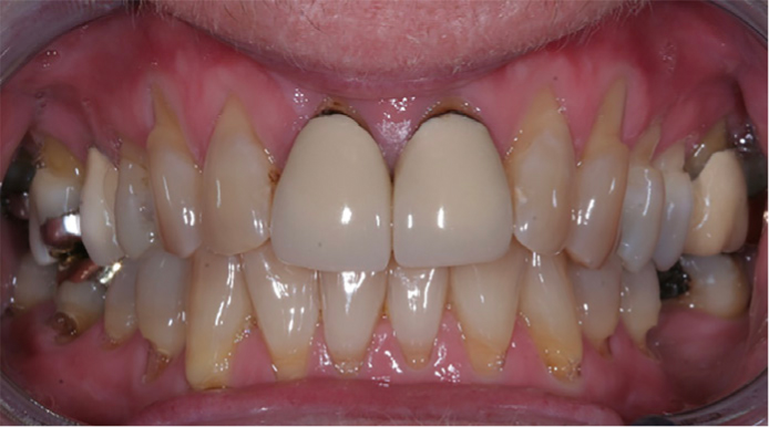
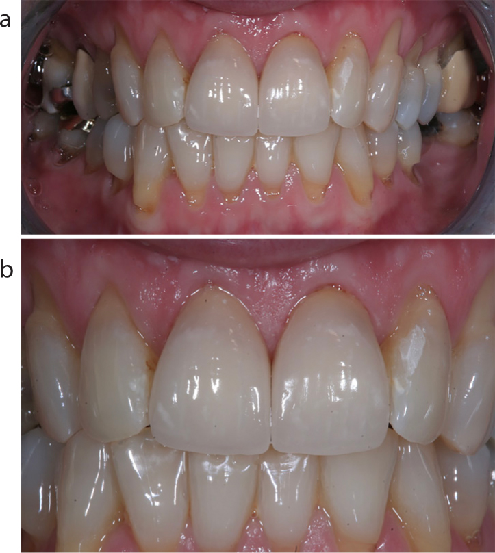
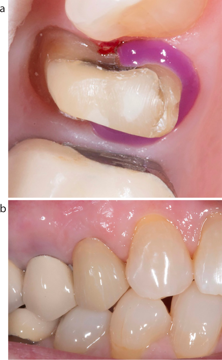
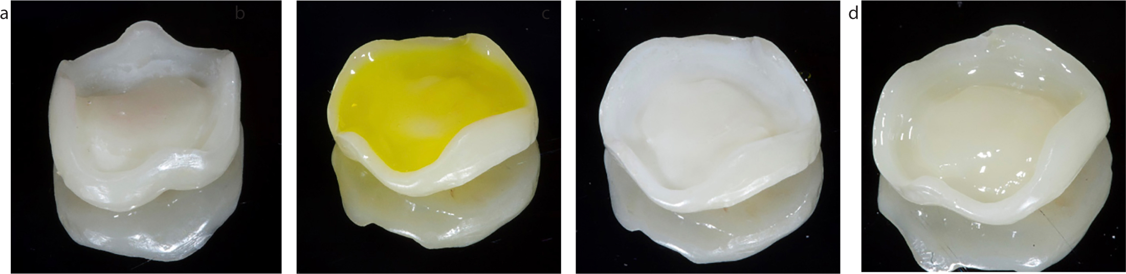
10-year performance of pressed lithium disilicate restorations (IPS e.max, Ivoclar Vivadent) placed in 556 patients (1960 complete coverage restorations) in a private clinic were examined. All restorations were constructed using the lost wax method and a glass-ceramic pressing system, and the fit surface of all was etched with 4.5% buffered HF and silane, coated with Monobond Plus (Ivoclar Vivadent). The crowns were bonded in place using a dentine bonding agent and resin cement Seven failures were recorded, with the average time to failure being 4.2 years, and these being confined to molar teeth. At 10.4 years, the mean annual failure rate was 0.2% The authors concluded that IPS e.max lithium disilicate crowns exhibited excellent survival, and tooth position or monolayered or bilayered structure had little effect on survival
Pieger et al20 Although not a recent publication (published in 2014), this systematic review is considered by the present authors to present useful insight into the performance of lithium disilicate when used for single crowns of FPDs. They identified 2033 studies, including 12 in their analysis The 5-year cumulative survival rate was calculated to be 97.8% for single crowns and 78.1% for FPDs, respectively The authors concluded that for lithium disilicate single crowns, excellent short-term survival rates are evident, while the evidence for medium-term survival of lithium disilicate FPDs is ‘not promising,’ adding that the majority of failures in both types of restoration were reported in the posterior region of the mouth and that caution is advised for the use of lithium disilicate for FPDs. This was confirmed by a critical review of this work by Coe in 201721
Brandt et al22 Kassel, Germany Retrospectively evaluation of 1058 full-coverage crowns and FPDs over 66 months, placed in a private dental practice, all being constructed in IPS e.max (Ivoclar Vivadent) There was a 5-year cumulative survival of 94.7% for crowns and 90.6% for FPDs The authors added that significantly superior outcomes were obtained when adhesive cementation was used and for vital versus non-vital teeth
Teichmann et al23 Prospective evaluation of 10-year clinical outcome of tooth- and implant-supported crowns, and FPDs formed in a lithium disilicate glass-ceramic framework material 184 restorations (106 single crowns, SCs; 32 implant crowns, ICs; and 33 FPDs) were placed in 73 patients. There were 14 patient drop-outs, and the final dataset included 87 SCs, 17 ICs and 27 FPDs. 10-year survival rate/chipping-free rate was 86.1%/83.4% for SCs, 93.8%/94.1% for ICs, and 51.9%/90.8% for FPDs The authors concluded that ‘the SCs had a lower outcome than expected, the ICs had a favourable outcome and the FPDs predominantly failed’
Figure 2. (a) Patient in Figure 1, crowns at UL1, UR1 replaced by IPS e.max (Ivoclar Vivadent) crowns (b) Close-up of IPS e.max (Ivoclar Vivadent) crowns at UL1 and UR1.

Figure 4 presents the chairside steps need to achieve synergism between a lithium disilicate onlay and tooth substance.

Figure 3. (a, b) IPS e.max (Ivoclar Vivadent) crown at UR4. The enamel margins were selectively etched prior to cementation using a dentine adhesive and resin cement.
Figure 4. (a) Fit surface of a lithium disilicate onlay as it might return from the laboratory, with minor imperfections on the surface. (b) Surface ‘cleansing’ with buffered HF acid. (c) Surface on rinsing and drying. Note the matt, homogeneous, clean surface. (d) Shiny fit surface after application of bonding agent. In this case, the bonding agent used incorporated a silane coupling agent.

Discussion

With regard to choosing appropriate indirect tooth-coloured restorative materials for the anterior and posterior region, biomaterial properties and the patient's oral condition are part of the consideration when choosing the most appropriate material. In Part 1,1 the material properties of lithium disilicate and zirconia were discussed and compared. When recent clinical studies are added to the ‘equation’, it is clear that there is little divergence between the recommendations made in the conclusion of that paper, namely:

  • While metal–ceramic remains the ‘gold standard’, all-ceramic crowns and FPDs formed with a zirconia framework hold promise, with results approaching those of metal–ceramic in a majority of the included studies;
  • That monolithic and veneered zirconia crowns and FPDs are capable of providing good clinical service; and,
  • That crowns formed in lithium disilicate provide good clinical survival in the anterior and premolar areas of dentitions in which where optimum aesthetics is an overriding factor. Therefore, as stated previously,1 in the anterior region where clinical performance and high aesthetics are required, lithium disilicate may be preferred to monolithic and layered zirconia.
  • It was stated at the start of this article that the correlation between laboratory and clinical studies was suboptimal. However, the reviews that we quoted related to dental adhesive systems2 and materials with polymer-based matrices,3 with the authors of these studies stating, respectively: that ‘the weak correlation of bond-strength test results with clinical parameters leads to the conclusion that one should not rely solely on bond strength tests to predict the clinical performance of an adhesive system but one should conduct other laboratory tests like tests on the marginal adaptation of fillings in extracted teeth and the retention loss of restorations in non-retentive cavities after artificial aging’, and: very few correlations of laboratory tests and clinical results are demonstrated and that ‘a large number of risk factors (operator, material, intraoral location, patient) may affect clinical outcomes and are not simulated well in dental laboratories’. It is also suggested3 that more long-term clinical trials, which involve 10–20 years of observations, are needed, but clinicians with clinical trial experience will understand that trials over such long periods are difficult to achieve in practice. This is reinforced in an earlier study by Heintze and co-workers24 in which they stated: ‘The standardized laboratory tests are important for the first material analysis. As opposed to other test methods, they correlate well in part with the clinical data. Laboratory tests do not replace clinical tests, but they do increase the safety of the patients who participate in controlled studies of the new materials’. While the results of the present and previous1 study are by no means exhaustive, it may be suggested that the correlation between laboratory and clinical studies on contemporary ceramic materials may be better than for resin-based materials.

    Finally, from the clinical perspective, occlusion and the parafunctional habits play a part in the choice of the materials, and also the aesthetic requirements of the patient. Perhaps Zarone and colleagues25 should be allowed to have the final words in this discussion, as they stated in their paper: ‘The noticeable properties and versatility make lithium disilicate and zirconia materials of choice for modern prosthetic dentistry, requiring high esthetic and mechanical performances combined with a minimal invasive approach, so that the utilization of such metal-free ceramics has become more and more widespread over time’.

    Conclusions

    Following this narrative review of clinical research on survival of restorations formed in lithium disilicate and zirconia, the following conclusions may be drawn:

  • In the posterior region, for a stronger material in the construction of crowns or bridges, monolithic zirconia is preferred. However, metal–ceramic remains ‘the gold standard’;
  • In the posterior region, in a patient without any parafunctional habits, monolithic lithium disilicate may perform well in the premolar region;
  • In the anterior region for single crowns where high aesthetic results are expected, lithium disilicate material is preferred;
  • In the anterior region for a bridge and a satisfactory aesthetic outcome is indicated, layered zirconia can be chosen where zirconia material as the framework core materials and hand-layered by feldspathic ceramic on the facial surface;
  • In the anterior region for a discoloured tooth or a tooth restored with a metal post, layered zirconia may be preferred as zirconia is opaque and hand-veneered feldspathic ceramic will improve the aesthetics.