Carey B, Setterfield J. Mucous membrane pemphigoid and oral blistering diseases. Clin Exp Dermatol. 2019; 44:732-739
Kanjanabuch P, Arporniem S, Thamrat S, Thumasombut P. Mucous membrane pemphigoid in a patient with hypertension treated with atenolol: a case report. J Med Case Rep. 2012; 6
Gaudin O, Seta V, Alexandre M Gliptin accountability in mucous membrane pemphigoid induction in 24 out of 313 patients. Front Immunol. 2018; 9
Porcelli B, Pozza A, Bizzaro N Association between stressful life events and autoimmune diseases: a systematic review and meta-analysis of retrospective case-control studies. Autoimmun Rev. 2016; 15:325-334
Stojanovich L, Marisavljevich D. Stress as a trigger of autoimmune disease. Autoimmun Rev. 2008; 7:209-213
Setterfield J, Shirlaw P, Kerr-Muir M Mucous membrane pemphigoid: a dual circulating antibody response with IgG and IgA signifies a more severe and persistent disease. Br J Dermatol. 1998; 138:602-610
Yancey KB, Egan CA. Pemphigoid: clinical, histologic, immunopathologic, and therapeutic considerations. JAMA. 2000; 284:350-356
Mihai S, Sitaru C. Immunopathology and molecular diagnosis of autoimmune bullous diseases. J Cell Mol Med. 2007; 11:462-481
Zillikens D. Diagnosis of autoimmune bullous skin diseases. Clin Lab. 2008; 54:491-503
Carey B, Joshi S, Abdelghani A The optimal oral biopsy site for diagnosis of mucous membrane pemphigoid and pemphigus vulgaris. Br J Dermatol. 2020; 182:747-753
Giurdanella F, Diercks GF, Jonkman MF Laboratory diagnosis of pemphigus: direct immunofluorescence remains the gold standard. Br J Dermatol. 2016; 175:185-186
Schmidt E, Zillikens D. Pemphigoid diseases. Lancet. 2013; 381:320-332
Taylor J, McMillar R, Shephard M World Workshop on Oral Medicine VI: a systematic review of the treatment of mucous membrane pemphigoid. Oral Surg Oral Med Oral Pathol Oral Radiol. 2015; 120:161-171
Mays JW, Carey BP, Posey R World Workshop of Oral Medicine VII: a systematic review of immunobiological therapy for oral manifestations of pemphigoid and pemphigus. Oral Dis. 2019; 25:(1)111-121
Stressful Life Events as a Trigger for Autoimmune Disease? A Case Report on Mucous Membrane Pemphigoid Robert Devine Melanie Simms Dental Update 2024 48:5, 707-709.
Authors
RobertDevine
BDS (QMUL), MJDF
Dental Core Trainee in Oral and Maxillofacial Surgery, Royal United Hospitals, Bath
This case discusses the acute presentation of a patient with mucous membrane pemphigoid to an emergency dental department. Mucous membrane pemphigoid is a rare condition, but its presentation can be severe and concerning for both the patient and clinician. The case presents the manifestations of florid desquamative gingivitis and extensive mucosal erosions due to burst bullae. We discuss the possible causes of the condition in this patient, likely to be the stress of recent cardiac surgery, as well as exploring the efficacy of diagnostic tools, treatment options and adverse effects of corticosteroid treatment.
CPD/Clinical Relevance: Correct and timely diagnosis of vesiculobullous disorders has notable impacts on patient outcomes and quality of life.
Article
Robert Devine
Mucous membrane pemphigoid (MMP) is a rare subepithelial bullous disorder predominantly involving the oral mucosa. Other mucous membranes that may be affected include the upper part of the aerodigestive tract, conjunctiva and genital mucosa. Occasionally the skin may also be involved.1 There is an inflammatory autoimmune response directed against the hemidesmosome adhesion complex, leading to subepithelial clefting and the clinical appearance of desquamative gingivitis, blisters or erosions. This case presents an acute presentation of MMP as florid desquamative gingivitis and extensive mucosal erosions. This article explores the efficacy of diagnostic tools available to identify the condition, discusses the immediate, short- and long-term treatment modalities as well as hypothesizes potential initiating stressor events and response to therapy.
Case report
Presenting complaint
A 64-year-old male presented to the dental emergency department at the University Hospital, Wales, complaining of painful, ‘peeling’ and bleeding gums. He found that brushing his teeth made the gums develop blood-filled blisters, prior to bursting and leaving a painful ulcerated area that did not heal. The patient reported one single episode of a blister on the skin of his nose that had since healed, and reported no other cutaneous, genital or ocular symptoms.
History of presenting complaint
The patient had undergone heart surgery with the placement of cardiac stents 5 weeks earlier and noted the presence of blisters on his gums 7 days after the surgery. Initially, his cardiologist believed this to be an adverse reaction to the recently prescribed clopidogrel, which was immediately substituted for ticagrelor as an alternative antiplatelet therapy. Despite this, the painful bleeding gingivae had not alleviated and had, in fact, worsened. The patient then presented to his general medical practitioner who prescribed 0.2% chlorhexidine mouthwash, along with high-concentration sodium fluoride toothpaste, and advised attendance with his dentist.
Medical history,
The patient's medical history including medications, is shown in Table 1. Ticagrelor had been started 3 weeks previously; however, all other medications were long-standing (several years).
Ticagrelor: 90 mg twice dailyGlyceryl trinitrate: 400 μg/dose aerosol sublingual spray, as neededLosartan: 25 mg, once a dayLansoprazole: 30 mg, once a dayAspirin: 75 mg, once a dayVitamin B12 injections
Clinical presentation
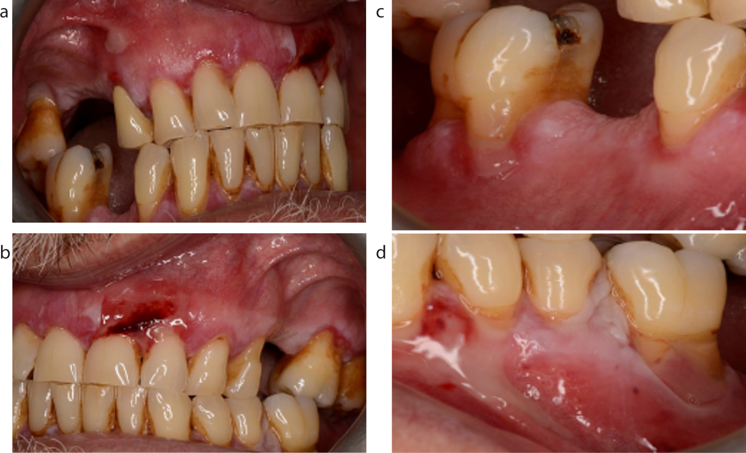
On examination, there was evidence of florid desquamative gingivitis: a marked erythematous appearance of the free and attached gingivae up to the mucogingival margin. Recently burst bullae resulting in extensive erosions were also present on the gingivae (Figure 1). Further examination showed that the other oral mucosal tissues were spared, and there was only mild erythema on the skin of the nose, where a previous blister was reported.
At this stage, MMP and pemphigus vulgaris (PV) were considered as the most likely diagnoses, with other blistering conditions such as linear IgA disease and erythema multiforme, or adverse drug reactions being considered as differential diagnoses.
Investigations
Following the clinical examination, serum was obtained for detection of circulating autoantibodies by indirect immunofluorescence (IIF) and assay of serum antibody titres by enzyme-linked immunosorbent assay (ELISA), and an oral mucosal biopsy was taken for direct immunofluorescence (DIF). Owing to the high clinical suspicion of a bullous disease, DIF was favoured over routine histopathology for gaining a definitive diagnosis, and therefore, a second biopsy specimen for routine histopathology was not obtained. The IIF results from the local immunology laboratory showed a weak positive result for pemphigus vulgaris; however, IIF and ELISA from a specialist immunodermatology laboratory were negative. DIF from the immunodermatology laboratory showed linear deposition of IgG (++), IgA (+), C3 (++) and fibrinogen (+) at the basement membrane zone, consistent with a definitive diagnosis of MMP.
Management and response
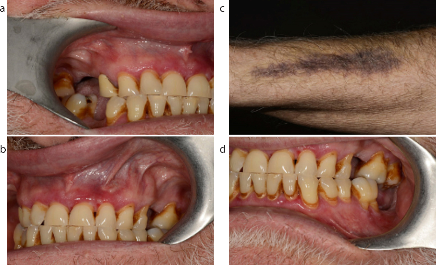
The patient was initially prescribed topical corticosteroids in the form of 500-μg betamethasone soluble tablets (made into a mouthwash, to be rinsed around for 2 minutes, up to four times daily) and 0.05% clobetasol cream (to be applied as a light smear to areas of sore oral mucosa, twice daily). This did not provide sufficient improvement in symptoms, and therefore systemic prednisolone was started at 40 mg/day with a tapering regimen, while awaiting biopsy results. Immediately following confirmation of the diagnosis from direct immunofluorescence, a second course of systemic prednisolone was started as symptom control was still poor, and mycophenolate mofetil (MMF) was introduced as a steroid-sparing immunosuppressant for long-term management. After 4 weeks of a therapeutic dose of MMF, there was resolution of the desquamative gingivitis and erosions. In the final week of the second course of systemic prednisolone (week 6), the patient developed bruising on his right forearm (Figure 2), suspected to be a side effect of prednisolone, particularly as he was also taking aspirin. Symptoms remained controlled after finishing the course of systemic prednisolone and continuing MMF.
Figure 2. (a–d) Clinical response showing a reduction in desquamative gingivitis after taking mycophenolate mofetil at the therapeutic dose for 4 weeks. Bruising on the right forearm developed in week 6 of the second course of systemic prednisolone.
The role of the general dental practitioner in MMP
The role of the general dental practitioner (GDP) should not be understated. Recognizing the oral manifestations, which may indicate bullous disease, and timely referral to oral medicine or oral and maxillofacial surgery, will have a significant effect on patient quality of life. Furthermore, undiagnosed or poorly controlled MMP can drastically reduce the ability of the patient to maintain good oral hygiene due to pain, and this may contribute to the onset or progression of periodontal disease. Therefore, the GDP also has a significant role in the long-term maintenance of oral health in MMP patients; these patients should receive relevant and personalized oral hygiene instruction and periodontal treatment where required (often under LA due to gingival soreness), recognizing that plaque accumulation will exacerbate gingival inflammation, in addition to that already caused by the bullous disease. For maximum symptom control, oral hygiene must, therefore, be excellent.
Both oral hygiene and disease control should be satisfactory before consideration is given to restoring edentulous spaces, and mucosa-borne prosthesis may be less favourable as they can traumatize the already fragile mucosa.
Medications to treat MMP can impact the oral cavity. Topical steroid use may cause oral candidosis, while immunosuppression can reduce healing and those on systemic steroids may require steroid cover for surgical procedures.
Discussion
MMP is a rare auto-immune bullous disorder that most commonly manifests in the oral cavity as desquamative gingivitis (80%), and oral mucosal blisters and erosions.1 Other mucous membranes may be involved, and occasionally there is involvement of the skin. As seen in this case, any delay in diagnosis from onset of symptoms results in significant detrimental effects on quality of life (inability to eat more than the softest of foods, severe burning and irritation caused by the mucosal erosions). Although it is known that MMP is an autoimmune condition, the exact trigger is uncertain in most cases. Despite this, the timeline of events in this patient points towards a drug-induced aetiology (symptoms beginning 1 week after commencing clopidogrel), but to the authors' knowledge, there are no reports of clopidogrel-induced MMP in the literature. Medications such as antihypertensives and gliptins for diabetes have been reported to cause MMP. 2,3 As the lesions did not resolve following the cessation of clopidogrel and the change to ticagrelor, it is postulated that rather than a drug-induced aetiology in this patient, the trigger was the event of cardiac surgery. It is recognized in the literature that autoimmune conditions may be triggered by life stressors.4,5
Diagnosis of MMP is based on history, clinical presentation and certain immunopathological features (present on serology, histology and immunofluorescence), but can be difficult, especially if a patient presents to a healthcare professional who is unfamiliar with bullous conditions. The contradictory results from this patient's diagnostic investigations highlights the less-than-perfect sensitivity, specificity and reliability of these investigations. IIF is only positive in approximately 50–84% of patients with MMP,6,7,8,9,10 and overall, a biopsy for DIF remains the gold standard for diagnosis of bullous disorders.10,11,12
Management of MMP is dependent on symptom severity and includes topical corticosteroids, topical calcineurin inhibitors, systemic corticosteroids, steroid-sparing immunosuppressants and possibly biologic agents such as rituximab. 12,13,14 Early assessment by an ophthalmologist and dermatologist is essential, to rule out ocular involvement and aid management of any cutaneous lesions.
Conclusion
This case report outlines the acute presentation, diagnosis and management of a case of MMP and highlights the importance of communication and timely referral between medical and dental specialties, to avoid the risk of delayed diagnosis, disease progression and adverse effects on patient quality of life. Diagnostic challenges stem from poor reliability of diagnostic tests, but rapid clinical improvement can be seen with appropriate treatment, including systemic prednisolone and MMF. Various aetiological factors have been proposed for MMP, in this case thought to be the precipitating stressor event of cardiac surgery that triggered or exacerbated subclinical autoimmune disease.