References

Dawson PE.St Louis: CV Mosby Publishing; 2007
Dyer K, Ibbetson R, Grey N. A question of space: options for the restorative management of worn teeth. Dent Update. 2001; 28:118-123
Yule PL, Barclay SC. Worn down by toothwear? Aetiology, diagnosis and management revisited. Dent Update. 2015; 42:525-532
Watt RG, McGlone P, Kay EJ. Prevention Part 2: Dietary advice in the dental surgery. Br Dent J. 2003; 195:27-31
Holbrook PW, Árnadóttir IB, Kay EJ. Prevention Part 3: Prevention of tooth wear. Br Dent J. 2003; 195:75-81
Moynihan PJ. Dietary advice in dental practice. Br. 2002; 193::563-568
London: Department of Health; 2014
Mizrahi B. A technique for simple and aesthetic treatment of anterior toothwear. Dent Update. 2004; 31:109-114
Chu F, Siu A, Newsome P. Restorative management of the worn dentition: 1. Aetiology and diagnosis. Dent Update. 2002; 29:162-168
Ricketts DNJ, Tait CME, Higgins AJ. Tooth preparation for post-retained restorations. Br Dent J. 2005; 198:463-471
Tait CME, Ricketts DNJ, Higgins AJ. Weakened anterior roots – intraradicular rehabilitation. Br Dent J. 2005; 198:609-617
Foxton R, Mannocci F, Melo L. Adhesive restoration of endodontically treated teeth-current research. Dent Update. 2006; 33:500-506
Hayashi M, Takahashi Y, Imazato S, Ebisu S. Fracture resistance of pulpless teeth restored with post-cores and crowns. Dent Mater. 2006; 22:477-485
Silva NR, Castro CG, Santos-Filho PC, Silva GR, Campos RE, Soares PV, Soares CJ. Influence of different post design and composition on stress distribution in maxillary central incisor: finite element analysis. Indian J Dent Res. 2009; 20:153-158
Okada D, Miura H, Suzuki C, Komada W, Shin C, Yamamoto M, Masuoka D. Stress distribution in roots restored with different types of post systems with composite resin. Dent Mater J. 2008; 27:605-611
Santos-Filho PC, Castro CG, Silva GR, Campos RE, Soares CJ. Effects of post system and length on the strain and fracture resistance of root filled bovine teeth. Int Endod J. 2008; 41:493-501
Giovani AR, Vansan LP, de Sousa Neto MD, Paulino SM. In vitro fracture resistance of glass-fiber and cast metal posts with different lengths. J Prosthet Dent. 2009; 101:183-188
Boschian Pest L, Guidotti S, Pietrabissa R, Gagliani M. Stress distribution in a post-restored tooth using the three-dimensional finite element method. J Oral Rehabil. 2006; 33:690-697
Jung SH, Min KS, Chang HS, Park SD, Kwon SN, Bae JM. Microleakage and fracture patterns of teeth restored with different posts under dynamic loading. J Prosthet Dent. 2007; 98:270-276
Gulamali AB, Hemmings KW, Tredwin CJ, Petrie A. Survival analysis of composite Dahl restorations provided to manage localised anterior tooth wear (ten year follow-up). Br Dent J. 2011; 211
Poyser NJ, Briggs PFA, Chana HS, Kelleher MGD, Porter RWJ, Patel MM. The evaluation of direct composite restorations for the worn anterior dentition – clinical performance and patient satisfaction. J Oral Rehabil. 2007; 34:361-376
Al-Khayatt AS, Ray-Chaudhuri A, Poyser NJ, Briggs PF, Porter RW, Kelleher MG, Eliyas S. Direct composite restorations for the worn mandibular anterior dentition: a 7-year follow-up of a prospective randomised controlled split-mouth clinical trial. J Oral Rehabil. 2013; 40:389-401

A Clinical Case: Restoration of Toothwear

From Volume 47, Issue 3, March 2020 | Pages 253-263

Authors

Janita Patel

BDS(Lond), MSc(RDP), MA(MedEd) FHEA, FFGDP(RCS Eng)

General Dental Practitioner, Northwood, Middlesex

Articles by Janita Patel

Email Janita Patel

Abstract

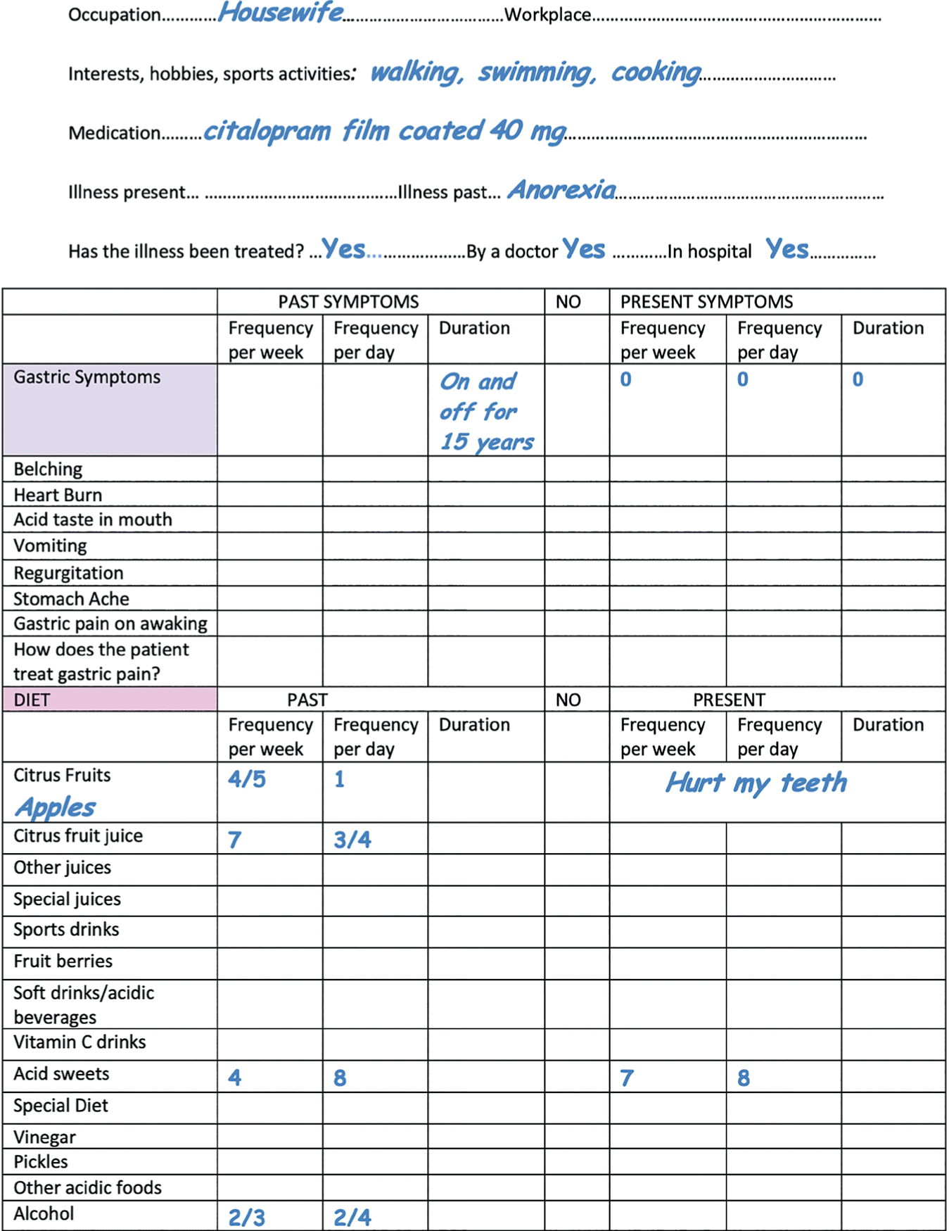
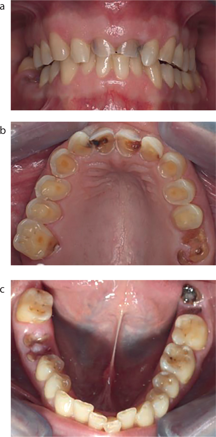
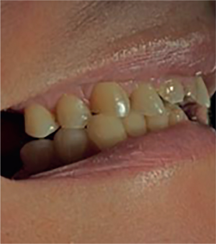
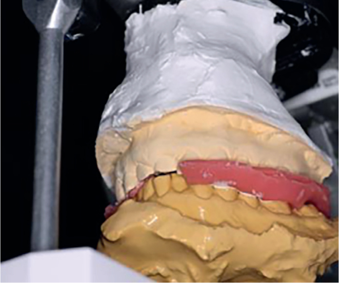
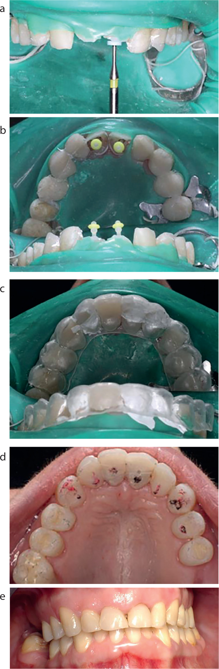
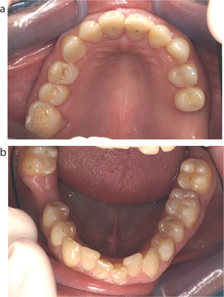
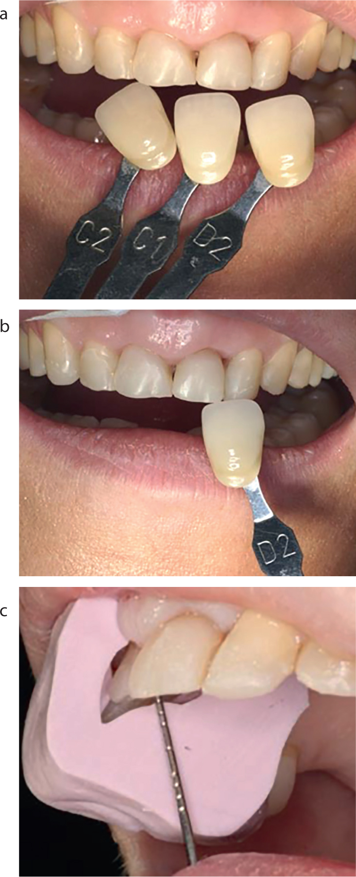
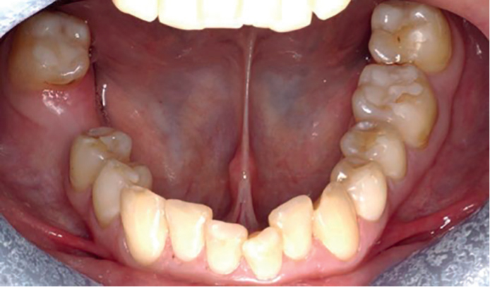
This paper presents a case demonstrating treatment of localized severe toothwear and generalized moderate toothwear, in a previously bulimic and anorexic patient. The treatment plan and rationale for treatment is discussed. The case shows the analytical diagnostic steps taken to formulate the treatment plan, which focused on prevention, treatment of disease and restoration of teeth affected by toothwear, in order to achieve the objective of establishing oral health, function and aesthetics.

CPD/Clinical Relevance: Using available interocclusal space created by repositioning the mandible into the retruded centric relation position, this case demonstrates minimally invasive direct additive composite placements to restore generalized toothwear in general practice.

Article

Clinical history

A 37-year-old female patient presented after complaining of a swelling she had experienced four weeks previously around the upper left side of her face, and that her upper left front tooth was symptomatic during this episode. She attended her general medical practitioner who had prescribed antibiotics and advised her to see a dentist. The swelling, pain and acute symptoms subsequently settled and, at the time of presentation, as there were no acute symptoms, the patient requested improved dental health. She complained of chronic symptoms of sensitivity, difficulty eating hard foodstuffs and of the poor appearance of her front teeth. The patient had not attended the dentist for six years, and was apprehensive of dental treatments.

Medical history revealed that the patient had suffered previously with anorexia nervosa and bulimia for 15 years. She was currently under medication for depression. The patient was a non-smoker and did not drink alcohol. There was no history of any habits or drug use.

Examination

A complete extra- and intra-oral examination was completed. There was no soft tissue or extra-oral pathology detected. There was no TMJ dysfunction or pathology detected.

Periodontal examination


1 1 1
2 2 1

BPE score

There was no pathological pocketing or mobility; however, there was gingival bleeding on probing from the lower lingual sextant with associated minor deposits of calculus. There was plaque stagnation and gingival inflammation adjacent to the carious roots.

Special investigations

Radiographs: Five periapical intra-oral radiographs were taken (Figure 1). Percussion: No teeth were TTP.

Figure 1. Radiographic report.

Vitality testing with BlueFreeze Cold Spray (Sterispray) revealed no response from the upper central incisors.

An erosion analysis sheet (Figure 2) and a diet sheet (Figure 3) were completed.

Figure 2. Erosion analysis sheet.
Figure 3. Diet analysis sheet.

Occlusal analysis

There was a loss of occlusal vertical dimension caused by the progressive toothwear. The patient was over-closed, with a reduced occlusal vertical dimension of 53 mm. The resting vertical dimension was 57 mm. The occlusal plane was irregular anteriorly and posteriorly. The LR7 and LL7 were unopposed and the first contact in the Retruded Contact Position (RCP) was the LL6. In Intercuspal Position (ICP), the patient postured her mandible anteriorly to achieve maximum contact of the upper and lower teeth due to the loss of palatal upper arch tooth tissue. The patient had developed an occlusion to maximize tooth-tooth contact by posturing the mandible, which she adapted due to progressive chronic toothwear. This maximized contact between her upper and lower anterior teeth, which helped the patient function. However, it was considered that this contact was accelerating the wear of the upper central incisors and causing lower incisor wear facets. There were irregular tooth contacts in ICP caused by the posturing, and also caused by the pattern of toothwear caused by erosion (Figure 4). Inter-occlusal space was created anteriorly when the mandible was guided in ICP,1 allowing restoration of the advanced anterior localized toothwear2,3 (Figure 5). There was group function in lateral and protrusive excursions. The patient presented with a Class I incisor relationship.

Figure 4. (a–c) Pre-operative view showing generalized toothwear.
Figure 5. Patient in ICP showing space created anteriorly for restoration of worn anteriors.

Diagnoses

  • Localized chronic marginal gingivitis;
  • Caries: UR621, UL126, LL8, LR6;
  • Early caries LL6 buccal surface;
  • Caries and chronic periapical infection UR1, UL1;
  • Advanced toothwear UR1, UL1 caused principally by primary erosive risk factors;
  • Moderate toothwear upper arch and early toothwear facets occlusal surfaces of the lower arch causing occlusal irregularities and compromised function.
  • Restorability assessment

    An assessment of residual tooth tissue remaining for restoration was made. For the lower arch there was a good prognosis for all teeth as there was sufficient tooth structure remaining. The upper teeth were affected by erosion, with cupping of the palatal tooth structure which had left a ring of enamel which improved the prognosis for restoration of these teeth if using adhesive materials and bonding. The carious roots UR6, LR6, LL8 required extraction. The prognosis of the UR1, UL1 depended on bonding for a core build-up to the remaining tooth tissue after caries removal and successful endodontic therapy.

    It was considered that good bonding techniques, using the peripheral enamel ring present for composite cores, and fibre posts within the canal for bonding to dentine, would enhance the biomimetic effect and improve the long-term prognoses of these restorations.

    Aims of treatment

  • Improve function;
  • Reduce sensitivity of exposed dentine;
  • Improve aesthetics.
  • Restoration of worn tooth tissue would require an increase in the vertical dimension. Once the mandible was in intercuspal position (ICP), space was created for restoration of toothwear. Additionally, the pattern of erosive wear caused by bulimia was advanced on the palatal surfaces of the upper arch. The ‘Dahl’ concept was not used as interocclusal space was created by restoring the worn dentition in the retruded position. In this retruded position the occlusal vertical dimension was increased by 2 mm without the intrusion and extrusion required from the Dahl concept to create space to increase the vertical dimension.

    Treatment plan

    1. Scaling, oral hygiene instruction, correcting brushing technique and preventive advice

    Erosion and caries analysis sheets were discussed to improve diet and reduce future risk of further caries and toothwear.4,5 Dietary risk factors that promote toothwear and caries were also discussed. Advice was given to reduce the frequency of juice intake, to dilute juice with water and to drink through a straw. The patient was advised to avoid eating sweets and reduced intake of acidic and sweet foods was recommended.6 A list of suggested alternative healthy snacks were given and discussed for the limitation of caries and toothwear and promotion of long-term oral health.7 A sensitive discussion was important, in consideration of the patient's medical history, to ensure a continued quality of nutrition by reducing dietary sugars and acids without dramatically reducing calorie intake or omitting meals.

    Topical fluoride therapy: Duraphat 2800ppm toothpaste (0.619% sodium fluoride Colgate) and a 0.05% sodium fluoride alcohol-free daily fluoride mouthwash was also discussed.7

    2. Extractions of carious roots UR6, LR6, LL8

    3. Removal of caries UR1, UL1 and assessment for restorability

    To proceed with root canal therapy over two visits of UR1, UL1 if sufficient tooth tissue remaining for successful restoration.

    4. Occlusal analysis

    Study casts mounted in ICP on a semi-adjustable articulator to analyse the restorative build-up of worn dentition (Figure 6).

    Figure 6. Occlusion in ICP recorded.

    5. Review of preventive advice

    Discussion of treatment options for long-term restoration and re-assessment of patient motivation regarding tolerance of various treatment options.

    6. Diagnostic wax-up of worn dentition

    Duplicate study casts mounted on a semi-adjustable articulator.

    7. Discussion of options for restoration of dentition

    Discussion of options for restoration of dentition with diagnostic wax-up.

    8. Restoration of upper central incisors

    Fibre posts were planned for the upper central incisors to retain a composite core.

    A ferrule and an enamel ring was present, enhancing the final bond strength, retention, restorative outcome and therefore long-term restorative stability. The possible risk of fracture of the tooth and/or post was explained to the patient.

    9. Upper acrylic matrix of diagnostic wax-up

    The upper acrylic matrix of the diagnostic wax-up was used to aid the provision of upper arch composite build-ups,8,9 including restoration of localized caries.

    10. Composite restorations in the lower arch wear facets

    11. Composite restorations in the upper arch UR5432, UL23456 to restore tooth tissue in centric relation increasing the occlusal vertical dimension by 2 mm

    12. Review of preventive advice and maintenance

    13. After 6 months long-term definitive restorations for upper central incisors

    Preparation of upper central incisors for porcelain-bonded crowns, using the existing composite build-up UR1, UL1 as cores. This would ensure long-term marginal coronal seal, and subsequent restorative longevity.

    Provision of these crowns depended on:

  • Success of root canal therapy;
  • Patient compliance in following of preventive daily routine and maintenance of oral health.
  • Factors influencing treatment plan decision to create composite build-ups to replace tooth tissue

    The lower arch required minimal restoration, and the most conservative method was to bond composite to these wear facets with no tooth preparation.

  • This also influenced the choice of material used to restore the upper arch. Composite in both arches would wear at the same rate, and not cause preferential wear of tooth structure or restorations. Ceramic indirect restorations could potentially cause preferential wear of opposing tooth structure and composite restorations causing further maintenance care in future.
  • It was decided to restore the upper centrals with a porcelain fused to metal crown, six months after the root canal therapy of UR1, UL1, in order to secure the long-term coronal seal for these root-treated incisors. Using metal on the palatal contact areas would reduce wear on opposing restorations.
  • A ceramic fit at the labial margins was appropriate as the patient had a high smile line, with visible labial margins. There was limited dentine present within the coronal core structure to consider a dentine-bonded crown.

  • The introduction of various types of glass fibre post may have advantages over metal alloy posts. They can be adhesively bonded to dentine and an adhesive core material, possibly creating a biomimetic effect and protecting the remaining tooth structure under vertical and oblique loading.10 Occlusal forces can be loaded with better stress distribution through the post system and remaining tooth structure with fibre posts than with metallic posts,11 resulting in fewer critical stresses that may cause root fracture.12 There is also a higher fracture resistance of glass fibre posts compared with metal cast post/cores.13,14 The elastic moduli of these materials are more similar to dentine than metal, which creates a restoration that is less damaging to the remaining tooth structure left,15 and also demonstrate less microleakage.16 The long-term clinical performance of these new materials, however, has to be proven before true clinical survival can be assessed.17,18,19
  • Composite material is easier to adjust, polish and to repair. Additionally, any initial or future occlusal adjustments are easier.
  • There was a ring of enamel around the attrition and erosion cavities. Composite bonding to a ring of enamel enhances the long-term restorative benefits.20,21,22
  • Conservation of tooth structure.
  • Minimal tooth preparation necessary.
  • Options for alternative treatment

    The patient was not concerned regarding replacement of missing posterior teeth. The remaining teeth and tooth tissue present in the upper and lower arch were adequate to create a satisfactory aesthetic and functional outcome after treatment.

    The financial cost was also considered. The patient was exempt from NHS charges and was unable to afford Private treatment for implants or other Private referral restorative services. All treatment was provided within Primary Care NHS Services. The patient declined a referral for NHS Specialist Restorative Services. She now felt confident and motivated to attend future appointments in Primary Care.

    Treatment details

    1. Root canal treatment of UR1, UL1

    K-flex files were used for initial canal negotiation and preparation ensuring patency (Figure 7a). A working length radiograph using a K-flex stainless steel file was taken. Copius 4% sodium hypochlorite irrigation was used and Canal + (Septodont) ethylenediaminetetra acetic acid (EDTA). Nickel-titanium rotary files (K3 Sybron-endo) were used to shape the canals, flared to size 40 with MAF size 25.06 taper. The canals were dressed with 98% calcium hydroxide paste Hypocal (Ellman) and temporized with poly F Plus cement (Dentsply), prior to the second stage root filling (Figure 7b) and future fibre post restoration.

    Figure 7. (a) Final root canal filling using gutta percha with cold lateral condensation and Sealapex sealant. (b) Final radiograph. Caries UL2 mesial was restored at the following visit.

    2. A diagnostic wax-up

    A diagnostic wax-up was carried out using a wax additive technique on casts mounted on a semi-adjustable Denar articulator. Interocclusal space was created for restorative build-up when the mandible was in the retruded contact position, ie centric relation. PKT wax carving instruments were used to build the occlusal morphology and contacts required for even occlusal contacts creating an effective ICP in this retruded contact position (Figure 8).

  • Anterior guidance was shared between the canines and lateral incisors, with no protrusive forces on the upper centrals, and only a light ICP stop created in the cingulum. This was to avoid overloading the upper centrals;
  • The occlusal vertical dimension was increased by 2 mm. The patient was informed of this change and advised to eat a soft diet initially;
  • Even axial occlusal loading of the teeth in the intercuspal position (ICP) was created which was coincident with the retruded contact position (RCP).
  • Figure 8. Diagnostic wax-up in the retruded contact position, using the wax additive technique.

    3. Composite restorations upper and lower arch

    A pre-formed lab-constructed matrix from the diagnostic wax-up was made and used to aid accurate chairside anatomical composite build-ups (Figure 9).

    Figure 9. (a–e) Pre-formed lab-constructed matrix from the diagnostic wax-up upper arch, upper and lower arch chairside direct composite placements.

    Fuji IX (GC) was used to restore the carious lesion in UL6 prior to composite restorations. Herculite XV (Kerr) was used to restore the upper anterior teeth and Heliomolar (Ivoclar-Vivadent) for the posterior teeth. Rubber dam was used for moisture control and PTFE tape was used as a non-stick separator interproximally. Heliobond (Ivoclar-Vivadent), dentine-bonding agent was used. The oxygen-inhibited layer was cured and composites polished using Shofu white stones with glycerine (Figure 10).

    Figure 10. Oxygen-inhibited layer cured through glycerine.

    Fibre posts (Parapost) were placed, after ensuring accurate post preparation within the canals, leaving the apical seal in-tact (Figure 11).10,11

    Figure 11. (a–c) Parapost preparation for direct fibre post UR1, UL1 prior to composite build-ups. A ‘split-dam’ was used for isolation to facilitate moisture control: protection of the coronal seal and improved bonding. (b) Parapost® Fibre (Coltène) placement: cemented with PanaviaTM f 2.0 (Kuraray). (c) Use of PTFE tape prior to composite placement. (d, e) Final composite restorations showing occlusion: anterior guidance UR23, UL23 and ICP anterior stops UR1, UL1.

    After the upper arch was restored the patient experienced improved function. For the first time in years she could eat hard foods. As a result, the patient gained in self-confidence following the dental treatment.

    After six months

    The patient had no complaints, had good function, and was happy with the appearance. No occlusal interferences were detected on checking the articulation, however, there was a composite fracture UR3 palatal surface that had occurred one week prior to this appointment. This was repaired along with a composite chip LL1 mesio-incisal edge (Figure 12). Porcelain-bonded crowns were used to restore the upper central incisors definitively to secure the long-term coronal seal for these endodontically-treated teeth (Figure 13). Figures 14, 15 and 16 present further follow-up illustrations.

    Figure 12. (a) Fractured composite UR3 palatal surface after 6 months. (b) Chip LL1 mesial-incisal edge.
    Figure 13. (a–c) Shade selection: Shade C2 neck, body mix of 50% C2 and 50% C1 shade. C1 tip with translucency 1 mm incisal edge. Reduction guides made from initial diagnostic wax-up.
    Figure 14. Lower arch 15 months after initial phase of treatment. Note: moderate composite wear LL6 buccal cusps and canine tips early wear. This was subsequently restored.
    Figure 15. Diagnostic and final radiographs showing root canal treatments and healing periapical areas of restored UR1, UL1.
    Figure 16. A 15 month follow-up.

    Conclusion

    A minimally invasive approach using composite resin has been used to treat this toothwear case. This case was completed eight years ago, hence the bonding system used.

    Only composite repairs have been necessary and completed since that time. The patient is stable, and composite restorations provide satisfactory longevity. However, some maintenance is required as there are risks of wear, chipping, marginal staining and fracture of the restorations, which is to be expected with time.

    Patients experience a high level of satisfaction from this method of treatment.20,21,22 The composite restorations can be repaired or replaced in a single visit and have acceptable maintenance.