Galbraith PJ, Richardson ML Permanently retained acupuncture needles: radiographic findings and case report. Radiol Case Rep. 2006; 1:120-122
Mizrahui B, Scully C Dental radiography: gold thread therapy. Br Dent J. 2014; 216
Alsaadi G, Jacobs R, Quirynen M, Van Steenberghe D Soft tissue augmentation of the cheeks detected on intra- and extra-oral radiographs: a case report. Dentomaxillofac Radiol. 2008; 37:117-120
Wang Y, Wang C, Liu K A women's secret. Ann Emerg Med. 2013; 62:224-234
Stark GB, Bannasch H The “Golden Thread Lift”: radiologic findings. Aesthet Plast Surg. 2007; 31:206-208
A 67-year-old female was referred to secondary care with asymptomatic foreign bodies visible on intra-oral radiographs. Clinical examination was unremarkable. Radiographic examination revealed nine pin-like radio-opaque objects throughout the face and head present on multiple previous imaging modalities. The patient was unaware of their presence and could not account for them. A review of available literature revealed similar objects have been used during acupuncture and cosmetic procedures without complication. As the objects were asymptomatic, the patient was reassured and discharged.
CPD/Clinical Relevance: Routine radiographic examination of patients may reveal the presence of unusual and unexpected findings.
Article
Ailish Clark
A 67-year-old female was referred to the Department of Oral Medicine at Glasgow Dental Hospital and School in late 2016 seeking a second opinion relating to foreign bodies visible on intra-oral radiographs. This referral was made by Practitioner Services, an NHS body responsible for approving certain courses of dental treatment in general dental practice within Scotland. This patient required a large course of dental treatment and it was felt that a specialist opinion regarding the origin of the objects identified on the radiographs was required.
Case history
On presentation, the patient did not have any complaints relating to the objects present in the head and neck region as they were entirely asymptomatic. The patient reported being unaware of their presence until alerted by her general dental practitioner. The patient described an episode of trauma in the 1980s; the exact nature of her injuries is unclear from this incident, however, it did result in some right-sided weakness. She also reported periods of significant illness in Malaysia. She stated that on one of these occasions she was taken for emergency treatment in Malaysia where she received a combination of hypnosis and acupuncture, using 22 carat gold needles. The patient reported that she had received this treatment to her head and neck region and other areas of her body.
Medical history
The patient had a previous diagnosis of Ramsay-Hunt syndrome resulting in a right facial palsy and numbness of her right cheek and lip. Also reported was a background of hypothyroidism, hayfever, osteoarthritis, anxiety and depression, benign paroxysmal positional vertigo and previous surgery for a mass in her stomach. Furthermore, due to persistent migraines, she had been investigated extensively by the Neurology Department in the past. Regular medications were unremarkable.
Social history
The patient was a regular dental attender in primary care, was retired, an ex-smoker, who drank no alcohol and lived alone.
Clinical examination
On extra-oral examination, there was no swelling, lymphadenopathy and no palpable objects noted in the soft tissues. There was a pre-existing lower motor neurone weakness of the right facial nerve affecting the temporal, buccal and marginal mandibular branches. The patient also reported numbness involving the lower right lip and border of the mandible. Intra-orally, the patient was partially dentate and was not wearing any removable prosthesis.
Examination of the soft tissues revealed a 3 mm white patch in the labial mucosa adjacent to a fractured lower left lateral incisor; the appearance was consistent with frictional keratosis. On palpation of the edentulous alveolar ridge over the area of the lower central incisors, there appeared to be a bony spur, likely related to recent extractions in this area. Otherwise there was nothing to note.
Investigations
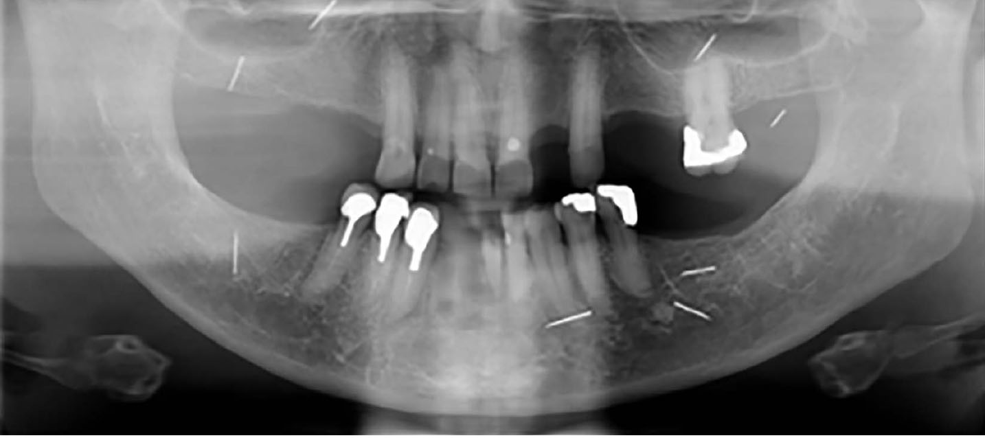
A Dental Panoramic Radiograph (DPT) was taken at the visit to evaluate the location and number of radio-opaque objects present (Figure 1).
Figure 1. Panoramic radiograph taken at initial consultation.
Nine straight, pin-like radio-opaque objects were located throughout the face and head. These measured approximately 8.0 mm in length and 0.3 mm in width. Caries was also present in the remaining dentition.
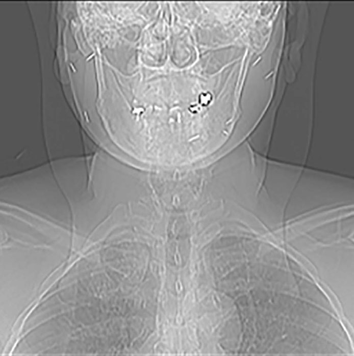
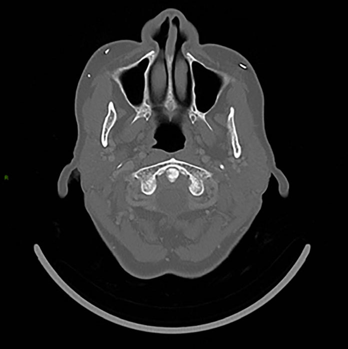
Figure 1 was compared to previous images available on an online database comprising five MRIs of the head taken over a 6-year period, a CT of the head and neck taken in 2015 and an additional full DPT taken in 2007. Images from the CT scan of the head in 2015 are shown in Figures 2 and 3.
Figure 2. AP scout image showing multiple bilateral linear radio-opaque objects.Figure 3. Axial view at level of pterygoid plates showing metallic objects within buccal subcutaneous tissues.
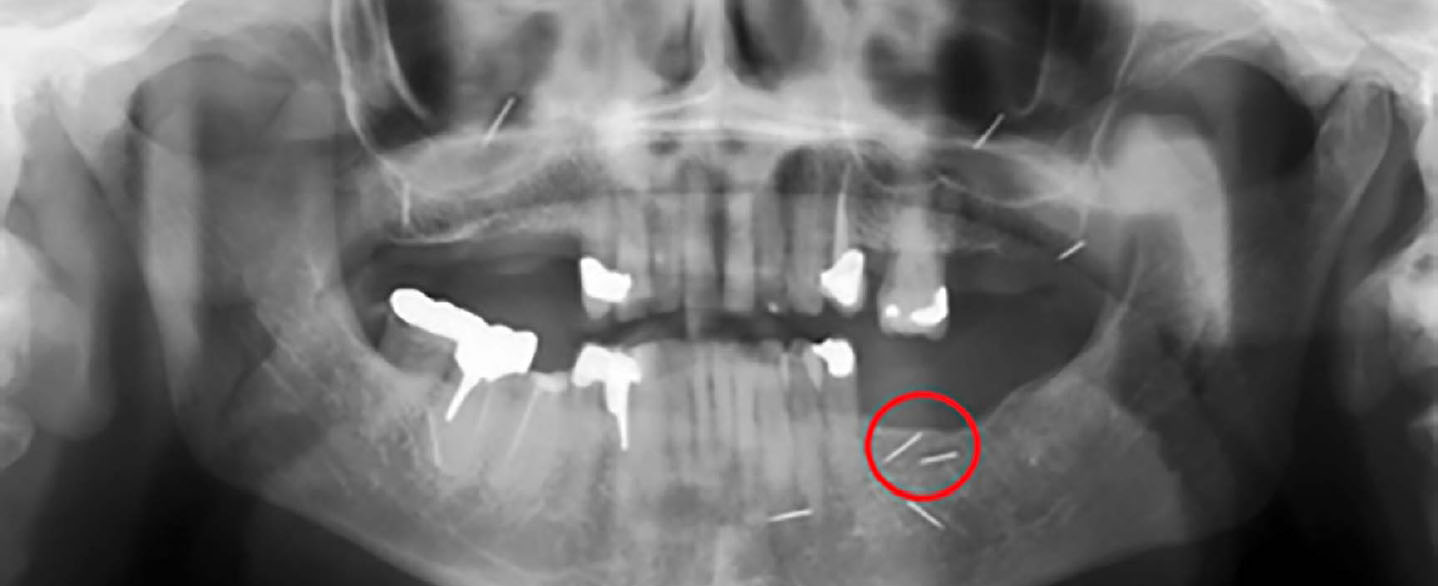
In the nine-year period between the two DPTs, the only noticeable difference was the disappearance of one object superimposed over the left body of the mandible (Figure 4).
Figure 4. OPT taken in 2007 with area of change between 2007 and 2016 images highlighted.
Differential diagnosis
It was decided that the foreign bodies were most likely present due to one of the following:
Implantation during acupuncture therapy;
Implantation during elective cosmetic procedure;
Debris from an accident.
Treatment
As the patient had not reported any cosmetic procedures but had received acupuncture therapy, it was thought that this was the most likely explanation for the origin of the objects. The patient was reassured and discharged.
Discussion
On reviewing the literature, the presence of radio-opaque objects of a similar appearance has been described in multiple case reports.1–5 Historically, these objects have apparently been used during both acupuncture and cosmetic procedures.1–5 In terms of cosmetic procedures, there are reports of radio-opaque objects being noted in the head and neck region due to ‘gold thread therapy’.2–5 During this practice, threads of 24 carat gold of 0.1 mm diameter are placed into the sub-dermal skin for the purpose of facial rejuvenation.2
This is reportedly used in plastic surgery practice in some areas in Europe to promote angiogenesis and is also used with chronic diseases such as arthritis.2–6 The case reports available describe the appearance of these objects as curvilinear and present in the soft tissues of the head and neck region.2–5
Other specialties have also reported this unusual finding on radiographic examination of other areas of the body such as the hands, chest and thorax.6–8 One report regarding radio-opaque objects (acupuncture needles), found in the chest region on radiographic examination, described the appearance and dimensions of the objects similar to those in our case and was thought to be due to the practice of Hari.1 Hari is a Japanese form of acupuncture where needles, usually gold, approximately 10 mm in length, are placed in the skin and intentionally left in situ for pain control.1 All the case reports mentioned related the objects to acupuncture therapy of some sort used for treatment of arthritis, other pain conditions or cosmetic treatment.1–8 Most cases did not report any adverse effects from the objects and it is thought that many people may be unaware of their presence until incidentally found. However, one report described previous instances of migration of the objects which could have detrimental effects.8
Our patient did not report any cosmetic procedures and additionally was unable to clarify details of the nature of the previous accident. Therefore, it was thought that these explanations were unlikely to explain the origin of the objects. Additionally, as the patient had undertaken multiple magnetic resonance imaging scans without movement of the objects, this lead to the conclusion that they were non-ferromagnetic or paramagnetic. Given the history and available literature, it was suspected that the objects were gold acupuncture needles which had not appeared to cause any adverse effects to date and so were left in situ. The patient was made aware that, should they migrate in future and cause symptoms, she may require removal of the objects.