Pitts N, Chadwick B, Anderson T. Child Dental Health Survey. Report 2.: Department of Health; 2013
Smith MC, Lantz EM, Smith HV. The cause of mottled enamel. Science. 1931; 74:(1914)
Black GV. Mottled teeth: an endemic developmental imperfection of the enamel of the teeth heretofore unknown in the literature of dentistry. 1916;
Chankanka O, Levy SM, Warren JJ, Chalmers JM. A literature review of aesthetic perceptions of dental fluorosis and relationships with psychosocial aspects/oral health-related quality of life. Community Dent Oral Epidemiol. 2010; 38:97-109
Neville BW, Damm DD, Allen CM, Chi AC. Oral and Maxillofacial Pathology.St Louis, USA: Saunders; 2015
Marshman Z, Rodd HD. The Psychosocial Impacts of Developmental Enamel Defects in Children and Young People. Planning and Care for Children and Adolescents with Dental Enamel Defects.Berlin Heidelberg: Springer; 2015
Dean T. Classification of mottled enamel diagnosis. J Am Dent Assoc. 1934; 21:1421-1426
Holloway PJ, Ellwood RP. The prevalence, causes and cosmetic importance of dental fluorosis in the United Kingdom: a review. Community Dent Health. 1997; 14:148-155
Pini NIP, Sundfeld-Neto D, Aguiar FHB, Sundfeld RH, Martins LRM, Lovadino JR Enamel microabrasion: an overview of clinical and scientific considerations. World J Clin Cases. 2015; 3:34-41
Rodrigues MC, Mondelli RFL, Oliveira GU, Franco EB, Baseggio W, Wang L Minimal alterations on the enamel surface by micro-abrasion: in vitro roughness and wear assessments. J Appl Oral Sci. 2013; 21:112-117
Sundfeld RH, Croll TP, Briso ALF, de Alexandre RS, Sundfeld Neto D. Considerations about enamel microabrasion after 18 years. Am J Dent. 2007; 20:67-72
Gugnani N, Pandit IK, Gupta M, Gugnani S, Soni S, Goyal V. Comparative evaluation of esthetic changes in nonpitted fluorosis stains when treated with resin infiltration, in-office bleaching, and combination therapies. J Esthet Restor Dent. 2017; 29:317-324
Gugnani N, Pandit IK, Goyal V, Gugnani S, Sharma J, Dogra S. Esthetic improvement of white spot lesions and non-pitted fluorosis using resin infiltration technique: series of four clinical cases. J Indian Soc Pedod Prev Dent. 2014; 32:176-180
Gupta A, Dhingra R, Chaudhuri P, Gupta A. A comparison of various minimally invasive techniques for the removal of dental fluorosis stains in children. J Indian Soc Pedod Prev Dent. 2017; 35:260-268
Wallace A, Deery C. Management of opacities in children and adolescents. Dent Update. 2015; 42:951-958
Kamp AA. Removal of white spot lesions by controlled acid-pumice abrasion. J Clin Orthod. 1989; 23:690-693
Balan B, Madanda Uthaiah C, Narayanan S, Mookalamada Monnappa P. Microabrasion: an effective method for improvement of esthetics in dentistry. Case Rep Dent. 2013; 2013
Castro KS, Clá Udia De Araú A, Ferreira J, Ngela R, Duarte M, Bio F Acceptability, efficacy and safety of two treatment protocols for dental fluorosis: a randomized clinical trial. J Dent. 2014; 42:938-944
Rogers HJ, Yesudian G, Rodd HD. Unusual extrinsic staining following microabrasion in a girl with amelogenesis imperfecta. Eur Arch Paediatr Dent. 2016; 17:271-275
Ashkenazi M, Sarnat H. Microabrasion of teeth with discoloration resembling hypomaturation enamel defects: four-year follow up. J Clin Pediatr Dent. 2000; 25:29-34
Azer SS, Hague AL, Johnston WM. Effect of bleaching on tooth discolouration from food colourant in vitro. J Dent. 2011; 39:e52-56
Karadas M, Seven N. The effect of different drinks on tooth color after home bleaching. Eur J Dent. 2014; 8:249-253
Boushell LW, Ritter AV, Garland GE, Tiwana KK, Smith LR, Broome A Nightguard vital bleaching: side effects and patient satisfaction 10 to 17 years post-treatment. J Esthet Restor Dent. 2012; 24:211-219
Marked extrinsic staining following microabrasion: a case report of a boy with dental fluorosis Nikolaos N Lygidakis Kathryn Harley Dental Update 2024 46:5, 707-709.
Authors
Nikolaos NLygidakis
BDS, DDent, MJDF, MPaedDent
Postgraduate in Paediatric Dentistry, Eastman Dental Hospital, 256 Gray's Inn Road, London WC1X 8LD, UK
A 12-year-old boy, born and raised in Iran, presented with dental fluorosis affecting all his teeth. The defects were predominantly opaque white in appearance with brown opacities on the maxillary central incisors. The treatment plan entailed microabrasion of the maxillary central incisors followed by vital bleaching, if required. Despite providing post-operative instructions, the patient had a drink containing turmeric later that day. He subsequently presented with yellow staining of his maxillary central incisors. A further course of microabrasion as well as vital bleaching provided a satisfactory final result. The teeth remained unchanged in a two-month review appointment.
CPD/Clinical Relevance: This case presents a rare complication following microabrasion for dental fluorosis which occurred because post-operative instructions were not followed.
Article
Developmental defects of enamel (DDE) are a common anomaly seen in teeth, with a prevalence of approximately 28% in 12-year-old children in the UK.1 One of the most common DDEs is dental fluorosis. Dental fluorosis is defined as hypomineralization of enamel resulting from an excessive intake of fluoride during tooth development.2
It is well documented that fluoride can have a beneficial impact on the dentition, with evidence going back to the early 1900s.3 The benefit comes primarily from the topical application of fluoride after the teeth have erupted by enhancing remineralization of the enamel and reducing the potential for demineralization following acid attacks. However, systemic absorption during tooth development leading to dental fluorosis is one of the most common dental anomalies. It is characterized by the presence of diffuse, thin, horizontal white striations and stained plaque areas.4 More severe cases may present with discoloration of enamel ranging from light yellow to dark brown, with associated pitting.5
This condition can cause cosmetic concern to young patients.6 Dental fluorosis was traditionally graded using the Dean's index which grades as mild, moderate and severe.7 In more recent years, the DDE index has also been used, however, this is not specific to fluorosis but rather it is used to describe all dental anomalies.8
There are several non-invasive treatments available for the restoration of appearance of anterior teeth. For superficial enamel defects or stains, microabrasion is a well-established technique as it is considered safe, conservative and cost-effective. There are various approaches, products and degrees of clinical success.9 Techniques involve the use of 18% hydrochloric acid and pumice, 10% hydrochloric acid with silica carbide particles and 37% phosphoric acid with pumice.10, 11 If the discoloration is present in the superficial layer of the labial enamel, then microabrasion can be effective and leave a smooth, glassy enamel surface as the finished result.
Other non-invasive techniques for the treatment of DDE include resin infiltration and vital bleaching. Resin infiltration is a relatively new procedure which is reported mostly through case reports.12, 13 In 2017, a comparative study of 80 patients comparing resin infiltration with bleaching for the treatment of dental fluorosis concluded that resin infiltration offers better results than bleaching.12 However, vital bleaching has been used for many years with good results.14, 15
This paper discusses a case of microabrasion that was complicated by failure to follow post-operative instructions with regard to the consumption of certain foodstuffs.
Case report
A 12-year-old boy attended the Department of Paediatric Dentistry for assessment of his discoloured teeth. He was fit and well. He did not like the appearance of his front teeth and, for that reason, he rarely smiled. He was born and raised in Iran before moving to the UK at 10 years of age. His sister, who was 13 years old, also had the same condition.
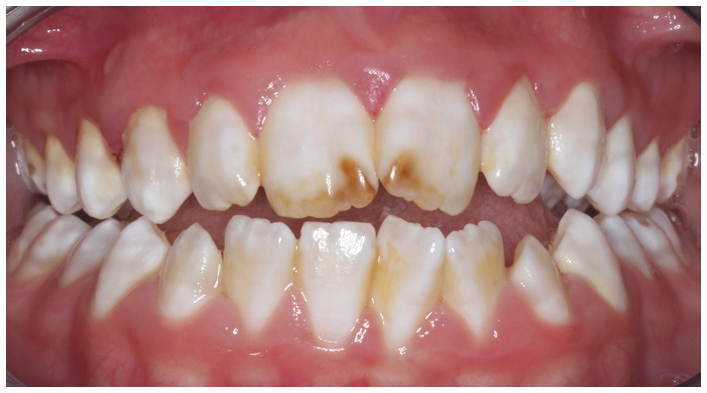
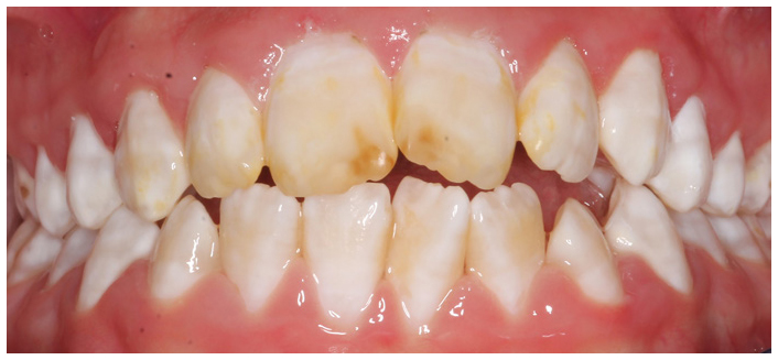
Clinical examination revealed that his oral hygiene was good and there was no caries. It was noted that he had generalized, symmetrical mottling of the enamel affecting all his permanent teeth. The maxillary central incisors also had significant brown discoloration towards the incisal edges (Figure 1). The clinical appearance, together with the history of his first 10 years spent in an area of water fluoridation, indicated a diagnosis of dental fluorosis.
Figure 1. Anterior view of dentition.
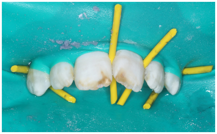
After discussion with his parents, it was agreed to carry out microabrasion of the maxillary central incisors and then, if needed, vital bleaching. The patient attended a few days later for the initial treatment of microabrasion. This was carried out on the labial aspect of the maxillary central incisors under rubber dam. No local anaesthesia was given thus the rubber dam was secured with Rubber Dam Wedgets® (Coltène) (Figure 2). Using a prophylaxis brush in a slow handpiece and a mixture of pumice and 37% phosphoric acid to create a slurry, the labial surfaces of the incisors were polished. The acid-etch 37% (SDI) and pumice (CR Minerals) mixture was used for 10 seconds, five times followed by washing, drying and polishing with fine Sof-Lex discs (3M). This technique was first described in 1989.16
Figure 2. Isolation of the maxillary anterior teeth under rubber dam.
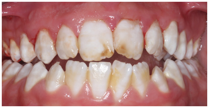
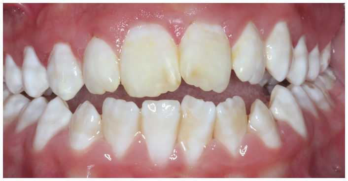
Following microabrasion, the brown opacities on the maxillary central incisors became less obvious, but were still visible (Figure 3). On the same day, an alginate impression for a maxillary bleaching tray was obtained in order for the patient to carry out home bleaching.
Figure 3. Post-operative view illustrating some improvement in the appearance of the anterior teeth.
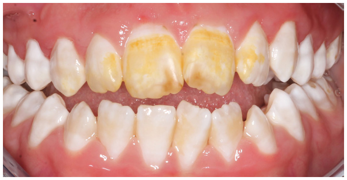
The patient and the parents were provided with verbal post-operative instructions emphasizing the need to avoid food and drinks with colouring for two days. He was reviewed after one week. Unfortunately, both the father and his son were unhappy when they returned as the microabraded teeth had developed a marked yellow discoloration (Figure 4). On questioning, the young boy stated that he had consumed a drink containing turmeric the same night that he had attended for his treatment of microabrasion. An attempt was made to polish the yellow stains with pumice, however, this failed. Thus, a second course of microabrasion was carried out using the same method as previously described.
Figure 4. Anterior view of dentition demonstrating yellow discoloration caused by tumeric.
Whilst the majority of the yellow staining was removed following a second course of microabrasion, a yellow hue still remained (Figure 5). On the same day, the patient was given a bleaching tray and two tubes of 10% carbamide peroxide gel (Opalescence) with clear instructions on its use. The patient was asked to clean his teeth last thing at night, apply the carbamide peroxide gel to the labial aspect of the bleaching tray and, following insertion of the tray to cover his maxillary teeth, wipe away any excess gel. The tray was worn overnight for 7 nights. In the morning, the tray was removed and cleaned, and the teeth brushed. Instructions were also given for managing sensitivity, however, the patient did not experience any during the treatment. Finally, advice was given on foods to avoid during the treatment, similar to the advice given for the microabrasion.
Figure 5. Appearance of maxillary anterior teeth following further microabrasion.
Follow up
The patient was reviewed after 2 weeks (Figure 6). The brown discolorations had almost completely disappeared and the staining from the turmeric was completely removed. However, on close examination, the teeth appeared ‘cream’ when compared to the opaque white colour of the adjacent and opposing teeth affected by fluorosis. Although this did not concern the patient, others may wish to continue bleaching the affected incisors for a longer period to reduce the contrast further. In a subsequent appointment 2 months later, the appearance of the teeth remained unchanged.
Figure 6. Anterior view of dentition following home bleaching of the maxillary incisors.
Discussion
The risks of microabrasion are minimal and side-effects rare. The main post-operative instructions following treatment are to avoid foodstuff with colouring for 48 hours. A review of microabrasion techniques in 2015 by Pini et al makes no reference to complications similar to that found in this case report.9
Microabrasion has been the technique of choice for managing dental fluorosis for many years. However, as demonstrated in this case, it may have complications if the post-operative instructions are not followed correctly. In recent years, bleaching materials, such as carbamide peroxide and hydrogen peroxide, have been used alone or in conjunction with microabrasion for the management of developmental defects of enamel, such as fluorosis and amelogenesis imperfecta.14, 15, 17 The reasons for that are the simplicity of the procedure, the potentially good results observed, and the minimal risk to the dentition. There remains no guidance as to whether microabrasion should be carried out before bleaching or bleaching ahead of microabrasion. This is best decided on an individual basis. It is, however, important to mention the guidelines regarding tooth bleaching for under 18s which, according to the EU regulation (Directive 2011/84 EU), is illegal. The GDC's position, however, is extremely helpful for cases such as that described, given that it states that the use of hydrogen peroxide to treat or prevent disease in individuals with defects of enamel is possible. In this case, bleaching was beneficial in reducing the discoloration of the enamel. The alternative, composite restorations would be very destructive and would not have benefited the patient.
A randomized control trial comparing microabrasion with microabrasion and bleaching for patients with fluorosis found that there was no difference in the outcome between the two techniques.18 However, they do conclude that the combination of the two techniques is a more effective means of reducing enamel fluorosis staining and observed that patient satisfaction was better when both techniques were used.18 This case demonstrated that bleaching did manage to remove the brown opacities towards the incisal edges of the maxillary central incisors, which microabrasion did not. This suggests that the combination of the two techniques resulted in a more successful result.
A literature search found only one published paper discussing similar complications following microabrasion for a child with amelogenesis imperfecta. In this case report, tomato sauce was ingested the same day as the microabrasion was performed and the teeth turned yellow.19 Management of the discoloured teeth was resolved by bleaching with 16% carbamide peroxide alone before placement of composite resin restorations. Apart from this study, there are no other documented cases of extrinsic staining following microabrasion. A study in 2000 found no instances of staining following a four-year follow-up of 18% hydrochloric acid and pumice treatment for the management of enamel discolorations.20 There are, however, studies linking extrinsic staining with vital bleaching, possibly because of its much wider use. There is an increased risk of extrinsic staining following bleaching with carbamide peroxide, which is due to increased surface roughness of the enamel following bleaching, which has been found to facilitate subsurface penetration of colourants.21, 22 However, these studies are in vitro. A literature search for clinical reports of discoloration following vital bleaching found no such cases, whereas other complications, such as sensitivity, are common.23 The changes in enamel structure following microabrasion are fundamentally different from that of bleaching, however, an increase in surface roughness is seen after both treatments.9 Additionally, a small amount of enamel is removed by microabrasion which, for 37% phosphoric acid with pumice, can reach 40 μm.10 Based on that assumption, the post-operative instructions following microabrasion are very similar to those of bleaching. These should include the avoidance of consumption of food and liquids with colourings for a minimum of 48 hours, as well as advising the patient to refrain from smoking for a minimum of 2 hours.
This case raises the question as to whether microabrasion should be the first treatment of choice for dental fluorosis. As seen here, vital bleaching provided a better result, by removing the yellow hue as well as the majority of the brown discoloration.
Conclusion
In the patient discussed, failure to follow post-operative instructions resulted in a complication, which is rarely seen. The consumption of a drink containing turmeric the same day as microabrasion of the maxillary incisors resulted in a yellow discoloration of the treated teeth. In order to resolve the acquired staining, it was necessary to provide a further course of microabrasion in addition to bleaching the teeth. The final result was satisfactory. This case illustrates the importance of post-operative instructions to inform patients of the need to avoid foodstuffs with colourings for at least 48 hours following treatment. Post-operative instructions for such cases should be written and given to both patient and parent, as complications can be avoided by carefully following them. This case also highlights the need for further research to understand how food and drinks with colourings can stain teeth with DDEs following microabrasion.