Echeverri EA, Wang MM, Chavaria C, Taylor DL. Multiple dens evaginatus: diagnosis, management, and complications: case report. Pediatr Dent. 1994; 16:314-317
Merrill R. Occlusal anomalous tubercles on premolars of Alaskan Eskimos and Indians. Oral Surg Oral Med Oral Pathol. 1964; 17:484-496
Kocsis G, Marcsik A, Kokai E, Kocsis K. Supernumerary occlusal cusps on permanent human teeth. Acta Biol Szeged. 2002; 36:71-82
Curzon M, Curzon J, Payton H. Evaginated odontomes in the Keewatin Eskimo. Br Dent J. 1970; 129:324-328
Palmer M. Case reports of evaginated odontomes in Caucasians. Oral Surg Oral Med Oral Pathol. 1973; 35:772-779
Siqueira V, Braga T, Martins MA, Raitz R, Martins MD. Dental fusion and dens evaginatus in the permanent dentition: literature review and clinical case report with conservative treatment. J Dent Child. 2004; 71:69-72
Yip W. The prevalence of dens evaginatus. Oral Surg Oral Med Oral Pathol. 1974; 38:80-87
Geist J. Dens evaginatus – case report and review of the literature. Oral Surg Oral Med Oral Pathol. 1989; 67:628-631
Senia E, Regezi J. Dens evaginatus in the etiology of bilateral periapical involvement in caries-free premolars. Oral Surg Oral Med Oral Pathol. 1974; 38:465-468
Stewart R, Dixon G, Graber R. Dens evaginatus (tuberculated cusps): genetic and treatment considerations. Oral Surg Oral Med Oral Pathol. 1978; 46:831-836
Davies P, Brook A. The presentation of talon cusp: diagnosis, clinical features, associations and possible etiology. Br Dent J. 1985; 159:84-88
Pearlman J, Curzon M. An evaginated odontoma in an Am Negro: report of case. J Am Dent Assoc. 1977; 95:557-572
Hattab F, Yassin O, Al-Nimri K. Talon cusp in permanent dentition associated with other dental anomalies: review of the literature and reports of seven. J Dent Child. 1996; 63:368-376
Stecker S, Diangelis A. Dens evaginatus a diagnostic and treatment challenge. J Am Dent Assoc. 2002; 133:190-193
Oehlers F, Lee K, Lee E. Dens evaginatus (evaginated odontome): its structure and responses to external stimuli. Dent Pract Dent Rec. 1967; 17:239-244
Darkner R, Harari D, Rotstein I. Dens evaginatus of anterior teeth – literature review and radiographic survey of 15,000 teeth. Oral Surg Oral Med Oral Pathol Oral Radiol Endod. 1996; 81:472-476
Mellon J, Ripa L. Talon cusp: a clinically significant anomaly. Oral Surg Oral Med Oral Pathol. 1970; 29:225-228
Ngeow W, Chai W. Dens evaginatus on a wisdom tooth: a diagnostic dilemma. Case report. Aust Dent J. 1998; 43:328-330
Oehlers F. The tuberculated premolar. Dent Prac Dent Rec. 1956; 6:144-148
Lau T. Odontomes of the axial core type. Br Dent J. 1995; 99:219-225
Hill FJ, Bellis WJ. Dens evaginatus and its management. Br Dent J. 1984; 156:400-402
Priddy W, Carter H, Auzins J. Dens evaginatus – an anomaly of clinical significance. J Endod. 1976; 2:51-52
Levitan MR, Himel VT. Dens evaginatus: literature review, pathophysiology, and comprehensive treatment regimen. J Endod. 2006; 32:1-9
Tsurumachi T, Suguro H, Ogata H, Hatori K, Kobayashi C, Ogiso B. Endodontic treatment of bilateral dens evaginatus premolars with large periapical lesions. J Oral Sci. 2009; 51:475-479
Yong SL. Prophylactic treatment of dens evaginatus. ASDC J Dent Child. 1974; 41:289-292
Koh ET, Pitt Ford TR, Kariyawasam SP, Chen NN, Torabinejad M. Prophylactic treatment of dens evaginatus using mineral trioxide aggregate. J Endod. 2001; 27::540-542
Chueh LH, Huang GTJ. Immature teeth with periradicular periodontitis or abscess undergoing apexogenesis: a paradigm shift. J Endod. 2006; 32:1205-1213
Bazan M, Dawson L. Protection of dens evaginatus with pit and fissure sealant. J Dent Child. 1983; 50:361-363
Chen R Conservative management of dens evaginatus. J Endod. 1984; 10:253-257
Pre-eruptive diagnosis and management of occlusal dens evaginatus in premolar teeth Farhad B Naini Ioannis Levisianos Lee Foo Daljit S Gill Dental Update 2024 45:9, 707-709.
Authors
Farhad BNaini
Consultant Orthodontist, St George's and Kingston Hospitals, London, UK
Dens evaginatus is an uncommon dental aberration that occurs primarily on the occlusal surfaces of premolar teeth. Developmentally, it is similar to a talon cusp, though the latter manifests on the palatal or lingual surfaces of anterior teeth. Its anatomical location is often the central fossa of the associated tooth; it can vary significantly in size and can be present symmetrically in the ipsilateral and contralateral quadrants. Histologically, the enamel protuberance may contain dentinal and pulpal tissue. If early, ideally pre-eruptive diagnosis is not made, there is a significant risk that, on eruption, these protuberances will shear off under occlusal forces, exposing the pulp, and potentially leading to loss of teeth in an otherwise healthy dentition. Therefore, its clinical implications are significant to the general dental practitioner, the paediatric dentist, the orthodontist and the restorative dentist.
CPD/Clinical Relevance: This review article aims to summarize the available dental literature and illustrate the appropriate management of a patient with dens evaginatus.
Article
Dens evaginatus (DE) is a developmental dental anomaly that presents as an enamel protuberance commonly associated with the tooth's anatomical fossa.1,2 This uncommon dental aberration may project superior to the neighbouring enamel tissue and histologically it is comprised of an external enamel shell, a dentine core and, occasionally, a pulp tissue horn. The pulp tissue may extend partially or entirely into the tubercle.3 DE most frequently arises from the occlusal fossa of posterior teeth and the palatal/lingual surface of anterior teeth. It is more commonly seen in premolars but it can present in molars and canines; its presentation being five times more common in the mandible than the maxilla.4 Typically, the anomaly will present bilaterally in a symmetrical fashion with a slight gender predilection for females.5 There is no evidence that the occurrence is related to any medical problems or syndromes. Indeed, the literature is awash with nomenclature that have been used to describe this morphological phenomenon, such as odontome, odontoma of the axial core type, evaginatus odontoma, occlusal enamel pearl, occlusal tubercle, tuberculum anomalus, accessory cusp, supernumerary cusp, interstitial cusp, tuberculated cusp, tuberculated premolar, Leong's premolar, dens evaginatus and talon cusp (in reference to the anterior teeth).5,6,7,8,9,10,11,12,13,14,15,16,17,18-19 Most of the literature is in agreement that the developmental processes that lead to the presentation of DE and talon cusp are very similar,1,6,19,20 to the extent that the terminology ‘talon cusp’ has traditionally been used to describe DE. The latter, coined by Oehlers (1967), was later universally adopted to allow differentiation between the anterior and posterior tooth presentations.18 Other developmental oddities, such as the cusp of Carabelli, with a reported prevalence of 17.4 to 90%, typically seen on the palatal surface of the maxillary first molar mesiopalatal cusp, seem to be morphologically distinct. The differentiation is primarily histological, as the enamel process rarely contains pulpal tissue and only occasionally dentinal tissue. In addition, the position of Carabelli's cusp is inconsequential and would not normally present an occlusal interference, in contrast to DE or a talon cusp.1
The most prevalent developmental hypothesis of the formation of DE is thought to be an irregular proliferation and enfolding of the inner enamel epithelium and subjacent ectomesenchymal cells of the dental papilla into the stellate reticulum of the enamel organ at an early stage of tooth formation (bell stage).21,22-23
Epidemiology
Dens evaginatus is much more common in the East Asian ethnic group populations, such as the Chinese, Thai, Japanese and Indian populations (Neo-Asiatics group) and the Indonesian-Malay populations (Filipino group), with a reported incidence of 0.5 to 4.3%.6 Case reports in other populations have shown an incidence of up to 15% in Alaskan Eskimos and native North, Central and South American Indians (Paleo-Asiatics group).5 A much smaller incidence has been reported in the Caucasian population group.24 There has been some suggestion in the literature that the inheritance of the DE trait could be closely associated to other ethnic-related morphological traits, such as ‘shovel-shaped’ incisors, which are often seen in East Asian populations,1 the presence of midline supernumeraries10,11 and three-rooted mandibular molars.12
Clinical appearance
Even though there can be a considerable differentiation of the DE tubercles, typically they can be up to 2.0 mm in diameter25 and up to 3.5 mm in height.8 There have been reports in the literature that the associated teeth can be larger mesiodistally; however, there is no evidence that the resulting tooth-size discrepancy is clinically significant. The morphology of the associated tooth is usually unaffected by the presence of the tubercle.3
As shown in Tables 1–3, there have been several classifications described in the literature used to categorize the DE tubercles. The classifications most commonly used are based on the size and location of the tubercle,6 the anatomical description of the tubercle,23 the extent of pulp tissue horn infiltration in the tubercle18 and, finally, the treatment required as a result of pulpal exposure of the protuberance.26
Type I
A cone-like enlargement of the lingual cusp
Type II
A tubercle on the inclined plane of the lingual cusp
Type III
A cone-like enlargement of the buccal cusp
Type IV
A tubercle on the inclined plane of the buccal cusp
Type V
A tubercle arising from the occlusal surface obliterating the central groove
Type 1
Wide pulp horns (34%)
Type 2
Narrow pulp horns (22%)
Type 3
Constricted pulp horns (14%)
Type 4
Isolated pulp horn remnants (20%)
Type 5
No pulp horn (10%)
Type I
Normal pulp, mature apex
Type II
Normal pulp, immature apex
Type III
Inflamed pulp, mature apex
Type IV
Inflamed pulp, immature apex
Type V
Necrotic pulp, mature apex
Type VI
Necrotic pulp, immature apex
Clinical significance
The cusp-like projections can become a diagnostic and treatment conundrum to the general dental practitioner (GDP) due to the tubercle's discreet appearance and often significant treatment sequelae. More commonly, the teeth with associated DE tubercles will erupt unimpeded in the dentition, at which point they will interfere with the static and functional occlusion. This will result in either the alteration of the natural occlusion in a less traumatic relationship or the occlusal wear or, more seriously, shearing fracture of the offending portion of the enamel (the tubercle's superior aspect).24,27 The sequelae of the tooth surface loss ultimately will depend on the extent of infiltration of the pulp horn (if any) towards the superior aspect of the protuberance. Microscopic exposure of the pulp horn following abrasion of the enamel and dentinal tissue will inevitably result in pulpal pathology requiring endodontic management.27
Several prophylactic restorative strategies have been described in the literature. The most conservative option is selective gradual reduction of the enamel outcrop in order to facilitate tertiary dentine formation and occlusion of the pulpal horn.24 Other management techniques include prophylactic pulp capping,28 conventional occlusal restorations and finally root canal treatment.29 The advent of Mineral Trioxide Aggregate (MTA) has greatly improved the prognosis of endodontically treated teeth by inducing apexification or apexogenesis, depending on the stage of the tooth's root development.30 Alternatively, in the instance where orthodontic treatment is planned, necessitating mid-arch extractions, a tooth affected by DE may be the preferred option. This would either obviate the need for future restorative work or reduce it substantially, depending on the number of teeth affected.
Case report
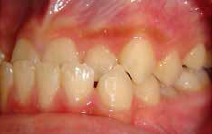
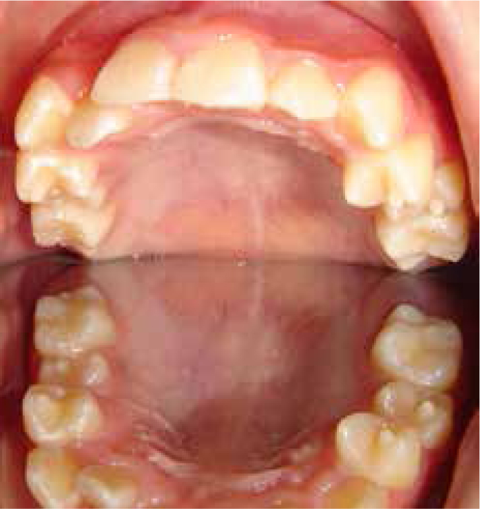
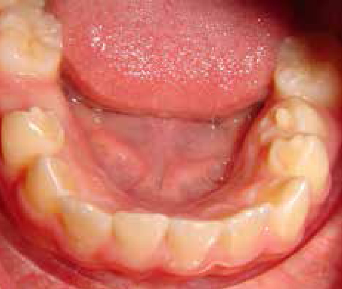
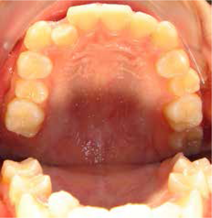
A 12-year-old male presented in the orthodontic clinic following a referral from his local GDP who was concerned about his anterior tooth relationship. The patient presented with no real concerns regarding his malocclusion. Clinical examination (Figure 1) revealed a Class III incisor relationship with non-displacing anterior crossbite on a Class III skeletal base in the early permanent dentition. As an incidental finding, enamel protuberances were observed on all the erupting premolars in the maxillary and mandibular arches (Figures 2 and 3). The diagnosis made was DE affecting all the maxillary and mandibular premolar teeth.
The orthodontic management of this patient included monitoring of the development of the malocclusion on an annual basis, as he or his parents were not aesthetically concerned with the anterior crossbite. Furthermore, due to the severity of skeletal discrepancy, his age and potential for further mandibular growth, it was deemed prudent to delay any orthodontic intervention up to the point that either the malalignment of the maxillary dentition became a concern or until skeletal maturity, when a comprehensive orthodontic-surgical approach could be implemented.
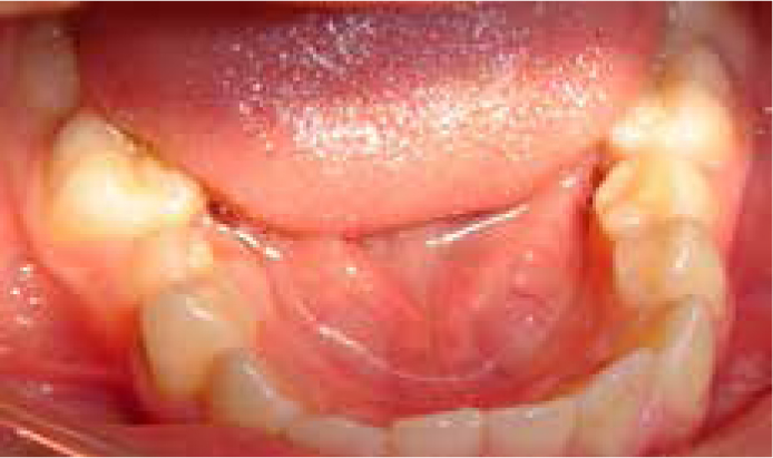
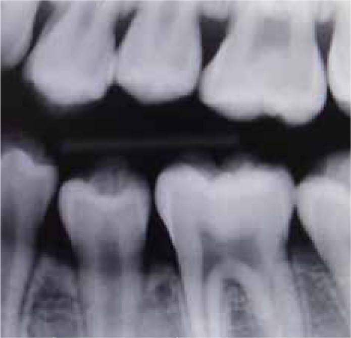
Management of the DE protuberances was thought to be of greater significance at the point of consultation. This was related to the patient's early presentation and the relative virginal state of the enamel tubercles, which implied that significant restorative treatment could be avoided in the future if an intervention could be employed at that time. After a consultation with a paediatric dentistry colleague, the proposed plan included gradual reduction of the evagination tubercles to allow for formation of reparative dentine over the pulpal horns. Following informed consent, there were several episodes of enamel reduction that were aimed at stimulating tertiary dentine formation. Approximately ¼ mm of enamel was removed per appointment, with appointments approximately one month apart. Topical fluoride was immediately applied on the reduced surface to increase the enamel's hydroxyapatite crystals resistance to acid dissolution between treatment episodes. Complete elimination of the protuberances was undertaken 4 months following the initial presentation that ensured that the pulpal horn had receded sufficiently. The exposed dentine was then covered with an acid-etched, light-cured composite resin. Clinical and radiographic examination showed no pathology or pulpal exposure associated with any of the premolar teeth (Figures 4–6). The patient was then advised to return on an annual basis to the orthodontic clinic to assess development of his malocclusion.
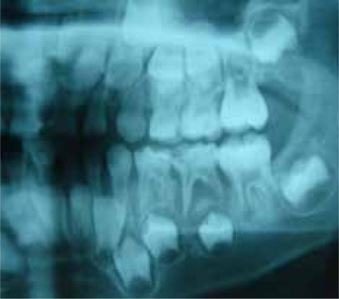
In most circumstances, the diagnosis of DE should be made by a simple oral examination. A clinical appearance of an unusual enamel anatomy associated with the tooth's fossa that features an enamel tubercle, is pathognomonic. The diagnosis can often be led astray by occlusal wear, depending on the dental age of the patient, or a lesser form of the tubercle, often not easily seen with the naked eye, or presence of caries. As the premolar teeth are commonly associated with DE, sound clinical practice dictates that a thorough clinical and radiographic investigation should involve all the premolar teeth, including the contralateral and ipsilateral quadrants. Ideally, this should include sensibility testing of erupted premolars, and periapical radiographs. Indeed, for this particular patient, a dental panoramic tomograph (DPT), that was kindly forwarded to the department by the referring practitioner, showed quite clearly the presence of fully formed tubercles on the unerupted premolars 3 years previously, whilst the patient was still in the early mixed dentition stage (Figure 7).
Figure 7. Pre-treatment dental panoramic tomograph (left side). The occlusal DE is evident on all the unerupted premolar teeth, but is particularly conspicuous on the unerupted LL5.
As illustrated previously, pulpal involvement originating from a DE tubercle can form extremely quickly following the eruption of the tooth in the oral cavity due to the masticatory occlusal forces. Therefore, early diagnosis may lead to preventive treatment that can minimize extensive future restorative treatment. In this case, an orthodontic referral led to the incidental finding of the DE tubercles, which precipitated a successful outcome. However, the DPT showed that the diagnosis ideally could have been made several years prior to the premolar eruption and would have helped in terms of patient education and future treatment planning.
Lastly, the preventive regimen that was implemented for this patient is commonly accepted to be the most successful in the literature. A number of alternative techniques have previously been used that have fallen out of favour due to the lack of convincing outcomes. Spot grinding has been previously used to disocclude the tooth carrying the tubercle without its complete removal. This has been found to produce unpredictable results as, even if pulpal involvement had been avoided with spot grinding, the enamel protuberance would still be susceptible to fracture, leading to enamel defects and dentine exposure.2 Another method has been the use of an unfilled resin sealant to provide protection for the tubercle; this was proven inadequate against occlusal forces.31 A third method involved reduction of the tubercle until the underlying dentine was barely exposed and then treating it with 8% stannous fluoride to precipitate formation of tertiary dentine.32 However, as the possibility of pulpal horn infiltration up to the dentino-enamel junction is high, there would be no guarantee that such a technique would not result in an accidental micro-exposure. Overall, it is accepted that the use of flowable (micro-fill) composite resins following the removal of the tubercle is the gold standard of treatment today.26,32 The newer, acid-etched, particle-filler resins have been found to be resistant to abrasion and occlusal loading, as well as precipitating a faster pulpal horn withdrawal and tertiary dentine formation.
Conclusion
Dens evaginatus is an uncommon dental aberration and, consequently, the majority of the available literature is case report-or case series-based. It can readily be diagnosed by clinical examination; however, as illustrated in this article, the importance of careful pre-eruptive reporting of any available radiographs cannot be over-emphasized. In conclusion, the dental practitioner should be familiar with the clinical presentation of DE, the radiographic features that can lead to a diagnosis in unerupted teeth and the possible consequences and treatment implications.