Murry T, Carrau R Clinical Management of Swallowing Disorders, 2nd edn. San Diego CA: Plural Publishing; 2006
Bramanti E, Arcuri C, Cecchetti F, Cervino G, Nucera R, Cicciu M Dental management in dysphagia syndrome patients with previously acquired brain damages. Dent Res J. 2012; 9:(4)361-367
Thexton AJ Mastication and swallowing: an overview. Br Dent J. 1992; 173:197-206
Legget R Review of transit times through major segments of the alimentary tract. Annals of the ICRP, ICRP 100: Human Alimentary Tract Model for Radiological Protection. 2006; 203-232
Orchardson R, Cadden S Mastication and swallowing 1: Functions, performance and mechanisms. Dent Update. 2009; 36:327-337
Orchardson R, Cadden S Mastication and swallowing 2: Control. Dent Update. 2009; 36:390-398
Nilsson H, Ekberg O, Olsson R Quantitative aspects of swallowing in an elderly non-dysphagic population. Dysphagia. 1996; 11:180-184
Kelly J, Wright D Administering medication to adult patients with dysphagia: part one. Nursing Standard. 2009; 23:(29)62-68
Shanley C, O'Loughlin G Dysphagia among nursing home residents: an assessment and management protocol. J Gerodontol Nurs. 2000; 26:(8)35-48
Gordon C, Hewer RL, Wade DT Dysphagia in acute stroke. Br Med J (Clin Res Ed). 1987; 295:411-414
Mann G, Hankey GJ, Cameron D Swallowing function after a stroke: prognosis and prognostic factors at 6 months. Stroke. 1999; 30:744-748
Rogers B, Stratton P, MK, Koerner P, Piazza J Long-term morbidity and management strategies of tracheal aspiration in adults with severe developmental disabilities. Am J Ment Retard. 1994; 4:490-498
Singh S, Hamdy H Dysphagia in stroke patients. Postgrad Med J. 2006; 82:383-391
Horner J, Massey EW Silent aspiration following stroke. Neurology. 1988; 38:317-319
Kreeft AM, Coen RN, Muller SH, Parmeijer FA, Hallo E, Balm AJM Cine MRI of swallowing in patients with advanced oropharyngeal carcinoma: a feasibility study. Europ Archiv Otorhinol. 2012; 269:(6)1703-1711
Crary MA, Carnaby-Mann GD, Groher ME Initial psychometric assessment of a functional oral intake scale for dysphagia in stroke patients. Archiv Phys Med Rehab. 2005; 86:1516-1520
Kunieda K, Ohno T, Fujishima I, Hojo K, Morita T Reliability and validity of a tool to measure the severity of dysphagia: The Food Intake LEVEL Scale. J Pain Symp Mgmt. 2013; 46:(2)201-206
Samuels R, Chadwick DD Predictors of aspiration risk in adults with intellectual disabilities and dysphagia. J Intellect Disab Res. 2006; 50:362-370
Geeganage C, Beavan J, Ellender S, Bath PMW Interventions for dysphagia and nutritional support in acute and subacute stroke. Cochrane Database of Systematic Reviews. 2012; (Issue 10) https://doi.org/10.1002/14651858.CD000323.pub2
Hyson HC, Johnson AM, Jog MS Sublingual atropine for sialorrhea secondary to parkinsonism: a pilot study. Mov Disord. 2002; 17:1318-1320
De Simone GG, Eisenchlas JH, Junin M Atropine drops for drooling: a randomized controlled trial. Palliat Med. 2006; 20:665-671
Lim M, Mace A, Nouraei R Botulinum toxin in the management of sialorrhoea: a systematic review. Clin Otolaryngol. 2006; 31:267-272
Porta M, Gamba M, Vaj P Treatment of Sialorrhoea with ultrasound guided botulinum toxin type A injection in patients with neurological disorders. J Neurol Neurosurg Psych. 2001; 70:538-540
Borg M, Hirst F The role of radiation therapy in the management of Sialorrhea. Int J Rad Oncol Biol Phys. 1998; 41:1113-1119
Consultant and Honorary Senior Lecturer in Special Care Dentistry; Clinical Lead, Department of Sedation and Special Care Dentistry, Guy’s and St Thomas’ NHS Foundation Trust, London
Dysphagia is defined as a ‘difficulty in swallowing’ and is commonly found in the general population, particularly in the elderly. This article gives an overview of the more frequently encountered swallowing disorders and provides advice on how to manage the dysphagic patient in the dental surgery.
Clinical Relevance: By identifying patients with dysphagia and being aware of the potential problems that the clinician may experience when treating them, the risk of aspiration, choking and healthcare-acquired upper respiratory tract infections may be reduced.
Article
Dysphagia is a medical term defined as a ‘difficulty in swallowing’ and can include saliva, liquids and foods of all consistencies.1 It derives from the Greek root dys meaning difficulty or disordered, and phagia meaning ‘to eat’. It describes difficulties in the oral preparation of food for swallowing or moving the bolus from the mouth to the stomach so may include difficulties in positioning of food in the mouth and oral movements. Dysphagia is distinguished from similar symptoms including odynophagia, which is defined as painful swallowing, and globus, which is the sensation of a lump in the throat.
Swallowing
Swallowing is a process by which food and liquid move from the mouth, down through the back of the throat, through the oesophagus and into the stomach. It is estimated that each individual swallows between 500 and 2000 times per day and it occurs during sleep and in utero.
Swallowing has been divided into three phases, which are defined by the position of the bolus at any one time:
Oral phase;
Pharyngeal phase;
Oesophageal phase.
Oral phase
In this phase, food is reduced to a bolus, chewed, mixed with saliva and then transported from the anterior to posterior oral cavity for passage into the pharynx.
Pharyngeal phase
This phase is characterized by involuntary acts and the activation of mechanisms which trigger peristaltic reflexes.2
The velopharyngeal opening completely closes, the hyoid and larynx ascend superiorly and the epiglottis downfolds. The tongue base makes contact with the pharyngeal wall to form a seal, then the top and bottom pharyngeal muscles start to contract. The hyoid-laryngeal complex continues to move superiorly and the laryngeal inlet closes. The epiglottis completely folds down to an inverted position and the vocal cords adduct. The cricopharyngeus muscle then relaxes and the upper oesophageal inlet opens.3 During the pharyngeal phase, which occurs rapidly, apnoea occurs to prevent food entering the airway.
Oesophageal phase
The upper oesophageal sphincter opens and peristalsis carries the bolus through the oesophagus to the stomach. As the bolus reaches the stomach, the lower oesophageal sphincter opens. The oesophageal phase is much slower and may take 10-12 seconds.4
The reader is referred to a series of articles by Cadden and Orchardson in Dental Update for a more comprehensive description of the mechanisms of swallowing.5,6
Incidence of dysphagia
It is believed that dysphagia is under-diagnosed; in 2007–2008 there were 22,510 patients admitted to hospitals in England and Wales with a primary diagnosis of dysphagia and its incidence increases with advancing age. One report suggests that 70–90% of older people have some degree of swallowing dysfunction.7 The increase in incidence may be due to conditions including cerebrovascular accidents (CVAs), Parkinson's disease or merely as a result of the ageing process, which is thought to influence all phases of swallowing negatively.8 One study found 40–60% of nursing home residents were found to have a diagnosis of dysphagia.9 It has been reported that 23–50% of individuals who sustain a CVA will develop dysphagia following the brain injury which may be permanent or temporary.10,11
People with learning disabilities have cognitive and physiological impairments which may result in dysphagia and a large proportion suffer from dysphagia.12 In 2004, the National Patient Safety Agency (NPSA) published a report ‘Understanding the patient safety issues for people with learning disabilities’13 which highlighted the risks of dysphagia. In the four years since the report was published, awareness of dysphagia has increased and the NPSA has received 605 reports of choking-related incidents involving adults with learning disabilities.14
Oropharyngeal dysphagia can be referred to as ‘high’ dysphagia and patients may have difficulty in initiating the swallow and usually identify the cervical area as the presenting problem;
Oesophageal dysphagia can be referred to as ‘low’ dysphagia and usually refers to a location in the distal oesophagus.
Causes of dysphagia
Dysphagia can be secondary to defects in any stage of the swallowing process so there are numerous causes. It is helpful to distinguish between the causes of oropharyngeal and oesophageal dysphagia.
Causes of oropharyngeal dysphagia
In younger patients, dysphagia is most commonly caused by muscular disease, webs and rings. The most frequent causes of dysphagia in elderly patients are CVAs, Parkinson's disease and dementia (Table 1).
The causes of oesophageal dysphagia may be divided into diseases causing narrowing of the lumen of the oesophagus, diseases which are caused by a physical obstruction to the oesophagus by lymph node enlargement or direct invasion, or diseases affecting the oesophageal smooth muscle or sphincters that interrupt the peristaltic wave (Table 2).
Diseases Causing Internal Narrowing of the Lumen of the Oesophagus
Oesophageal tumours
Peptic strictures
Oesophageal webs and rings, eg Plummer Vinson syndrome
Chemical injury
Post-radiation therapy
Infectious/eosinophilic oesophagitis
Diseases Causing External Narrowing of the Lumen of the Oesophagus
Tumours, eg lymphomas/lung tumours
Infections, eg Tuberculosis
Cardiovascular, eg aortic aneurysm
Diseases Affecting Smooth Muscle
Achalasia
Scleroderma
Medications which may cause dysphagia
Many medications can cause dysphagia. They can be divided into:
Medications affecting the musculature of the oesophagus, for example oxybutynin, tolterodine;
Medications (or combinations of medications) that may produce a dry mouth; these impair the ability to move food within the oral cavity. Common examples include diuretics, calcium channel blockers, antihistamines;
Antipsychotic/neuroleptic drugs can cause a dry mouth and, additionally, some can cause movement disorders which may affect the muscles of the face and tongue used in swallowing: examples are haloperidol, risperidone, clozapine;
Local anaesthetics used for dental treatment may cause a temporary loss of sensation which may affect the ability to swallow;
Drugs affecting the central nervous system can decrease voluntary muscle control which may affect swallowing. Examples are anti-epileptic medication, benzodiazepines, narcotics and smooth muscle relaxants used to relieve severe muscle spasms.
Assessment of swallowing
Swallowing assessment may be divided into clinical examination and examination using instrumentation. Clinical examination can be carried out by clinicians, speech and language therapists or nurses in a hospital setting. The patient is presented with a small amount of food or water and the assessor watches for signs of dysphagia and aspiration. The signs include leakage of water from the mouth, facial weakness, poor muscular co-ordination, delayed pharyngeal/laryngeal elevation, choking, breathlessness and changes in voice quality after swallowing.16
Videofluoroscopy (VFS)
The ‘gold-standard’ for swallowing assessment is videofluoroscopy (VFS),17 also known as the modified barium swallow. Radio-opaque barium liquid of varying consistencies is swallowed by the patient and moving images of the swallowing process are captured. The ‘swallow’ is recorded and can be played back at varying speeds to show the passage of the radio-opaque material through the oral cavity, pharynx and oesophagus. The motion picture produced will illustrate whether any of the barium has entered the airway. This procedure takes approximately 15 minutes and is available at most hospitals. It is carried out by speech therapists, radiographers and radiologists.
Fibre-optic endoscopic evaluation of swallowing (FEES)
This examination has been developed over the last 20 years and has the advantage over VFS in that it does not expose the patient to radiation. It is carried out at the bedside and involves the insertion of a nasoendoscope to the level of the soft palate to give a view of the hypopharynx and larynx when the patient is eating. It requires a highly skilled operator and technical equipment.
Dynamics magnetic resonance imaging
Dynamics magnetic resonance imaging, commonly referred to as Cine MRI, is a new technique that has been used to visualize the dynamic structures of the oral cavity during swallowing.
Cine MRI directly visualizes the dynamics of swallowing of patients with oral and oropharyngeal cancer and has been shown to yield additional information compared to standard examination of swallowing. It is not intended to replace other swallowing evaluation techniques but is considered as a useful adjunct, showing particular tongue muscles, the floor of the mouth, tongue mobility and the extent and location of the tumour.18
Other tools used to measure the severity of dysphagia
Two scales have been described which are used to reflect the oral intake of patients with dysphagia:
The Functional Oral Intake Scale (FOIS);
The Food Intake Level Scale (FILS).
The Functional Oral Intake Scale (FOIS)
This scale was developed to assess the oral intake of food and liquid in dysphagic patients who had suffered CVAs. It describes the functional level of the patient's actual daily oral intake of food or liquid.19 Patients are assigned a level of 1–7 relating to their degree of oral feeding. Levels 1–3 relate to varying degrees of non-oral feeding; levels 4–7 relate to varying degrees of oral feeding with non-oral supplementation (Table 3).
Tube Dependent (Levels 1–3)
Level
Oral intake
1
No oral intake
2
Tube dependent with minimal/inconsistent oral intake
3
Tube supplements with consistent oral intake
Total Oral Intake (Levels 4–7)
Level
Oral intake
4
Total oral intake of a single consistency
5
Total oral intake of multiple consistencies requiring special preparation
6
Total oral intake with no special preparation, but must avoid specific food or liquid items
7
Total oral intake with no restrictions
To score the functional oral intake using this scale, information is obtained from various sources including medical charts, dietary journals and patient/carer reporting. This scale is useful for documenting changes in oral feeding function following a CVA.
The Food Intake Level Scale (FILS)
The food intake level scale is a scale described in 2012. It comprises a 10 point observer-rating scale to describe the severity of dysphagia20 (Table 4). Although the scale shows similarities to the FOIS, it is believed to be more sensitive to changes in the oral intake of food than the FOIS as it has 10 items rather than 7.
No Oral Intake
Level 1
No swallowing training is performed except for oral care.
Level 2
Swallowing training not using food is performed.
Level 3
Swallowing training using a small quantity of food is performed.
Oral Intake and Alternative Nutrition
Level 4
Easy to swallow food less than the quantity of a meal (enjoyment level) is ingested orally.
Level 5
Easy to swallow food is orally ingested in one or two meals, but alternative nutrition is used as a complement.
Level 6
The patient is supported primarily by ingestion of easy to swallow food in three meals, but alternative nutrition is used as a complement.
Oral Intake Alone
Level 7
Easy to swallow food is orally ingested in three meals. No alternative nutrition is given.
Level 8
The patient eats three meals by excluding food that is particularly difficult to swallow.
Level 9
There is no dietary restriction, and the patient ingests three meals orally, but medical considerations are given.
Level 10
There is no dietary restriction, and the patient ingests three meals orally (normal).
The risks of swallowing
Choking may occur during the normal process of swallowing. Choking occurs when the passage of air to the lungs is blocked by a foreign body and is a precursor to asphyxiation. Signs of choking include coughing, gagging, the inability to speak, breathe or cry, loss of consciousness and cyanosis.21 If the obstruction is not successfully removed, then asphyxiation and ultimately death may result.
Swallowing problems may lead to inhalation of either the oropharyngeal or gastric contents into the airway. If the oropharyngeal or gastric contents enter the lower respiratory tract or larynx (below the true vocal cords), then aspiration is said to have occurred.13 People without swallowing abnormalities routinely aspirate microscopic amounts of food and liquid but gross aspiration is abnormal and can lead to respiratory complications.
In addition to the syndromes listed above, dysphagia may lead to malnutrition, dehydration and a poor quality of life. Chadwick et al reviewed dysphagia in people with learning disabilities and stated that aspiration frequently leads to chest infections which are the leading cause of death in this group.13
Sialorrhoea (drooling) is described as the unintentional loss of saliva from the mouth and is frequently seen in patients with dysphagia. It is thought to be due to a tendency to swallow less frequently and weakness of the orofacial musculature rather than the production of excessive saliva. It is worsened by poor posture, impaired head control and excitability.
Sialorrhoea frequently leads to the development of dry, sore, inflamed skin in the mouth, chin and neck region, which may lead to infection. Dehydration may occur owing to the loss of fluid and eating may become difficult.
The management of dysphagic patients
Interventions for treating dysphagia are often administered by speech and language therapists (SLTs). Basic interventions involve the modification of fluid and food consistencies, postural techniques, swallowing exercises, and stimulation of oral and pharyngeal structures. Local stimulation techniques include thermal and electrical stimulation. Transcranial direct current stimulation (TDCS) and transcranial magnetic stimulation (TMS) are also under investigation. Acupuncture techniques are used routinely in some countries and show some success.
A number of types of pharmacological agents (capsaicin, black pepper oil, cabergoline, angiotensin-converting enzyme (ACE) inhibitors, and nifedipine) have also been studied in patients, mostly with chronic dysphagia, but their effects are questionable.22
The nutritional intake of patients with dysphagia may be managed by using modified diet consistencies or tube feedings if swallowing is severely impaired. For the latter, tube feedings can be inserted into the nose and positioned in the stomach (nasogastric tube (NGT)), jejunum (nasojejunal tube (NJT)) or surgically placed in the stomach (percutaneous endoscopic gastrostomy (PEG)), radiologically inserted gastrostomy (RIG) tube feeding, or parenteral (intravenous (iv)) feeding.
Insertion of an NGT is relatively easy and readily reversed, but still carries significant risks, which have been highlighted by safety alerts from the UK National Patient Safety Agency.23 PEG insertion is an invasive procedure and can be complicated by bleeding, local infection, peritonitis, perforation, and aspiration leading to pneumonia and increased mortality in older stroke patients. PEG is more acceptable to patients and is more effective in the delivery of feed and maintaining nutritional status in long-term dysphagic patients, however, its insertion requires an invasive procedure carried out under general anaesthesia.
The dental management of patients with swallowing disorders
The dental team treating someone with dysphagia should be aware of the problems that may present:
Frequent choking;
Gagging;
Inability to control saliva;
Nasal regurgitation;
Wet/hoarse voice following swallowing;
Coughing;
Chronic heartburn;
Sensation of inhalation of food while swallowing;
Food stuck in the throat or chest;
Recurrent chest infections;
Unexplained weight loss.
The major concern when treating patients with dysphagia is the risk of aspiration during dental treatment which may lead to choking or aspiration pneumonia. In order to avoid this:
Treat the patient sitting upright;
Allow the patient frequent breaks during treatment;
Protect the airway with rubber dam or use high volume suction with a competent, experienced dental nurse;
Reduce the flow of water from the fast handpiece;
Use the slow speed handpiece as much as possible;
Use fast-setting dental materials which are not moisture sensitive if the patient is unable to control his/her saliva;
Take care that trays are not overloaded when taking impressions;
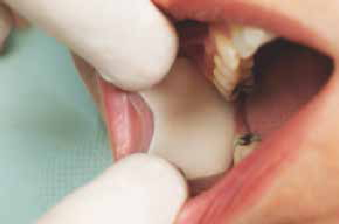
Use saliva ejectors as much as possible and other aids, such as carboxymethylcellulose pads, in the buccal sulcus (Dry Tips, Molynlycke Healthcare Ltd, Dunstable, Bedfordshire) (Figure 1) or cotton wool rolls.
Take care when placing restorations or cementing cast restorations to remove excess material;
Ultrasonic scalers can be used with caution provided that there is effective high volume suction;
Use local anaesthesia with caution in the posterior parts of the oral cavity as it may further impair the process of swallowing; infiltration anaesthesia may be more appropriate than nerve blocks;
Sedate the dysphagic patient with caution using intravenous sedation in slowly titrated doses. Many patients with dysphagia have additional medical problems which may render them unsuitable for sedation in a primary care setting and they should be referred to a specialist centre.
Figure 1. Molnlycke ‘Dry Tips’ saliva absorbents.
In addition to the dental management of the patient with dysphagia, the dental team should be aware of other problems that commonly arise in the dysphagic patient. These are:
Dysphagic patients tend to have poor oral hygiene with increased levels of plaque and calculus throughout the oral cavity. They have a tendency to develop caries, in part due to the poor oral clearance of sugars and in part due to their increased susceptibility to a dry mouth. Many are elderly and have disabilities, so poor manual dexterity is common;
High calorie nutritional supplements given to the patient to aid weight gain are usually cariogenic;
Difficulties in the wearing of dentures as the oral cavity is dry;
Attending appointments can be difficult due to frequent respiratory tract infections; domiciliary care may be necessary.
The management of sialorrhoea
Many attempts have been made to manage sialorrhoea and have met with varying success rates. Input may be required from speech and language therapists and occupational therapists (who work with patients to improve their swallowing mechanics and support their posture with devices such as the head-back wheelchair). Ear nose and throat surgeons may be required to identify and correct causes of airway obstruction which contribute to drooling and neurologists may assess the patient for particular neuropathies.
After a thorough assessment, treatments are usually offered in a stepwise fashion from non-invasive, non-surgical therapies to most invasive:
Programmes aimed at improving motor control, including input from speech and language therapists and occupational therapists;
Correction of any situational factors, eg tonsillectomy, adenoidectomy, correction of relevant oral malocclusions and provision of suitable wheelchair/braces to improve posture;
Provision of orthodontic appliances, eg customized plates formed to fit the palate to aid better lip closure;
Biofeedback and automatic cueing techniques to aid the development of swallowing reflexes;
Acupuncture;
Medications: The use of anti-cholinergic medications has been reported. The most widely used drugs are glycopyrrolate, benzatropine, amitryptiline and hyoscine hydrochloride. In the UK, atropine ophthalmic drops 1% are suggested in the NICE clinical guideline for the management of Parkinson's Disease.24 They are administered sublingually twice daily to reduce sialorrhoea, although studies have shown variable success rates with this therapy, possibly due to difficulties in the administration using a small pipette.25,26
Intraglandular injection of botulinum toxin A has been used to reduce sialorrhoea. It is injected, with ultrasound guidance, into bilateral parotid and submandibular glands. Reports suggest that the effects of the toxin last only a few months, so retreatment is necessary for long-term control. The effect of repeated injections of botulinum toxin over time, or the risk of developing antibodies, are not known.27,28
Radiation therapy to salivary glands has been described as a reasonable treatment option in elderly patients who would not tolerate other forms of therapy. The effects of the radiation may last years, however, this management option is controversial as the radiation may induce malignancies, which typically would be expected to develop 10–15 years after treatment. It is therefore only suggested in the elderly and debilitated.29
Surgical therapies: Various surgical options have been described including surgery to denervate the salivary glands and surgery on the salivary glands and ducts.
Transtympanic neurectomy is a process of denervation of the salivary glands. It is carried out through the middle ear where the chorda tympani and tympanic plexus pass through en route to the salivary glands. The procedure is quick and easy to perform under local anaesthesia. Salivary function returns within 6–18 months as the nerve fibres regenerate.
Submandibular duct relocation distally within the oral cavity may be performed or the submandibular gland(s) may be excised. Parotid ducts may be relocated or ligated.
Oral care for the dysphagic patient
The following instructions should be given to the carer/patient:
Teeth must be brushed in an upright position using a small amount of non-foaming, fluoride toothpaste and a small headed, soft toothbrush;
Teeth and dentures should be brushed after meals to remove food debris using a small-headed soft toothbrush;
If there is considered to be a high caries risk, a high fluoride toothpaste should be used such as sodium fluoride toothpaste 1.1% (Colgate Duraphat 5000ppm, produced by Colgate-Palmolive, Guildford, Surrey) which can be prescribed by the dental team;
Chlorhexidine gel/spray may be used to reduce the oral bacterial flora. If the patient's periodontal condition is poor, chlorhexidine 1% oral gel (Corsodyl Gel produced by GlaxoSmithKline, Harlow, Essex) can be brushed on the teeth once or twice daily with a toothbrush to inhibit plaque formation or applied to the fitting surface of partial dentures to reduce the caries rate twice daily after brushing the teeth;
The patient should be reviewed by the dental team at three monthly intervals for the application of high fluoride varnish (eg Duraphat 22,600ppm, Colgate Palmolive, Guildford, Surrey).
Aspirating/suction toothbrushes
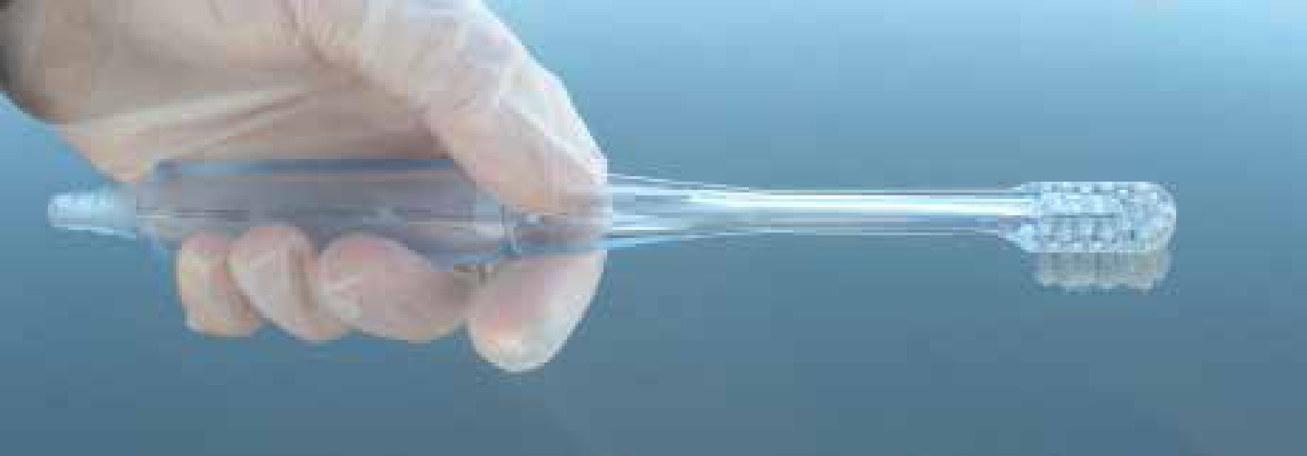
These are recommended for patients in nursing homes where suction is readily available. There are several examples on the market, such as the ‘Plak-vac’ (Trademark Medical, St Louis, Missouri) which is a re-usable combination yankauer sucker and toothbrush. It can be used with wall suction onwards or with portable suction pumps. The ‘Vac-U-Brush’ (PR Medical, Steamboat Springs, Colorado) is another alternative which is designed for use with portable suction units (Figure 2).
Figure 2. ‘Vac-U-Brush’ PR Medical.
Single use suction toothbrushes called ‘Toothettes’ (Sage Products, Cary, Illinois) which attach to standard suction lines are available and commonly used in North America.
Kimberley Clark has developed an oral care system largely for use with the ventilated critically ill patient to reduce the incidence of ventilator associated pneumonia. Their system is called ‘KimVent VAP Solutions’ and consists of a variety of suction toothbrushes, swabs and suction catheters which may be interchanged, depending on which component is most suitable for individual patients.
Conclusion
Dentists will frequently see patients with dysphagia in their practice. Careful assessment is required but the majority of patients can receive dental treatment in a primary care setting with some modifications.