Steele JG, Wassell RW, Walls AWG. Changing patterns and the need for quality. Br Dent J. 2002; 192:144-148
Silva GR, Roscoe MG, Ribeiro CP, Mota ASd, Martins LRM, Soares CJ. Impact of rehabilitation with metal-ceramic restorations on oral health-related quality of life. Braz Dent J. 2012; 23:403-408
Zarone F, Russo S, Sorrentino R. From porcelain-fused-to-metal to zirconia: clinical and experimental considerations. Dent Mater. 2011; 27:83-96
Michalakis KX, Stratos A, Hirayama H, Kang K, Touloumi F, Oishi Y. Fracture resistance of metal ceramic restorations with two different margin designs after exposure to masticatory simulation. J Prosthet Dent. 2009; 102:172-178
Mizrahi B. The anterior all-ceramic crown: a rationale for the choice of ceramic and cement. Br Dent J. 2008; 205:251-255
Eliasson A, Arnelund C-F, Johansson A. A clinical evaluation of cobalt-chromium metal-ceramic fixed partial dentures and crowns: a three- to seven-year retrospective study. J Prosthet Dent. 2007; 98:6-16
Kimmich M, Stappert CF. Intraoral treatment of veneering porcelain chipping of fixed dental restorations: a review and clinical application. J Am Dent Assoc. 2013; 144:31-44
Goodacre CJ, Bernal G, Rungcharassaeng K, Kan JY. Clinical complications in fixed prosthodontics. J Prosthet Dent. 2003; 90:31-41
Sailer I, Pjetursson BE, Zwahlen M, Hämmerle CH. A systematic review of the survival and complication rates of all-ceramic and metal–ceramic reconstructions after an observation period of at least 3 years. Part II: fixed dental prostheses. Clin Oral Implants Res. 2007; 18:86-96
Anusavice KJ. Standardizing failure, success, and survival decisions in clinical studies of ceramic and metal–ceramic fixed dental prostheses. Dent Mater. 2012; 28:102-111
Shadid RM, Sadaqah NR, Abu-Naba'a L, Al-Omari WM. Porcelain fracture of metal-ceramic tooth-supported and implant-supported restorations: a review. Open Journal of Stomatology (OJST). 2013; 3
Friedman M. A 15-year review of porcelain veneer failure – a clinician's observations. Compend Contin Educ Dent. 1998; 19:625-628
Haselton DR, Diaz-Arnold AM, Dunne JT. Shear bond strengths of 2 intraoral porcelain repair systems to porcelain or metal substrates. J Prosthet Dent. 2001; 86:526-531
Özcan M. Fracture reasons in ceramic-fused-to-metal restorations. J Oral Rehabil. 2003; 30:265-269
Malhotra N, Acharya SR. Conservative approach for esthetic repair of fractured ceramic facing in ceramic-fused-to-metal crowns: a case series. Compend Contin Educ Dent. 2012; 33:E123-129
Steele J, Nohl F, Wassell R. Crowns and other extra-coronal restorations: occlusal considerations and articulator selection. Br Dent J. 2002; 192:377-387
Mizrahi B. The Dahl principle: creating space and improving the biomechanical prognosis of anterior crowns. Quintessence Int. 2006; 37:245-251
Torbjörner A, Fransson B. Biomechanical aspects of prosthetic treatment of structurally compromised teeth. Int J Prosthodont. 2004; 17:135-141
Karl M, Fischer H, Graef F, Wichmann MG, Taylor TD, Heckmann SM. Structural changes in ceramic veneered three-unit implant-supported restorations as a consequence of static and dynamic loading. Dent Mater. 2008; 24:464-470
Johansson A, Omar R, Carlsson GE. Bruxism and prosthetic treatment: a critical review. J Prosthodont Res. 2011; 55:127-136
Brägger U, Aeschlimann S, Bürgin W, Hämmerle CH, Lang NP. Biological and technical complications and failures with fixed partial dentures (FPD) on implants and teeth after four to five years of function. Clin Oral Implants Res. 2001; 12:26-34
Kukiattrakoon B, Hengtrakool C, Kedjarune-Leggat U. Effect of acidic agents on surface roughness of dental ceramics. Dent Res J (Isfahan). 2011; 8:6-15
Rosenstiel SF, Land MF, Fujimoto J., 4th edn. St Louis, Missouri: Mosby Elsevier Inc; 2006
Newsome P, Owen S. Improving your margins. Int Dent SA. 2009; 11:36-42
Boksman L. Optimizing occlusal results for crown and bridge prostheses. Dent Today. 2011; 20:154-157
Prakash P, D'Souza D, Kumar M, Viswambaran M. Effect of firing cycle and surface finishing on the sag resistance of long-span metal ceramic framework using base metal alloys – an in vitro study. Med J Armed Forces India. 2012; 68:145-150
Taskonak B, Griggs JA, Mecholsky JJ, Yan JH. Analysis of subcritical crack growth in dental ceramics using fracture mechanics and fractography. Dent Mater. 2008; 24:700-707
Kinsel RP, Lin D. Retrospective analysis of porcelain failures of metal ceramic crowns and fixed partial dentures supported by 729 implants in 152 patients: patient-specific and implant-specific predictors of ceramic failure. J Prosthet Dent. 2009; 101:388-394
El-Sheikh AM, Hobkirk JA, Howell PG, Gilthorpe MS. Passive tactile sensibility in edentulous subjects treated with dental implants: a pilot study. J Prosthet Dent. 2004; 91:26-32
Özcan M, Niedermeier W. Clinical study on the reasons for and location of failures of metal-ceramic restorations and survival of repairs. Int J Prosthodont. 2002; 15:299-302
Bulbule N, Motwani BK. Comparative study of fracture resistance of porcelain in metal ceramic restorations by using different metal coping designs – an in vitro study. J Clin Diagn Res. 2014; 8:ZC123-ZC127
Rashid H. The effect of surface roughness on ceramics used in dentistry: a review of literature. Eur J Dent. 2014; 8
Zhang Y, Griggs JA, Benham AW. Influence of powder/liquid mixing ratio on porosity and translucency of dental porcelains. J Prosthet Dent. 2004; 91:128-135
Cheung K, Darvell B. Sintering of dental porcelain: effect of time and temperature on appearance and porosity. Dent Mater. 2002; 18:163-173
Deepak K, Ahila S, Muthukumar B, Vasanthkumar M. Comparative evaluation of effect of laser on shear bond strength of ceramic bonded with two base metal alloys: an in-vitro study. Indian J Dent Res. 2013; 24:610-615
All dental restorations are liable to failure during function. Failure could be biologic, aesthetic, mechanical or a combination. Ceramic restorations in particular, including metal-ceramics, are prone to mechanical fracture, especially the fracture of veneering porcelain. Fracture of a metal-ceramic restoration jeopardizes function as well as aesthetics. It is equally onerous to manage for both patient and dentist. Optimal management of such cases requires a detailed knowledge of the aetiology behind this phenomenon. The current paper aims to highlight possible causative factors involved in the mechanical failures of metal-ceramic restorations.
CPD/Clinical Relevance: Ceramic fracture in metal-ceramic crowns and fixed partial dentures is routinely encountered in dental clinics. Knowledge of the aetiology is required to diagnose and manage such cases accurately as well as to avoid these errors in future.
Article
Rehabilitation of teeth with crowns has increased greatly over the last three decades.1 Despite rapid advancements in the development of newer and stronger ceramic systems,2 metal-ceramic restorations still remain the ‘gold standard’ in prosthodontics since their introduction in the 1960s.3 Metal-ceramic systems combine the biomechanical advantages of metals with the aesthetics of ceramic materials,4 resulting in restorations with considerable clinical longevity.5 A survival rate of 97% for metal-ceramic restorations was reported by Eliasson et al6 after a period of ten years in clinical service.
All dental restorations are liable to failure during function. Failure may be biologic, aesthetic, mechanical or a combination. Ceramic restorations in particular, including metal-ceramics, are more prone to mechanical fracture (Figure 1), especially the fracture of veneering porcelain.7 A systematic review carried out by Goodacre et al8 revealed that fracture of veneering porcelain is a common complication associated with metal-ceramic prostheses. However, a review on the survival rate and complications of metal-ceramic restorations reported a mean chipping rate of 2.9% after a 5-year observation period.9 Such paradoxes in survival rate values exist because of a lack of detailed reporting systems for describing fractures of ceramic-based restorations.10
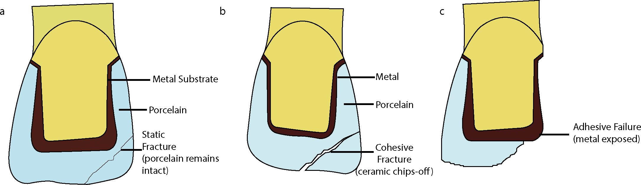
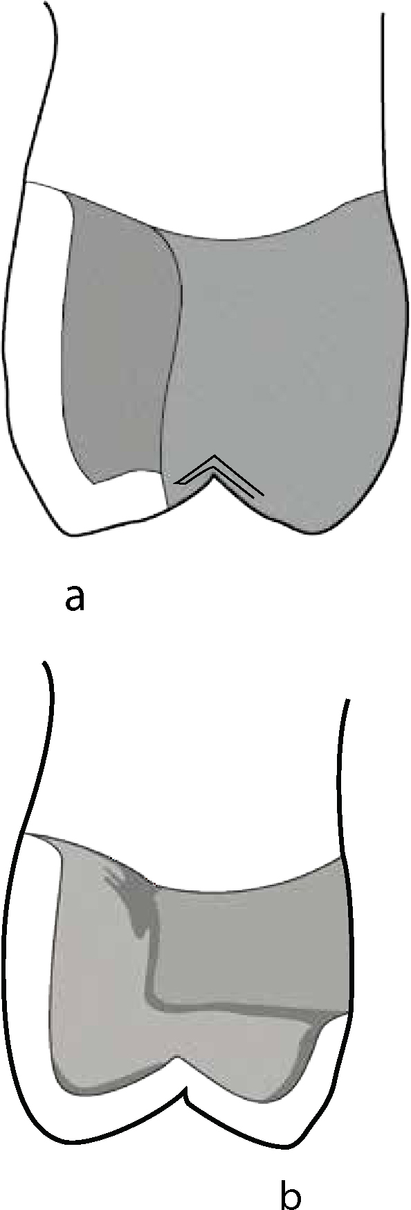
Restoration failures are often a multi-factorial phenomenon. A number of different factors may be responsible for the mechanical failure of metal-ceramic restorations,11 and factors may also vary depending on the type of fracture that has occurred. Friedman12 classified porcelain fractures into three types, namely:
Static fracture: where a segment of porcelain fractures but remains intact;
Cohesive fracture: that occurs within the body of porcelain due to tensile loads; and
Adhesive fracture: where failure of the bonding interface between the porcelain and the substrate is seen (Figure 2).
Figure 2. Pictorial representation of Friedman's classification of porcelain fractures: (a) Static fracture; (b) Cohesive fracture; (c) Adhesive fracture.
Haselton et al13 specifically described metal-ceramic restoration fractures, classifying them as:
Simple: involving only porcelain;
Mixed: involving both metal and porcelain; and
Complex: where a large area of metal framework is exposed.
Fracture of a metal-ceramic restoration jeopardizes function as well as aesthetics. Optimal management of such cases requires a detailed knowledge of the aetiology behind this fracture phenomenon.14 Unfortunately, there is a paucity of studies available that provide a comprehensive review of the reasons leading to the failure of metal-ceramic restorations. The current paper aims to highlight the possible causative factors involved in the mechanical failures of metal-ceramic restorations, thereby helping clinicians avoid them in clinical practice.
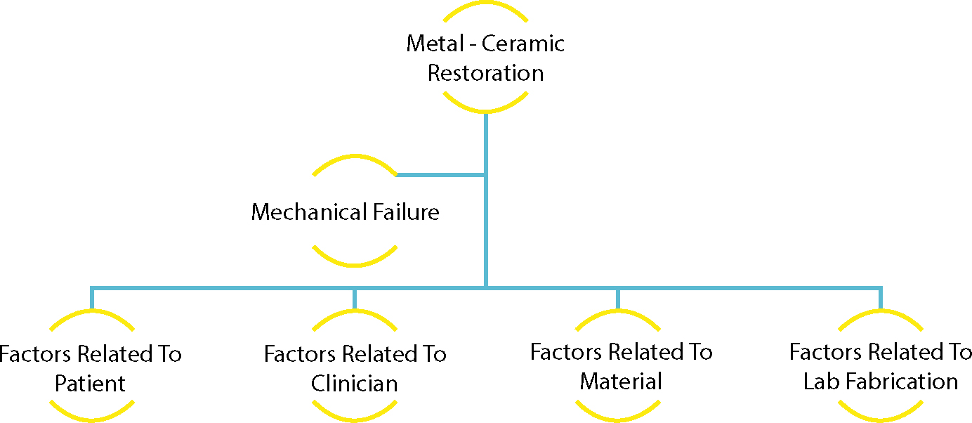
Factors affecting failure
The success or failure of metal-ceramic restorations can be attributed to a variety of factors. These can be divided into different categories (Figure 3).15
Figure 3. Factors associated with mechanical failure of metal-ceramic restorations.
Factors related to patient
Trauma
Physical trauma is one of the major causes of porcelain fracture.16 Low fracture toughness makes porcelain a brittle material.17 Any blow to the restoration, whether due to a fall, a fight, a road-traffic or sports accident, will result in the immediate fracture of the porcelain.
Occlusal interferences
Occlusal interferences have important implications when contemplating anterior crowns.18 Any premature contacts in centric and eccentric movements generate increased localized stresses in the porcelain. These stresses create ‘Hertzian cone cracks’ which may lead to chipping fracture of surface porcelain.16
Increased overbite
In increased overbite cases, where the patient exhibits a great amount of vertical overlap with only a moderate amount of horizontal overlap, non-axial stresses are generated.19 Excessive non-axial forces may lead to fracture of the restoration. Designing a prosthesis, such that the non-axial forces are reduced, will increase the longevity of the restoration and the restored tooth.20 However, this factor affects anterior restorations only.
Parafunctional habits
Parafunctional habits, namely clenching and bruxism, are characterized by dynamic repetitive loading.21 Parafunctional habits expose the restorations to greater and often unfavourably directed occlusal loads, thereby increasing the risk of mechanical failure.22 The risk is significantly higher in patients who do not use a protective occlusal device.23 Abnormal biting habits, such as nail-biting or biting a pen or pencil, exploit the brittle nature of porcelains and can lead to their fracture.
Injudicious use
If the patient, even after being instructed otherwise, uses the prosthesis injudiciously such as to crack hard nuts, to bite harder foods such as sugar cane or bones, it is likely to result in the failure of the restoration.16
Acidic beverages
Common beverages with low pH ranges have been shown to promote the breakdown of glass-containing dental restorations.11,14 This occurs because of release of basic ions which are less stable in the glassy-phase. Such a breakdown results in surface roughening of dental ceramics, thereby decreasing strength and promoting failure.24
Factors related to clinicians
Insufficient tooth reduction
An uneven tooth preparation may result in a porcelain layer of uneven thickness, creating areas of stress concentration and eventual fracture.14 Insufficient tooth reduction yields too little space to accommodate both the metal substructure and porcelain.25 The result may be an over-contoured, bulky, opaque-looking crown,26 or, if the porcelain is too thin, it will be more liable to failure.
Knife-edge margins
Knife-edge margin designs have been shown to be more susceptible to chipping and fracture, especially during the try-in and cementation.15
Inadequate impression recording
This factor affects all restorations, and not just metal–ceramic ones. An impression of the prepared tooth that has been poorly recorded, with no attention to details, will result in a restoration more likely to fail, both in aesthetics and in function.25 In addition to impressions, occlusal registration may also affect the accuracy of a restoration. Dental laboratories receive a large number of unreliable and poorly recorded bite registrations.27 Incorrect registration of occlusion and articulation yields premature contacts. Premature contacts, if not detected and relieved, act as stress-bearing zones on ceramic.11
Factors related to the selected dental material
Use of weak material with low fracture toughness
Fracture toughness is the ability of the material to resist crack propagation when subjected to tensile stress.15 Materials with low fracture toughness are more prone to fracture. Of all the ceramics, traditional feldspathic porcelain has the lowest fracture toughness of 0.7 MPa.m1/2.17
Elastic modulus of the metal
The support available for porcelain by the framework is directly proportional to the elastic modulus of the metal.11 The higher the elastic modulus, the stiffer will be the material and better able to resist deformation under loading. An alloy with low modulus of elasticity will flex under loading, yield poor support to porcelain and increase the risk of porcelain fracture.28
Presence of scratches or pits on ceramic
Scratches, pits or similar flaws present on the surface of the ceramic material behave as sharp notches with narrow tips. Tensile stresses, generated during occlusal loading, are concentrated at the tips of these defects, leading to crack propagation and fracture.14,15
Thermal incompatibility of materials
A large difference in the coefficient of thermal contraction of metal and ceramic, where ceramic contracts more than the metal, can generate excessive tensile stresses in the ceramic layer, thus promoting fracture.14 Moreover, if the veneering porcelain has a coefficient of thermal contraction lower than that of the core porcelain, tensile stresses will be generated at the framework surface, making the material more prone to fracture.7,11 Such a thermal mismatch between the core porcelain and the veneer porcelain may lead to increased failure of metal-ceramic systems.
Fatigue failure
All intra-oral restorations are exposed to small alternating forces during mastication. Such repeated loading may lead to fatigue failure of the restoration.11,14
Low thermal conductivity
Low thermal conductivity of core porcelain, as compared to that of veneering porcelain, creates a temperature difference between the core and the veneering porcelain.15 Tensile stresses arise in the deeper layers of the material and facilitate crack propagation.
Ageing
Premature failure of restorations may ensue in the humid oral cavity. The oral environment expedites the ageing of dental ceramics, reducing flexural strength and lowering fracture toughness. Studies have shown that silicate bonds present in ceramics are susceptible to hydrolysis by moisture present in the oral environment.17 The phenomenon, termed as ‘static fatigue’, is further exaggerated in the presence of mechanical loading.11 Ceramics undergo ‘stress corrosion cracking’ in the presence of water.29 This results in a reduced metal-ceramic bond strength, leading to crack propagation and eventual failure of the restoration.
Type of prosthesis
Metal-ceramic restorations on implant-supported prostheses are more prone to fracture as compared to the ones on tooth-supported prostheses.30 This is probably because implants lack the resilient periodontal ligaments and their associated neurosensory mechanisms that help in the detection of excessive occlusal loads or occlusal interferences.31
Factors related to laboratory fabrication
Coping design
Metal coping design is paramount to the success of metal-ceramic restorations and yet it is often overlooked. The coping must be designed so as to allow the porcelain to remain in compression by supporting the incisal region, the occlusal table and the marginal ridges32 or the unsupported porcelain will fracture.
A metal–ceramic restoration is likely to fail if the coping does not meet six important design features including:
Thickness of the porcelain veneer;
Support of the porcelain veneer;
Thickness of metal underlying and adjoining the porcelain;
Placement of occlusal and proximal contacts;
Extent of the area to be veneered; and
Design of the facial margin.
Porcelain thickness: Relatively thin porcelain, of uniform thickness and supported by rigid metal, is strongest. The absolute minimum thickness of porcelain is 0.7 mm, and the desirable thickness is 1.0–1.5 mm. Extensions of porcelain beyond 2.0 mm are prone to fracture, even if these thick areas of porcelain are not in areas of force concentration.11 Stresses generate in the thick bulk of porcelain during initial firing and cooling.
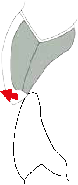
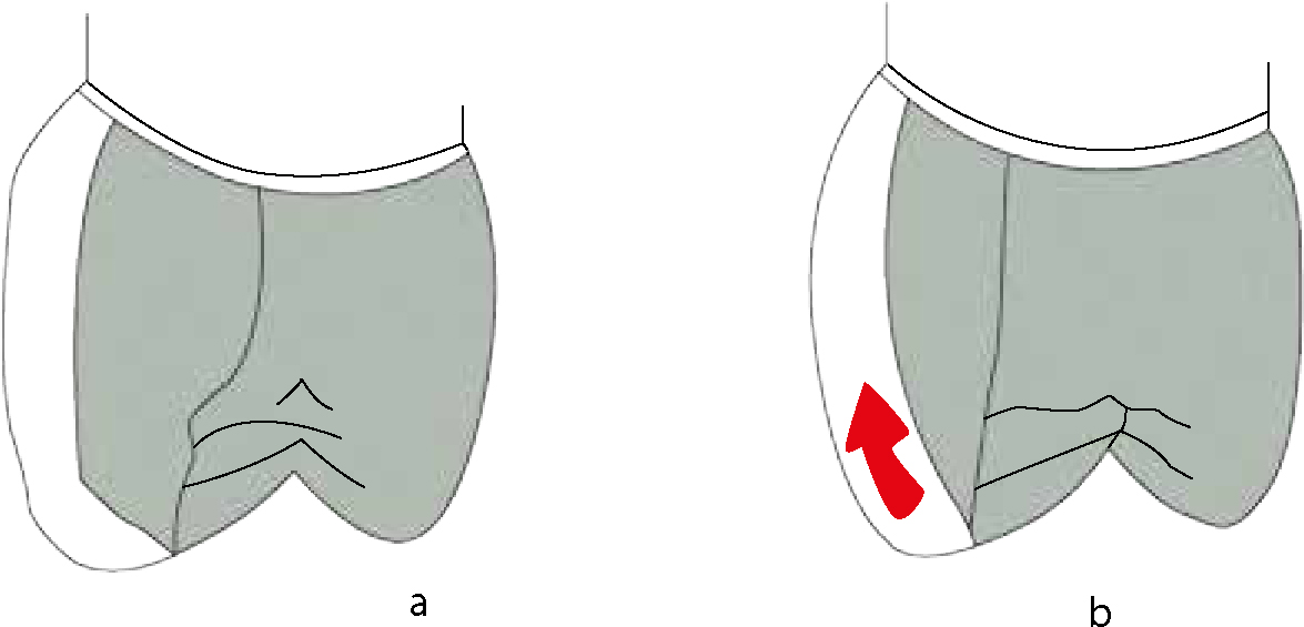
Support of porcelain: Porcelain veneer that is supported by an evenly contoured metal framework is better able to withstand stress. The metal should be contoured so that the overlying veneering porcelain will be subject to compressive forces when a load is applied. Examples of this consideration include avoiding the extension of lingual metal to the incisal edge of a maxillary anterior restoration (Figure 4) and establishing a supporting ledge under the facial cusp of a maxillary premolar or molar metal-ceramic restoration (Figure 5).32 Failing to meet these criteria will expose the brittle porcelain veneer to shearing forces and may lead to premature porcelain fracture.
Figure 4. Metal extending too far incisally makes the unsupported porcelain prone to fracture.Figure 5. Maxillary posterior metal–ceramic coping with (a) proper metal support and (b) without proper metal support under facial cusp.
Metal thickness: Copings should be rigid to avoid flexing during seating or under occlusal loads. Rigidity of the coping is directly proportional to the thickness of the metal used. A noble metal coping should be at least 0.3–0.5 mm thick, while a base metal alloy with a higher yield strength and higher melting temperature may be as thin as 0.2 mm.14 A metal coping with lesser thickness is likely to flex and cause porcelain fracture.
Occlusal/proximal contacts: Contact near the metal–ceramic junction can lead to metal flow and subsequent porcelain fracture. The porcelain-metal junction should be placed 1.0 mm from occlusal contacts at the position of maximal intercuspation.25,33 Proximal contacts for anterior teeth should be on porcelain and the metal should be placed more lingually. Placing the porcelain-metal junction lingual to the proximal contact areas leads to improved stress distribution.11,32
Extent of area to be veneered: The porcelain on the facial surface usually extends over the cusp tip and about halfway down the palatal incline of the facial cusp on posterior teeth (Figure 6a). There must be a rounded ledge of metal under the facial cusp to support the porcelain. Without a supporting ledge, the ceramic will fracture.11 This design is more resistant to fracture than those in which the porcelain extends to the central groove or covers the entire occlusal surface (Figure 6b).25
Figure 6. Coping design for a metal–ceramic restoration with (a) buccal cusp in porcelain and (b) full porcelain occlusal coverage.
Design of facial margin: Porcelain-covered metal margin design requires either a heavy chamfer or a beveled shoulder finish line with the metal coping extending to the cavo-surface margin and thinned to the minimum thickness possible.25 Porcelain is extended to cover this metal. This design may lead to metal distortion during firing and metal flexure with resulting porcelain fracture as a result of excessive thinning of the coping.32
Lack of aesthetics with the conventional metal collar led to the use of the all-porcelain facial margins.34 They utilize special shoulder porcelains that are stronger in flexure than conventional porcelains,15 making the margin more resistant to fracture.
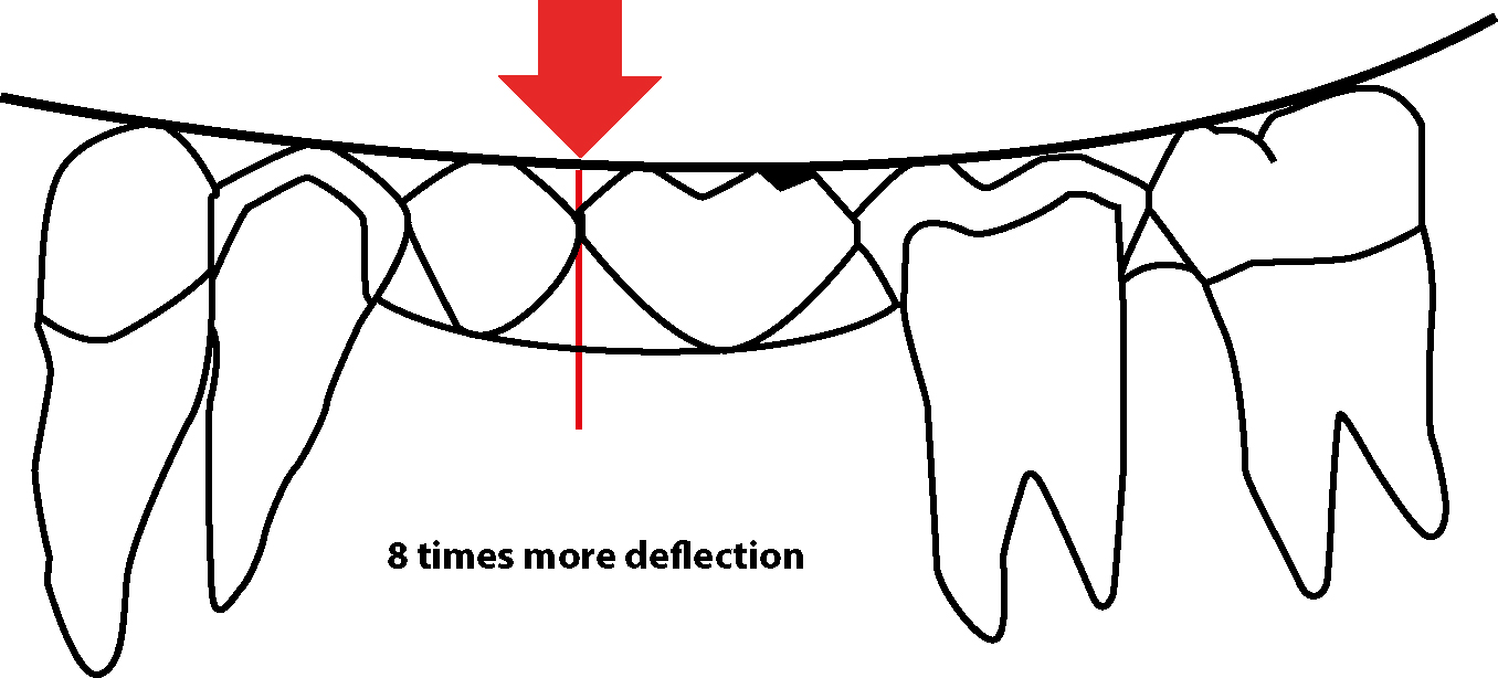
Antero-posterior span of fixed partial denture
Long span fixed partial dentures flex more under heavy occlusal loads, leading to the fracture of porcelain. If all other parameters are kept constant, a four-unit fixed partial denture replacing two teeth will bend eight times more as compared to a three-unit fixed partial denture with a single pontic (Figure 7).11,15,25 Where a large number of missing teeth need to be replaced, considering an implant-supported prosthesis may help decrease the span length and yield better results.11
Figure 7. Deflection of a fixed dental prosthesis (FDP) is directly proportional to the cube of the length of its span. An FDP with 2 pontics will deflect 23 ie 8 times as much as an FDP with 1 pontic.
Thickness of pontics
Occluso-gingival thickness of the pontic affects the deflection of framework. Deflection varies inversely with the cube of the occluso-gingival thickness of the pontic. If the pontic thickness is halved, the framework deflection will be increased eight times.24,32
Design of connectors
For clinical longevity, connectors of a fixed partial denture should be thick enough to resist the occlusal loads.25,32 However, for optimal aesthetics, occlusal and gingival embrasures must be created.11
Poor porcelain adaptation
One of the most common fabrication flaws is the incorporation of air in the ceramic mix.35 If air is entrapped within the ceramic particles, porosities occur in the final restoration, reducing its strength and increasing the chances of fracture.25,33 Porosities may be introduced in the dental porcelain due to faulty condensation, incorrect powder/liquid ratio or firing time and temperature disparities.35-37 This factor is applicable to all restorations that use feldspathic porcelain.
Method of adding the veneering layer
Hot isostatically pressed (HIP) glass ceramic materials are less prone to chipping and fracture as compared to hand layered veneering porcelains.11,25
Poor metal-ceramic bond
Clinical success of a metal-ceramic restoration largely depends upon the bond formed between the metal and the porcelain. A metal-ceramic bond results from the interplay of a number of different factors including mechanical bonding, chemical bonding, Van der Waals forces and compression fit due to a difference in coefficient of thermal expansion.38 A poor metal-ceramic bond can be due to poor choice of metal, eg one that does not form oxides, or by inadequate preparation of metal to be bonded to porcelain.15 When the metal-ceramic bond fails, it leads to delamination of porcelain from the metal or adhesive failure.
Firing protocols
During firing procedures, a material with low thermal conductivity will be incompletely baked, hence becoming more prone to chipping fracture.17 Also, any mismatch between the coefficient of thermal expansion of the core porcelain and the veneering porcelain will generate residual stresses and promote fracture. During the cooling phase, the difference in thermal conductivity of core and veneer porcelain results in residual stresses. These stresses bring about adhesive failure of the restoration.15
Faulty polishing and glazing
Glazing helps reduce the depth and width of flaws present in the ceramic surface,15 hence is considered a ceramic-strengthening method.35 Any fault in glazing or polishing would hamper the strength of ceramic.
Conclusion
Failure of a metal-ceramic restoration is a complex phenomenon. A critical review of the available literature revealed a multitude of factors that may play a role in the mechanical failure of metal-ceramic restorations. These factors may be as simple as a single episode of blunt trauma or may be complex, involving a combination of issues related to material properties, restoration design and fabrication. To optimize the performance of metal-ceramic restorations, the clinician needs to understand all the factors affecting the restoration's longevity. This will enable the clinician to exploit the material's strengths and compensate for its flaws.