
Abstract
This series of three papers reviews the causes, diagnosis and differential diagnosis, and outlines the management of sore and/or swollen lips.
From Volume 43, Issue 10, December 2016 | Pages 971-980
This series of three papers reviews the causes, diagnosis and differential diagnosis, and outlines the management of sore and/or swollen lips.


This is an irritant contact cheilitis characterized by a diffuse erythema, swelling, exfoliation, and/or even blisters which break leaving superficial ulcerations in the lips and may arise within a few hours or days after the drug intake. The severity of the lesions depends on the drug type, its dose and the individual patient's response. Cheilitis can occur as a result of allergy or as a pharmacological effect (Table 1).
| Angular stomatitis (cheilitis, cheilosis) |
| Candidiasis (denture-induced stomatitis) |
| Staphylococcal, streptococcal or mixed infections |
| Herpetic infections |
| Anaemia |
| Ariboflavinosis (rarely), iron, folate or B deficiency |
| Crohn’s disease and orofacial granulomatosis |
| Down’s syndrome |
| HIV disease |
| Blisters |
| Herpes labialis |
| Herpes zoster |
| Erythema multiforme |
| Pemphigus/Pemphigoid |
| Epidermolysis bullosa |
| Burns or frostbite |
| Drugs (especially fixed drug eruption) |
| Allergic cheilitis |
| Mucoceles |
| Impetigo |
| Desquamation and crusting |
| Dehydration |
| Exposure to hot dry winds |
| Exfoliative cheilitis |
| Acute febrile illness |
| Mouth-breathing |
| Actinic cheilitis |
| Chemical or allergic cheilitis |
| Drugs (Retinoids) |
| Atopic cheilitis |
| Psoriasis (very rarely) |
| Candidal cheilitis Erythema multiforme Psychogenic (self-induced) Kawasaki syndrome |
| Dryness |
| Sjögren’s syndrome |
| Lip-licking |
| Mouth-breathing |
| Diabetes |
| Drugs |
| Radiation mucositis |
| Diffuse swellings |
| Angioedema (allergic or hereditary) |
| Oedema (trauma or infection or insect bite) |
| Allergic cheilitis |
| Contact cheilitis |
| Chemical cheilitis |
| Crohn’s disease |
| Orofacial granulomatosis |
| Cheilitis granulomatosa |
| Sarcoidosis |
| Cheilitis glandularis |
| Cold urticaria |
| Ascher’s syndrome |
| Haemangioma |
| Lymphangioma |
| Recurrent erysipelas |
| Chronic herpes simplex labialis |
| Candida cheilitis |
| Ulcerations |
| Infective Herpes labialis Herpes zoster Syphilis Tuberculosis Kawasaki disease |
| HIV |
| Leishmaniasis |
| Blastomycoses |
| Histoplasmosis |
| Actinomycosis |
| Orf |
| Tumours |
| Squamous cell carcinoma |
| Basal cell carcinoma |
| Keratoacanthoma |
| Melanoma |
| Kaposi’s sarcoma |
| Lymphomas |
| Salivary gland tumours |
| Gut diseases |
| Pyostomatitis vegetans |
| Others |
| Burns |
| Trauma dermatoses |
| Erythema multiforme |
| Toxic epidermal necrolysis |
| Lichen planus |
| Lupus erythematosus |
| Pemphigoid |
| Pemphigus |
| Leukocytoclastic angiitis |
| Drug-induced |
| Gold |
| Sulphonylureas |
| Phenylbutazone |
| Chlorpromazine |
| Phenobarbitone |
| Methyldopa |
| Thiazides |
| Statins |
| Retinoids |
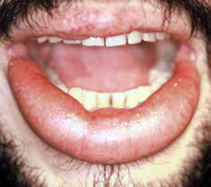
Many drugs can be implicated but the aromatic retinoids, such as etretinate and isotretinoin, cause dryness and cracking of the lips in almost all patients1,2 (Figure 1). The mechanism of this pharmacological effect is unknown, but is dose-related.
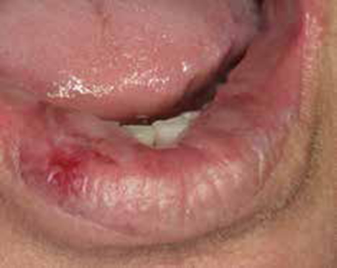
Life-threatening anaphylactic reactions have been reported in patients who have applied chlorhexidine gluconate topically to the lips.3 Erythema multiforme, which is commonly caused by drugs, can produce haemorrhagic crusting of the lips (Figure 2). Many other drug reactions have been reported occasionally (Table 1). Statins,4 antineoplastic agents (busulphan), antivirals (indinavir, a proteases inhibitor),5 antibiotics (tetracycline or streptomycin),6 clofazimine and psoralens, vitamins like A or B127 or even gold salts and methyldopa sometimes cause cheilitis.8
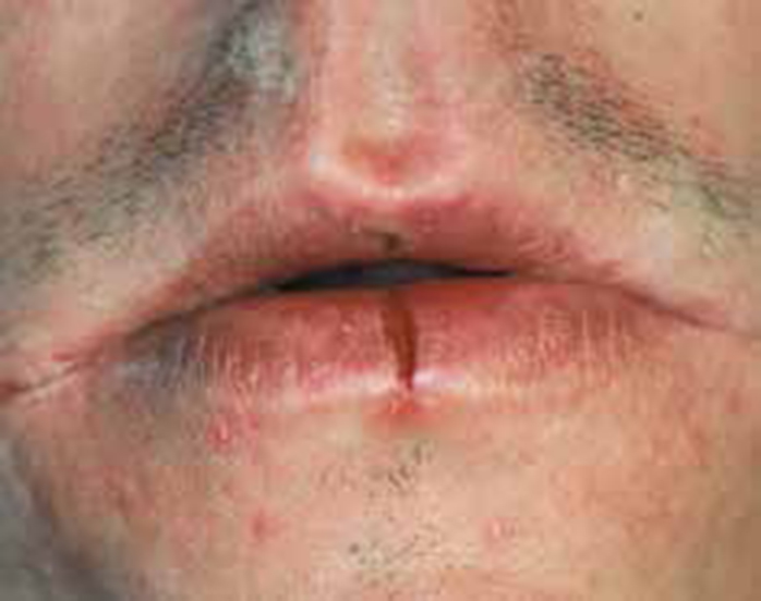
This is a chronic superficial inflammatory disorder characterized by hyperkeratosis and desquamation of the vermillion epithelium, with persistent scaling (Figure 3). The diagnosis is restricted to those few patients whose cheilitis cannot be attributed to other causes, such as contact sensitization or UV light.9
Most cases occur in girls or young women, the majority of whom seem to have a personality disorder10 and, indeed, a psychogenic cause was proposed by the French researchers, designating this ‘le tic des lèvres’ to indicate manipulation as being the basis. A preoccupation with the lips is prevalent in some individuals. Many cases are thus thought to be factitious, caused by repeated self-induced trauma such as biting, picking, lip-sucking, chewing or other manipulation of the lips.11,12 Exacerbations have been associated with stress. In some cases the condition appears to start with chapping or with atopic eczema, and develops into a habit tic.
There appears to be no association with dermatological or systemic diseases, though some cases are infected with Candida species13 and rare cases are seen in HIV disease. In one large Russian series, almost half the cases had been associated with thyroid disease,14 but this observation has not been confirmed.
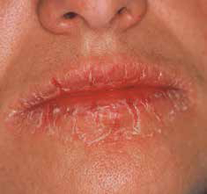
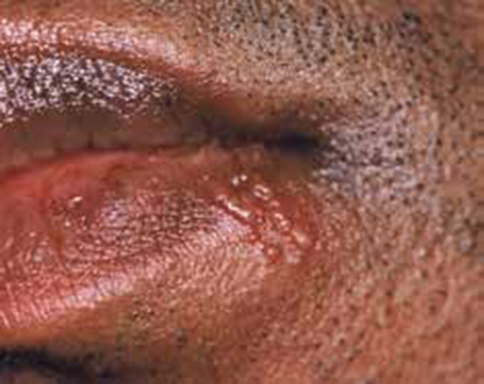
Exfoliative cheilitis often starts in the centre of the lower lip and spreads to involve the whole of the lower or of both lips. The keratin scales appear as scattered white or yellowish plaques, which easily detach with friction against the teeth or the patient's fingers, leaving a normal lip surface beneath.10,12 The patient may complain of irritation or burning sensation and can be observed frequently with biting or sucking the lips. Lip scaling and crusting is more or less confined to the vermillion border, persisting in varying severity for months or years. There may be bizarre yellow hyperkeratotic or thick haemorrhagic crusts (Figure 4). The sloughing of sheets of epithelium is another feature in some. Some cases have improved with psychotherapy and antianxiolytic or antidepressant treatment.
This is a rare, chronic inflammatory disorder of minor labial salivary glands in which there is mucus hypersecretion. This is present as a diffuse painful swelling of the lower rather than the upper lip in Caucasian rather than Asian men, associated with erythema, ulcerations, crusting and prominent dilated salivary duct openings where muco-purulent fluid exudes on palpation. It may have a malignant tendency.15,16
Granulomatous cheilitis is an uncommon, chronic swelling of the lip due to granulomatous inflammation within the submucosa, seen in isolation or in other granulomatous disorders (eg Crohn's disease, Sarcoidosis or TB) and appearing initially as a painless diffuse or restricted swelling, often of the upper lip, with a tendency to resolve within a few days. After weeks or months, however, the lower lip starts gradually to enlarge (Figure 5) and this swelling can remain for months or even years and may be associated with hypertrophic gingivitis and fissured tongue. There may be splitting of the lips and angular stomatitis. Ulcers classically involve the buccal sulcus where they appear as linear, often with granulomatous masses flanking them. Mucosal lesions also include thickening and folding of the mucosa to produce a ‘cobblestone’ type of appearance and mucosal tags. Purple granulomatous enlargements may appear on the gingiva. Facial palsy of the lower motor-neurone type occurs in some 30% of cases. This may precede the attacks of oedema by months or years, but more commonly develops later. Though intermittent at first, the palsy may become permanent. It may be unilateral or bilateral, and partial or complete.17 Other cranial nerves (the olfactory, auditory, glossopharyngeal and hypoglossal) may occasionally be affected. Involvement of the central nervous system has also been reported, but the significance of the resulting symptoms is easily overlooked as they are very variable, sometimes simulating multiple sclerosis but often with a poorly defined association of psychotic and neurological features. Autonomic disturbances may occur.
Cheilitis granulomatosa may be part of syndromes like the Melkersson-Rosenthal syndrome.18,19 It can also be a manifestation of systemic granulomatous diseases affecting the mouth (orofacial granulomatosis) intestine (Crohn's disease), lungs or skin (sarcoidosis). Miescher's cheilitis is the term used when the granulomatous changes are confined to the lip only. Melkersson-Rosenthal syndrome is the term used when there is cheilitis with facial palsy and plicated tongue. Melkersson described recurrent facial palsy in association with labial oedema20 and Rosenthal added plicated tongue to the syndrome.21 Miescher's cheilitis is generally regarded as a monosymptomatic form of the Melkersson-Rosenthal syndrome, although the possibility remains that these may be two separate diseases. A localized granulomatous cheilitis is also a feature of a foreign body reaction to a cosmetic filler material (Figure 6). Some patients with granulomatous cheilitis are predisposed to Crohn's disease,22 since regional ileitis may follow some years later.23,24,25 Reports of granulomatous cheilitis preceding gastrointestinal involvement have suggested that orofacial granulomatous involvement actually represents a continuum in the presentation of Crohn's disease.
Orofacial granulomatosis appears to develop because of an adverse reaction to various food additives, such as cinnamic aldehyde,26,27 butylated hydroxyinosole or dodecyl gallate (in margarine), or menthol (in peppermint oil), though these reactions are by no means always relevant: for example, only one of nine patients had a relationship to food intake28 or to cobalt.29 Non-caseating granulomas and lymphoedema may be seen, but the granulomas tend to be sparse and deep, close to the muscle. It is unclear where in the spectrum of Crohn's disease/sarcoidosis/allergy/infections these lesions (and related conditions such as Melkersson--Rosenthal syndrome and granulomatous cheilitis) lie.
This is a rare inflammatory disorder of the lips with a characteristic bandlike infiltrate of plasma cells in the upper lamina propria/dermis. Plasma cell cheilitis has similar clinical features to allergic cheilitis (erythema, exfoliation and ulceration of the vermillion), but it differs as it has other intra-oral lesions such as in the tongue (plasma cell glossitis) or gingivae (plasma cell gingivitis).30,31 The cause is unknown, presumably immunological. Precipitants may include a range of substances such as additives to cosmetics or dentifrices.
A similar lesion which tends to form a warty mass with a hyperkeratotic surface, known as plasma-acanthoma32 reportedly after trauma, and needs to be differentiated from extramedullary plasmacytoma. Extramedullary plasmacytomas (EMPs)33 are rare plasma-cell tumours of the soft tissues that occur predominantly in the paranasal sinuses and oropharynx. Subcutaneous and cutaneous plasmacytomas of the lip are rare. Biopsy reveals sheets of monoclonal plasmablasts and plasma cells with anaplastic features. The possibility of a plasma cell dyscrasia, such as an underlying myeloma, or extramedullary plasmacytoma, should be excluded.
Nutritional cheilitis, especially pellagra, can cause the vermillion zone to become shiny and cracked, sometimes even eroded. Milder degrees of deficiency cause angular cheilitis (Part 1), oral ulcers and sometimes glossitis.34
Deposits may be foreign bodies, such as silicone or other materials for iatrogenic lip augmentation, or amyloidosis which typically form discrete swellings35 (Figure 6).
Fissures in the lip may develop when a patient, typically a child, is mouth-breathing. Most lip fissures are seen in males and are typically found in the middle lower lip and can be chronic, causing discomfort and bleeding from time to time.36 Contrary to the clinical impression that fissures are seen only in the lower lip (Figure 7), there is also a high prevalence in the upper lip. Though sun, wind, cold weather and smoking are thought to predispose, the aetiology remains obscure. A hereditary predisposition for weakness in the first branchial arch fusion seems to exist. Lip fissures are common in Down's syndrome and in Cheilitis granulomatosa.
Giant cell arteritis (temporal) is a rare cause of ulceration, especially of the upper lip.37,38 This is an emergency as steroids are needed to prevent eye damage in temporal cell arteritis. Ulceration of the lips is occasionally seen as a result of leukocytoclastic angiitis.
Graft-versus-host disease (GVHD) following bone marrow transplantation (BMT) is common. Acute GVHD may consist of painful mucosal desquamation, erythema and ulcerations are most pronounced at 7–11 days after BMT, and may be associated with obvious infection. The ventrum of the tongue, buccal and labial mucosae and gingivae may be affected by ulcerations (mucositis).39,40 Lichenoid plaques or striae (small white lesions) affect the buccal, labial and lingual mucosa early on, but clear by day 14 after BMT. The oral lesions in chronic GVHD are considered as one of the criteria for establishing the diagnosis,41 and cheilitis may be sometimes the only manifestation of this disease, or coincident with skin lesions. Lip biopsy is useful in the diagnosis of chronic GVHD and should include both mucosae and underlying minor salivary glands.42
Odontogenic infections arising from maxillary anterior teeth may cause upper lip swelling while those from mandibular anterior teeth lower lip swelling.43 Cutaneous infections may spread and involve the lip(s).44
Herpes simplex virus, that may occasionally produce primary, peri-oral lesions, affects up to 15% of the population in relation to an interferon defect.45
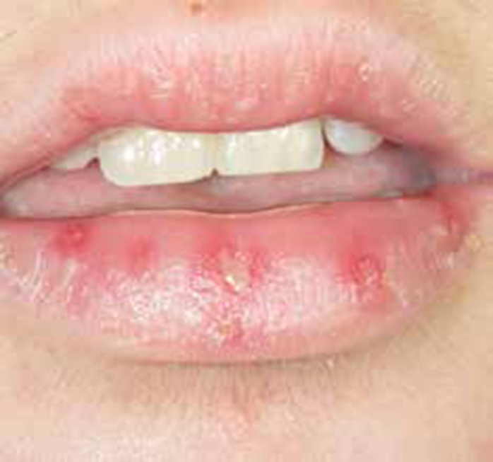
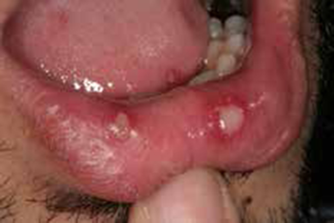
These lesions heal within one or two weeks and the HSV remains latent in the trigeminal (geniculate) ganglion. Factors such as fever, sunlight (ultraviolet B), trauma or immunosuppression can reactivate the virus with clinical recrudescence to produce Herpes labialis (cold sores) (Figure 8) or stomatitis.46
Herpes varicella zoster virus infection may affect the lips and other mucosae causing ulcerations in varicella (chickenpox). Reactivation of the virus is common among patients with various immunodeficiencies and presents with a discrete cluster of papulovesicular exanthema along to a specific neurodermatome. Zoster of the maxillary division of the trigeminal nerve affects the upper lip, (Figure 9),47 while the lower lip is affected by mandibular zoster.48
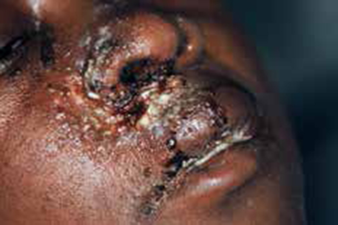
Human immunodeficiency virus (HIV) may cause candidosis, or aphthous-type ulcers (Figure 10), especially of the major type. Infections from opportunistic viral pathogens, such as the herpesviruses (herpes simplex, varicella zoster, Epstein-Barr virus, cytomegalovirus), pathogenic fungal infections such as histoplasmosis or cryptococcosis, syphilis, mycobacteria such as tuberculosis or non-tuberculous mycobacteria (NTM), protozoa such as leishmaniasis, or neoplasms such as KS or lymphoma, may be seen.49 Histoplasmosis, blastomycosis and paracoccidioidomycosis are uncommon causes of chronic ulceration affecting the lips, producing very similar clinical lesions to Leishmaniasis,50 and may mimic a neoplasm. A wide spectrum of other orofacial lesions can be seen in HIV/AIDS, including human papillomavirus (HPV) infections and, in particular, warts (condyloma acuminata) on lips or in the mouth.
Kawasaki disease is a rare vasculitis of apparently infectious aetiology, initially reported in Japan and manifests mainly in childhood, with fever, oedematous lips and pharyngitis generalized lymphadenopathy, and a predominantly truncal erythematous rash with desquamation of hands and feet. It may be complicated by myocarditis.51
Other viral infections of the lips are rare, but occasionally Coxsackie viruses orf52 molluscum contagiosum53 and vaccinia virus may be seen.
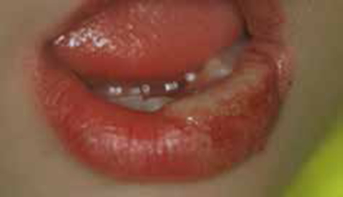
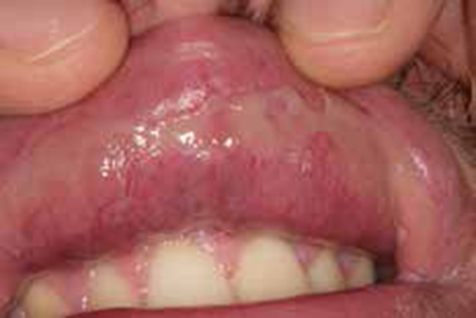
The lips are not infrequently traumatized by a deliberate or accidental blow in some occupations or from habits such as lip-licking (Figure 11). The use of various musical instruments may cause cheilitis. Bites by assailants are uncommon but typically involve the lower lip.54 Child abuse must always be borne in mind.55
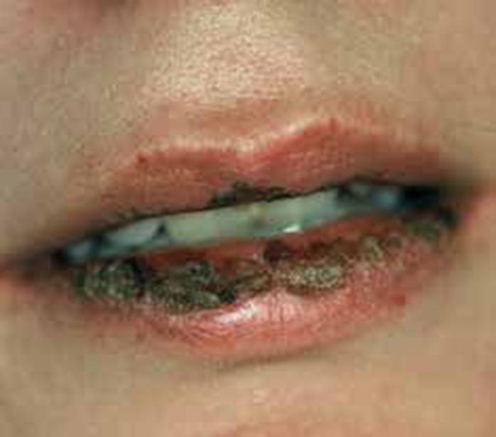
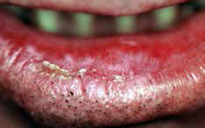
Burns may be caused by heat or cold, irradiation, chemicals or electricity. Thermal burns can result from hot foods, liquids or instruments.56 Cold burns most commonly follow cryosurgery. Irradiation burns result from ultraviolet light or ionizing radiation (mainly). Chemical burns are due, for example, to holding mouthwashes in the mouth or drugs against the buccal mucosa, and can cause white sloughing lesions of the mucosa (Figure 12).
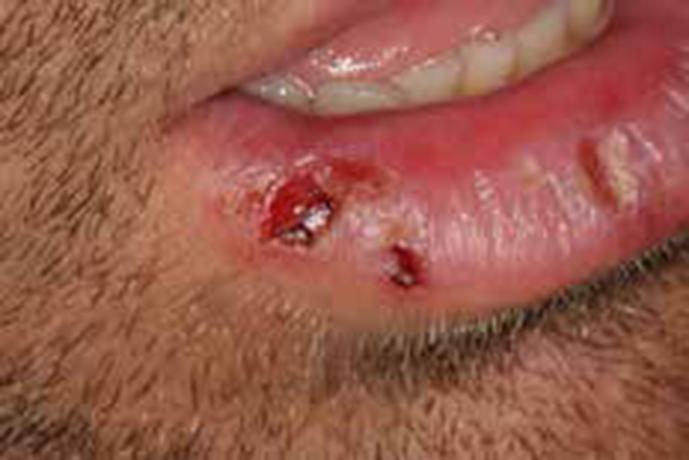
In self- or accidental-harm, the lower rather than the upper lip may also be bitten accidentally, particularly when anaesthetized57 (Figure 13), or deliberately,58,59 as in the mentally challenged,60 in congenital indifference to pain,67 in Munchausen's syndrome,62 or occasionally in Lesch-Nyhan syndrome.63
This is a common mucocutaneous disorder characterized in some patients by oral white lesions or bullae64 (Figure 14) and sometimes with genital lesions and/or an itchy rash.
Lichen planus lesions are often seen in the lip vermillion border together with other areas of the oral mucosa, pharynx, oesophagus, stomach, anus, larynx and genitals, and in the skin, mainly on the wrists and ankles, palms and soles and scalp and nails. In most cases of labial lichen planus there are characteristic intra-oral lesions, but some are said to be seen in isolation.65,66
Lupus erythematosus is a chronic systemic or cutaneous disorder, which manifests especially with a rash (Discoid-DLE or Systemic-SLE).67,68 Lupus cheilitis is characterized clinically by single or multiple lip lesions consisting of a white reticular network at the periphery and a central erythematous area intermingled with small capillaries.69 Lupus cheilitis is rare (up to 4%) of DLE70 and associated with facial erythema, pericarditis and polyserositis in patients with SLE.71 Lupus resembles, clinically, other skin diseases. Discoid lupus erythematosus sometimes clinically overlaps lichen planus cheilitis.72 Labial lesions of DLE have a small premalignant potential.
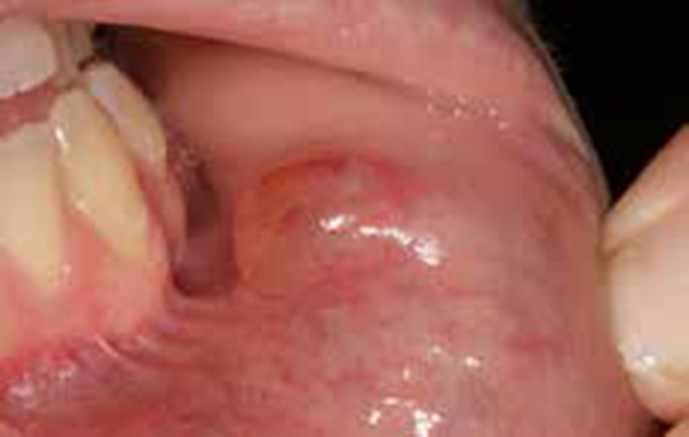
Mucoceles is a salivary extravasation or retention cyst and may be sore if traumatized73 (Figure 15).
Neoplasms can arise from the skin, mucosa, muscles, salivary glands or other tissues. They can either be benign or malignant. The most important malignant neoplasms are: carcinomas, sarcomas (particularly Kaposi sarcoma) or lymphomas.
Squamous cell carcinoma is the most common malignancy to affect the lip and is seen mostly in middle-aged or older men employed in outdoor activities with chronic sun exposure, such as farming and fishing.74 Squamous cell carcinomas of the lip occur predominantly on the lower lip (89%), with 3% on the upper lip and 8% at the commissures,75 and are especially seen in fair-skinned outdoor workers in sunny climates.76,77
The single most significant predisposing factor for the development of lip squamous cell carcinoma is chronic ultraviolet exposure.78 Other risk factors may include low social class, tobacco smoking, syphilis, neglected dentition and infection with herpes simplex virus79 or human papillomavirus (HPV), especially in immune suppression such as in transplant patients. Genetic disorders such as xeroderma pigmentosum, recessive dystrophic epidermolysis bullosa, oculocutaneous albinism and Fanconi anaemia predispose toward the development of squamous cell carcinoma of the lip,80 as do discoid lupus, actinic cheilitis and cheilitis glandularis.81
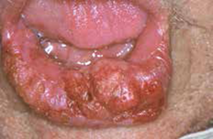
Lip carcinoma presents with thickening, induration, crusting or chronic ulceration, usually at the vermillion border of the lower lip just to one side of midline82 (Figure 16).
Kaposi sarcoma is a multifocal cutaneous and extracutaneous vascular proliferative disorder associated with Herpes Virus type 8 (HHV-8 or Kaposi sarcoma herpes virus, KSHV).83,84,85 Four types exist: Classical KS affecting middle-aged patients with Mediterranean origin; African endemic; iatrogenic immunosuppressed and finally AIDS-related KS.
Kaposi sarcoma often presents as an erythematous, violaceous nodule or plaque in the upper or lower lip. The diagnosis is based on biopsy. Local treatment includes surgical excision, cryotherapy, or intralesion injections of vinblastine alitrenoin gel or even radiotherapy. Systemic treatment with antivirals such as HAART prevent and sometimes can regress oral KS.85
Lymphomas are malignant neoplasms of the lymphoreticular system and uncommonly affect the oral cavity and even rarely the lips.86 Lip lymphomas affect patients of any age and present as a chronic swelling associated sometimes with pain or paraesthesia. Lip lesions are seen in patients with Burkitt lymphoma together with swollen jaws and loose teeth, in Sjögren's syndrome87 or HIV/AIDS.88 Biopsy is required for the diagnosis. The treatment of choice is surgery together with chemotherapy.
Salivary gland neoplasms can arise from the labial salivary glands. Lip salivary gland neoplasms are rare but often malignant89 and present as a solitary chronic swelling in the upper rather than the lower lip of middle-aged or older patients who are chronic smokers or alcohol drinkers, or exposed to carcinogens at work, or having radiotheraphy for head or neck tumours. Local excision is the only treatment and palliative radiotherapy can be given if distant metastases are found.90
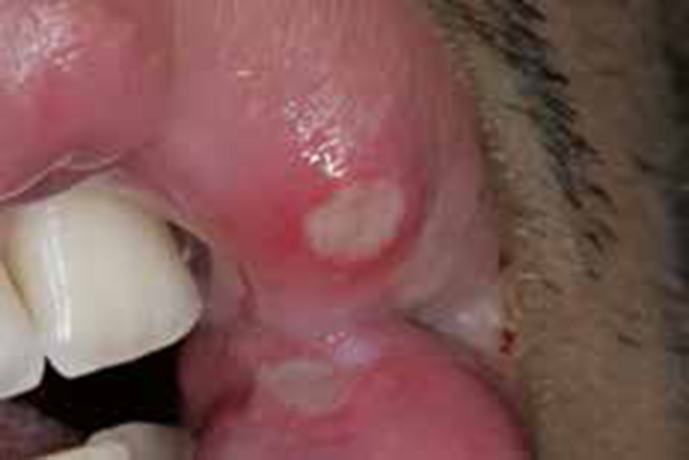
These are common recurrent mouth ulcers which typically start in childhood and have a natural history to improve with age. Recurrent aphthae typically occur within the mouth but may affect the labial mucosa, and occasionally extend onto the vermillion.91
The ulcers characteristics are:
They recur and have a yellowish depressed floor and a pronounced red inflammatory halo.
Aphthae may present different clinical appearances and behaviours.
Characterized by the presence of single or multiple bullae which sometimes break easily leaving painful ulcerations in the vermillion border in the labial, oral and other mucosae but with various pathogeneses.
Epidermolysis bullosa acquisita may involve the labial mucosae.92
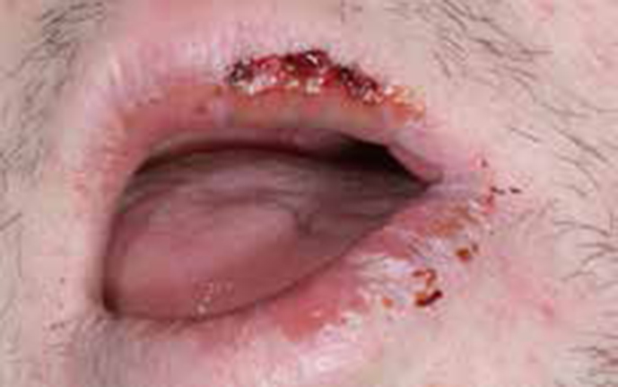
This is an acute, often recurrent disorder affecting mucocutaneous tissues, is characterized by serosanguinous exudates on the lips (Figure 18), and sometimes target-like lesions on the skin, and lesions on other mucosae.93,94 A range of factors trigger what appears to be an immunologically related reaction with sub- and intra-epithelial vesiculation. In most patients the trigger is infection (such as herpes simplex or mycoplasma), but drugs (sulphonamides, cephalosporins, barbiturates, hydantoins, cimetidine and others), or food additives such as benzoates, may also be implicated.95
This is a cause of chronic bullous cheilitis (a group of skin diseases which cause an inflammation of one or both lips due to the formation and rupture of bullae, leaving extensive painful erosions or superficial ulcerations covered by whitish-yellowish pseudo-membranes or bloody crusts. However, intact bullae are seldom seen in the vermillion border, ano-genital mucosa, eyes and skin as they break easily with friction (Figure 19).96 Pemphigoid is the term given to a group of autoimmune disorders which affect stratified squamous epithelia and mimic pemphigus, but has a better prognosis and is characterized by sub-epithelial, rather than intra-epithelial, vesiculation.
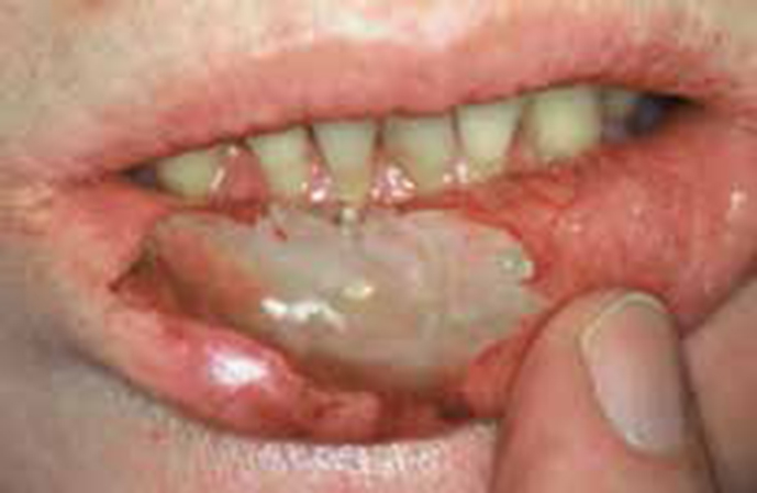
Pemphigus is a rare life-threatening autoimmune disease which affects stratified squamous epithelia, producing intra-epithelial vesiculation. Lesions appear anywhere, but especially where there is trauma, and therefore blisters or erosions are common on the palate97,98,99 and floor of the mouth and on the lips100 (Figure 20).
Pyostomatitis vegetans is typically seen in ulcerative colitis, sclerosing cholangitis and other liver diseases and may cause erosions and superficial ulcers in the labial mucosa and on the vermillion.101
Toxic epidermal necrolysis is clinically characterized by extensive mucocutaneous epidermolysis preceded by a macular or maculopapular exanthema and enanthema. There is widespread painful blistering and ulceration of oral mucosa, including all labial surfaces.102 Toxic epidermolysis is typically induced by drugs and may be associated with antimicrobials (sulphonamides, thiacetazone rifampicin, fluconazole and vancomycin), analgesics (phenazones), antiepileptics, allopurinol and chlormezanone.103 TEN can be an occasional feature in HIV disease or Graft-versus-host disease.104
Lips are the site of primary or secondary manifestations of various systemic infections; neoplasms and immune-related disorders. A plethora of bacteria, fungi and viruses can affect the lips causing innocent but sometimes contagious lesions which can later become dangerous, especially in high risk patients (eg pregnant women, children and older patients). Malignant lip neoplasms (mainly carcinomas) are common and strongly related to solar irradiation. Lip ulcerations, either recurrent (aphthae; erythema multiforme) or persistent (bullous disorders), cause patients distress while chronic swelling (granulomatous diseases) can cause facial disfiguration. Drugs for the treatment of systemic diseases may cause severe side-effects on the lips.