Oyajobi BO, Mundy GR Pathophysiology of myeloma bone disease, 2nd edn. In: Gahrton G, Durie BGM, Samson DS London: Arnold; 2004
Oyajobi BO Multiple myeloma/hypercalcemia. Arthrit Res Ther. 2007; 9
Ariyaratnam S, Sweet C, Duxbury AJ Low grade multiple myeloma that presented as a labial swelling – a case report. Br Dent J. 2005; 199:433-435
Goldschmidt H, Lannert H, Bommer J, Ho AD Multiple myeloma and renal failure. Nephrology, dialysis, transplantation. Nephrol Dial Transplant. 2000; 15:301-304
Ludwig H, Pohl G, Osterborg A Anaemia in multiple myeloma. Eur J Haematol. 1994; 53:207-212
Dammacco F, Luccarelli G, Prete M, Silvestris F The role of recombinant human erythropoietin alpha in the treatment of chronic anemia in multiple myeloma. Rev Clin Exp Hematol. 2002; 1:32-38
Mittelman M The implications of anemia in multiple myeloma. Clin Lymphoma. 2003; 4:S23-29
Kariyawasan CC, Hughes DA, Jayatillake MM, Mehta AB Multiple myeloma: causes and consequences of delay in diagnosis. Int J Med. 2007; 100:635-640
Westermark P, Benson MD, Buxbaum JN Amyloid: toward terminology clarification. Report from the Nomenclature Committee of the International Society of Amyloidosis. Amyloid. 2005; 12:1-4
Rajkumar SV, Gertz MA, Kyle RA Primary systemic amyloidosis with delayed progression to multiple myeloma. Cancer. 1998; 82:1501-1505
Gertz MA, Lacy MQ, Dispenzieri A Amyloidosis. Hematol Oncol Clin North Am. 1999; 13:1211-1233
Desikan KR, Dhodapkar MV, Hough A Incidence and impact of light chain associated (AL) amyloidosis on the prognosis of patients with multiple myeloma treated with autologous transplantation. Leuk Lymphoma. 1997; 27:(3–4)315-319
Bhalis NJ, Lazarus HM Multiple myeloma-associated AL amyloidosis: is a distinctive therapeutic approach warranted?. Bone Marrow Transplant. 2006; 38:7-15
Rajkumar SV, Gertz MA, Kyle RA Prognosis of patients with primary systemic amyloidosis who present with dominant neuropathy. Am J Med. 1998; 104:232-237
Durie BGM, 1st edn. North Hollywood: International Myeloma Foundation; 2011
Grosbois B, Decaux O, Azais I Current treatment strategies for multiple myeloma. Eur J Intern Med. 2002; 13:85-95
Smith D, Yong K Multiple myeloma. Br Med J. 2013; 346
Henley E, Houghton N, Bucknall R Localized amyloidosis of the palate. Clin Exp Dermatol. 2008; 33:100-101
Pentenero M, Davico Bonino L, Tomasini C Localized oral amyloidosis of the palate. Amyloid. 2006; 13:42-46
Viggor SF, Frezzini C, Farthing PM Amyloidosis: an unusual case of persistent oral ulceration. Oral Surg Oral Med Oral Pathol Oral Radiol Endod. 2009; 108:e46-50
Gupta R, Perry M Lesson of the week: digital examination for oral cancer. Br Med J. 1999; 319:(7217)1113-1114
Kyle RA, Gertz MA, Witzig TE Review of 1027 patients with newly diagnosed multiple myeloma. Mayo Clin Proc. 2003; 78:21-33
Berenson JR Myeloma bone disease. Best Pract Res Cl Ha. 2005; 18:653-672
Sakalova A, Herrmann Z, Gazova S Osteoporosis in multiple myeloma. Vnitrni lekarstvi. 1998; 44:649-653
Epstein JB, Voss NJ, Stevenson-Moore P Maxillofacial manifestations of multiple myeloma. An unusual case and review of the literature. Oral Surg Oral Med Oral Pathol. 1984; 57:267-271
Scutellari PN, Orzincolo C Mandibular lesions in multiple myeloma. La Radiologia medica. 1992; 83:219-223
Witt C, Borges AC, Klein K, Neumann HJ Radiographic manifestations of multiple myeloma in the mandible: a retrospective study of 77 patients. J Oral Maxillofac Surg. 1997; 55:450-453
Vieira-Leite-Segundo A, Lima Falcao MF, Correia-Lins Filho R Multiple myeloma with primary manifestation in the mandible: a case report. Med Oral Patol Oral Cir Bucal. 2008; 13:E232-234
Elias HG, Scott J, Metheny L, Quereshy FA Multiple myeloma presenting as mandibular ill-defined radiolucent lesion with numb chin syndrome: a case report. J Oral Maxillofac Surg. 2009; 67:1991-1996
Glaspy JA Hemostatic abnormalities in multiple myeloma and related disorders. Hematol Oncol Clin N. 1992; 6:1301-1314
Mozaffari E, Mupparapu M, Otis L Undiagnosed multiple myeloma causing extensive dental bleeding: report of a case and review. Oral Surg Oral Med Oral Pathol. 2002; 94:448-453
Smith A, Wisloff F, Samson D Guidelines on the diagnosis and management of multiple myeloma 2005. UK Myeloma Forum. Nordic Myeloma Study Group, British Committee for Standards in Haematology. Br J Haematol. 2006; 132:410-451
Badros A, Weikel D, Salama A Osteonecrosis of the jaw in multiple myeloma patients: clinical features and risk factors. Am J Clin Oncol. 2006; 24:945-952
: SDCEP; 2011
Bird J, Owen R, d'Sa S: British Committee for Standards in Haematology in conjunction with the UK Myeloma Forum (UKMF); 2010
Huang TT, Liu TC, Chen PR Deep neck infections: analysis of 185 cases. Head Neck. 2004; 10:854-860
Amyloidosis presenting as macroglossia and restricted tongue movement Basim ES Dawoud Jennifer Taylor Senathirajah Ariyaratnam Michael N Pemberton Dental Update 2024 43:7, 707-709.
Authors
Basim ESDawoud
MBChB, BDS, MFDS RCS(Ed)
Dentally Qualified Medical Student, University of Leeds Medical School (basim.d@gmail.com)
Amyloidosis (AL) is a well recognized cause of macroglossia. A case of localized deposition of systemic amyloidosis secondary to multiple myeloma is reported in a 63-year-old female presenting with restricted tongue movement in the dental setting. Amyloidosis secondary to multiple myeloma is well documented in the literature, with amyloid deposits being found at various sites throughout the oral cavity and systemically. This case emphasizes the importance of a full oral soft tissue examination, including the need to examine mobility of the tissues, as it was the restricted movement of the tongue that ultimately alerted the clinician to the diagnosis. This report also highlights other dental implications of managing patients with multiple myeloma and amyloidosis.
CPD/Clinical Relevance: To provide an understanding of what multiple myeloma and amyloidosis are whilst, importantly, relating this to how these conditions can impact on routine dental treatment.
Article
Multiple Myeloma (MM) is a malignant neoplasm of the bone marrow characterized by proliferation of bone marrow plasma cells producing monoclonal immunoglobulins or light chains. These light chains are known as Bence-Jones proteins, which are excreted in the urine and are a diagnostic feature of multiple myeloma. This uncontrolled production can lead to renal failure, immunosuppression, skeletal destruction and anaemia of chronic disease. Prognosis is related to the staging of the disease. The disease predominantly affects older adults and, because of the multiple manifestations of this disease, patients can present with vague and confusing symptoms.1 It is thought that 5–15% of patients with multiple myeloma will develop amyloidosis. Amyloidosis is the localized and systemic extra-cellular deposition of insoluble fibrous protein aggregates known as amyloid. Amyloidosis can present intra-orally with various signs and symptoms including macroglossia.
Case report
A 63-year-old female was referred to the Oral Medicine Clinic at the University Dental Hospital of Manchester by her general dental practitioner (GDP).
The patient complained of difficulty swallowing, restricted tongue movement and a sore mouth. These problems had been ongoing for over a year and were slowly worsening in severity. She had mentioned these oral problems to the medical team managing her multiple myeloma, however, they felt that the visual appearance of the tongue was essentially normal and had not investigated further. On mentioning the same symptoms to her GDP, referral had been initiated.
Her past medical history showed that she had a five-year history of kappa light chain secretory multiple myeloma. Initial treatment with chemotherapy (‘VAD Therapy’ vincristine, doxorubicin (adriamycin), dexamethasone) and autologous stem cell transplant had resulted in remission of the myeloma for three years. Recurrence had been treated with further chemotherapy. There was no other significant medical history. Medication at presentation consisted of thalidomide (for myeloma) and sodium clodronate (for bone loss).
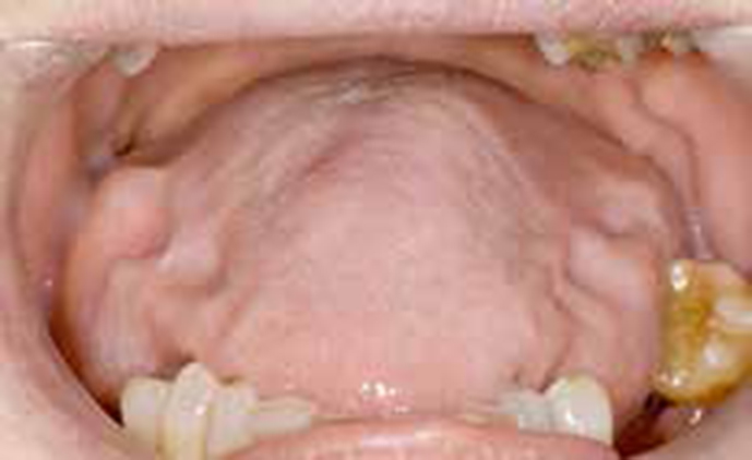
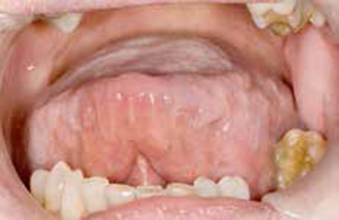
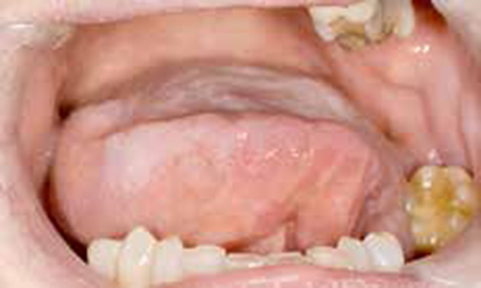
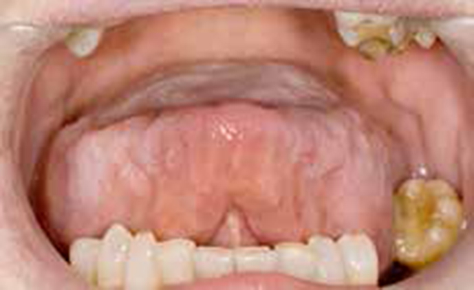
Extra-oral examination showed bilateral non-tender swelling of the cervical lymph nodes. Intra-oral examination showed an enlarged tongue that had crenulations on the lateral borders, however, the mucosal surface appeared otherwise unremarkable (Figure 1). On requesting the patient to move her tongue there was clear limitation of the range of movement in all directions (Figures 2–4). Palpation revealed an abnormally firm, thick and nodular tongue. Also noted was a mildly enlarged labial mucosa of the lower lip, which was also firm on palpation.
Figure 1. Crenulations evident on the bilateral borders of the tongue.Figure 2. Maximum range of tongue movement to the patient's right.Figure 3. Maximum range of tongue movement to the patient's left.Figure 4. Maximum range of tongue movement when asked to raise her tongue to the palate.
In view of the macroglossia, enlarged firm lower lip and significant limitation in movement of the tongue, amyloidosis secondary to multiple myeloma was suspected. Biopsies from the lower labial mucosa and tongue showed deposition of rounded globules of amorphous hyalinized material, separated by collagen fibres and ‘plump’ fibroblastic cells. The material stained positive with Congo red and showed pale green birefringence. Immunohistochemical staining was strongly positive for immunoglobulin kappa light chain and variably positive for lambda light chains. Electron microscopy showed fine filaments with a measured mean width of 11.0 nm in size. Overall, the features were consistent with amyloid. The biopsy material was sent to the National Amyloidosis Centre, University College Hospital, London for review.
They reported the features to be diagnostic of amyloid. The patient was referred for further assessment to the National Amyloidosis Centre and their final diagnosis was of systemic amyloidosis. The assessment showed predominantly soft tissue involvement, confirming that the macroglossia was due to amyloid deposition. There was no evidence of visceral amyloid deposition.
The patient initially commenced seven cycles of treatment with bortezomib and prednisolone, with a partial clonal response to treatment. The macroglossia remained with no change in the mobility of the tongue. The patient's oral symptoms were managed with benzydamine-containing mouthwash (Difflam®) for pain relief and chlorhexidine gel to aid oral hygiene. Her health subsequently deteriorated and she died two years post diagnosis due to cardiac complications related to her amyloidosis.
Discussion
Multiple myeloma
Myeloma is the 14th most common cancer amongst men and the 17th most common amongst women, accounting overall for more than 1% of all cancers and 11% of all haematological cancers.2,3,4,5 The incidence of the disease increases with age, with 71% of cases being in people aged 65 and over and the incidence peaking at 85 years of age.2,5. Myeloma is a disease of the bone marrow characterized by uncontrolled proliferation of plasma cells in the bone marrow, however, there are other aspects to the disease, which involve:6
Monoclonal protein (monoclonal means that all proteins produced by this cell line have exactly the same identity and the same impaired function);
Osteolytic bone lesions and hypercalcaemia;
Renal disease;
Immunodeficiency;
Anaemia.
A pathognomic feature of multiple myeloma is the Bence-Jones protein found in two-thirds of affected patients. These are immunoglobulin light chains produced by the neoplastic plasma cells, in either ‘kappa’ or ‘lambda’ forms, however, they are never produced simultaneously.7 They are found in urine and are contributory to renal failure.
Osteolysis leads to osteoporosis and is caused by tumour-induced bone resorption due to an increased osteoclastic activation via over-production of potent cytokines such as RANKL, TNFs and MIPs (macrophage inflammatory protein) by the neoplastic cells.8 This increased bone resorption leads to hypercalcaemia. However, not all patients develop hypercalcaemia; it is usually a feature late in the disease and also contributory to renal failure.9 With increased bone resorption, patients are also at risk of bone fracture.
Even with the large production of IgG antibodies by the neoplastic plasma cells, patients still suffer from immunosuppression. This is because the IgG proteins tend to be incomplete molecules and therefore dysfunctional. In combination with this, the level of normal immunoglobulins is typically reduced, therefore contributing to the increased susceptibility to infections.10
Renal failure is often a complication of advanced disease and approximately 20% of all patients with multiple myeloma will develop progressive renal failure during the course of the disease.11
Anaemia is a common complication in patients with multiple myeloma. It has been found to occur in two-thirds of all patients with the disease.12 The underlying pathophysiology is anaemia of chronic disease, with relative erythropoietin deficiency due to renal impairment in combination with any suppressive effects of chemotherapy.12 Other factors contributing to the development of anaemia include a reduced red blood cell survival and production of inflammatory cytokines.13 The neoplastic plasma B cells secrete Interleukin-1 and tumour necrosis factor, which can suppress erythropoiesis (red blood cell production) and thereby reduce the oxygen-carrying capacity of the blood.14 There has been a range of case reports describing the delay of the diagnosis of MM in symptomatic patients due to clinicians overlooking patients' signs and symptoms. A retrospective review in the UK found that more than half of symptomatic patients presenting to their general medical practitioners (GMP) had a delay of 6 or more months before specialist referral.15
Amyloid and amyloidosis
Amyloidosis can be a manifestation of a disease rather than a disease in itself. Amyloid is composed of β-pleated sheet protofilaments, which deposit in tissues. These consist of an eosinophilic hyaline protein with a fibrillar structure when viewed by electron microscopy.16 There are 25 different human proteins that have been shown to be precursors to these amyloid deposits, causing a variety of different amyloid-related diseases. These include ‘AL amyloidosis’, usually associated with multiple myeloma.17
It has been reported that 12–15% of patients with multiple myeloma will develop overt clinical amyloidosis through the course of their disease.18 However, up to 30% of myeloma patients are found to have subclinical amyloid deposits. These may be found in subcutaneous fat pads, bone marrow and vital organ biopsies. Nevertheless, little is known about the management of these patients with subclinical amyloidopathy, as they are usually unrecognized.19,20
The diagnosis for AL amyloidosis requires histological confirmation with biopsy staining positive with Congo red and demonstrating apple green birefringence under polarized light.21
Amyloid deposits systemically and can involve all organs, including the heart, causing heart failure; the kidney causing renal failure; and the peripheral nervous system causing paraesthesia, carpal tunnel syndrome and pain. Involvement of the gastrointestinal system may cause bleeding, pseudo-obstruction and diarrhoea.22
Prognosis, treatment and classification of disease
Both the number and the properties of the neoplastic cells determine prognosis in myeloma. These include the multiplication rate of the neoplastic cells, the rate of cytokine production causing cellular/organ damage and the production rate of monoclonal proteins. Myeloma has historically been staged by the Darie/Salmon staging system which, up until now, has been the most commonly used staging system worldwide.23 The staging goes from stage I through to stage III. Patients are classified by their haemoglobin value, serum calcium in combination with bone X-rays examining bone structure, and the value of IgG and IgA to give an indication of the amount of monoclonal protein. The staging allows optimal treatment to be targeted specifically to the severity of disease. More recently, serum β2 microglobulin, serum albumin, platelet count, serum creatinine and age have been shown to be better prognostic indicators for survival and this has developed into the International Staging System (ISS).23 Multiple myeloma is not a curable disease and not all stages require treatment. For example, general consensus is to adopt a ‘wait and see’ policy for most stage I patients.24
First line therapy for multiple myeloma includes high-dose chemotherapy followed by autologous stem cell transplantation, which is presently the treatment of choice for those who are young (under the age of 60–65) and fit enough.24 Drug therapy is used for the treatment of symptomatic myeloma. Advances in treatment have been made with the introduction of bortezomib (a protease inhibitor), thalidomide and lenalidomide (immunomodulatory drugs), and these agents are now the main stay of therapy.25 Most patients respond to initial therapy and enter a period of disease stability. Relapse, however, is inevitable, with each relapse likely to become increasingly less responsive to treatment, until refractory end stage disease ensues.
Oral manifestations
Amyloidosis can present in various ways in the mouth. The most featured sign of intra-oral deposition of amyloid is in the tongue, typically causing macroglossia. Deposition of amyloid can also cause localized intra-oral swellings, which can present in combination with macroglossia or they can occur independently of each other.26 Deposits are usually localized to the buccal mucosa, gingivae or tongue, however, there have been reports of deposition in the soft palate.27,28 Amyloid deposition should be considered in the differential diagnosis of patients presenting with macroglossia (Table 1). Amyloidosis has also presented in a dental context due to persistent intra-oral ulceration.29 Other documented signs include intra-oral purpura due to the induced thrombocytopenic status resulting from the multiple myeloma.26 Furthermore, sufferers of multiple myeloma may also present with progressively mobile teeth due to root resorption.16
Amyloidosis
Acromegaly
Down's syndrome
Cretinism
Lingual thyroid
Congenital haemangioma or Lymphangioma
It is important to examine the soft tissues carefully in order to detect any clear or subtle abnormalities. Clearly, any oral lesion must not only be investigated by sight but also by palpation.30 This simple investigation may reveal an abnormal texture, which should alert the clinician for referral.
Patients with myeloma develop a progressively ‘osteoporotic’ condition of the bone, with 70% developing either unexplained spontaneous deep bone pain and/or pathologic fracture.31,32 This is due to an increased level of bone remodelling whereby osteoclastic bone resorption supercedes osteoblastic deposition; therefore the net effect is a gradual weakening bone structure.33 Despite the wide documented range of incidence, up to 30% of myeloma patients are thought to develop osteolytic bone lesions in either the maxilla or mandible, or both.34 One particular study which examined 237 patients with multiple myeloma found mandibular lesions to be present in 10.54% of cases.35 These osteolytic bone lesions may appear radiographically as ‘punched-out’ or ill-defined radiolucencies, multiple radiolucent areas or generalized bone rarefaction, with the majority found in the molar region of the mandible.36,37 Although detected radiographically, these lesions may manifest clinically by involvement of the inferior dental or mental nerve. ‘Numb chin syndrome’ has been reported as the presenting feature in a patient who was later diagnosed with osteolytic bone lesions secondary to multiple myeloma.38
It is important to remember that, since multiple myeloma patients can be placed on long-term immunosuppressants, this predisposes them to other sinister conditions, such as lymphomas or even oral cancer.16 It is therefore essential that the GDP routinely screens the oral soft tissue for any abnormalities and refers accordingly.
Implications for dental treatment
Impaired bone marrow function, long-term steroid therapy (which further suppresses the bone marrow) and chemotherapy may all induce thrombocytopenia.39 This lack of circulating platelets results in a higher likelihood for bleeding. One particular case report highlighted a patient with undiagnosed multiple myeloma undergoing a minor oral surgical procedure which, following treatment, resulted in continuous intra-oral bleeding, which was ultimately only controlled with significant difficulty.40 Patients classified as ‘late stage’ multiple myeloma are at a higher risk of haemorrhage following extraction. It is therefore essential to determine whether treatment can be carried out in primary care; liaison with the patient's haematologist is necessary to determine whether referral to secondary care for treatment would be preferable.
Bisphosphonate therapy is recommended for all multiple myeloma patients requiring chemotherapy, whether they exhibit osteolytic bone lesions or not.41
A recent retrospective review of 90 multiple myeloma patients found that there was a 9% risk of developing ONJ, with invasive procedures such as extractions ‘9 times’ more likely to cause ONJ.42 Current guidelines state that individuals classified as ‘high risk’ of developing ONJ are those who take bisphosphonates with concurrent use of systemic corticosteroids or immunosuppressants.43 High-risk patients are also those undergoing chemotherapy or radiotherapy with concurrent bisphosphonate treatment. Furthermore, malignant/non-malignant conditions affecting bone are considered high risk; this therefore includes patients with bone lesions associated with their multiple myeloma.43 Consideration of referral of these patients to secondary care is preferable and recommended. GDPs play an essential role in the management of multiple myeloma sufferers, as guidelines for treatment of the disease identifies dental evaluation prior to IV bisphosphonate treatment as an essential prerequisite.44 Identifying dental problems early in this way can significantly reduce the incidence of ONJ.
Consideration should be given to the immunological status of these patients. With a significantly reduced number of circulating functional IgG antibodies, which is compounded by long-term systemic steroid therapy, a state known as immune paresis can be induced. GDPs should check for conditions typically affecting the immunocompromised, such as candida or even herpes simplex infections.16 Furthermore, potential exists for the rapid spread of local infections, leading to potentially life-threatening sepsis, as identified in an analysis of 185 cases which found immune paresis as a common feature in 35% of patients admitted to hospital with sepsis.45
Dental prescribing should be undertaken with precaution. Most multiple myeloma patients are subject to long-term steroid therapy and it is wise to avoid the prescription of, or advise the use of, NSAIDs or aspirin, as there could be an increased risk of uncontrolled haemorrhage typically from a peptic ulcer.16 The risk of bleeding is further compounded by the patients' already reduced circulating platelet count.39
Conclusions
Macroglossia is an important physical sign and requires investigation. The differential diagnosis should include systemic amyloidosis deposition and this is more likely when there is also restricted tongue movement, as described here. This case emphasizes the need for a look, feel and move approach when examining patients and not just relying on an ‘inspection only’ approach. This patient's diagnosis was delayed owing to a failure to palpate and check tongue mobility. On appearance, the tongue did not show any significant mucosal surface abnormality. Nevertheless, when the tongue was palpated, it felt firm and when the patient was asked to move her tongue there was clear restriction upon range of movement. The importance of palpation in the diagnosis of oral soft tissue lesions has been highlighted previously and the case presented here once more emphasizes this message.30
This case illustrates an unusual presentation of amyloidosis, with few documented cases of restricted tongue movement reported in the literature. It is important that clinicians are aware that such a serious disorder can present in such a way and the importance of full examination of the oral soft tissues is emphasized.