World Health Organisation (www.who.int/en/). Tuberculosis Fact Sheet No 104 copyright 2011 [updated November 23rd 2010].
Health Protection Agency UK. Tuberculosis in the UK: Annual Report on Tuberculosis Surveillance in the UK 2009 copyright 2011 [updated November 2010].
National Institute for Health and Clinical Excellence UK (www.nice.org.uk). Tuberculosis clinical diagnosis and management and measures for its prevention and control NICE clinical guidelines 117 copyright 2011 [updated March 2011].
Department of Health UK (www.gov.uk/dh). Stopping tuberculosis in England. An Action Plan from the Chief Medical Officer October 2004 crown copyright 2011 [updated 2004].
National Institute for Health and Clinical Excellence UK. Improving Outcomes in Head and Neck Cancer copyright 2011 [updated March 2010].
Control and prevention of tuberculosis in the United Kingdom: recommendations 2000. Thorax. 2000; 55:887-901
Chemotherapy and management of tuberculosis in the UK: recommendations 1998. Thorax. 1998; 53:536-548
Non-pulmonary tuberculosis – a case report: importance and pitfalls of diagnosis Hannah Pepper Rebecca Davies Ceri Hughes Steve Thomas Miranda Pring Martin Hetzel Dental Update 2024 43:5, 707-709.
Professor and Consultant in Oral and Maxillofacial Surgery, Division of Oral and Maxillofacial Surgery, University of Bristol, Lower Maudlin Street, Bristol, BS1 2LY, UK
Consultant in Respiratory and General Medicine, Department of Oral and Maxillofacial/Head and Neck Surgery, Bristol Dental Hospital and Respiratory Medicine, Bristol Royal Infirmary, University Hospitals Bristol NHS Foundation Trust, Lower Maudlin Street, Bristol BS1 2LY, UK
A case of tuberculosis presenting as a neck lump is highlighted. Tuberculosis is on the increase. There are national and international strategies to improve the management of tuberculosis in the United Kingdom, and raising clinical awareness of tuberculosis is an important part of that strategy. Neck lumps can present in the dental setting and the differential diagnosis should include tuberculosis, with referral to an appropriate multidisciplinary team. Special tests to aid diagnosis are helpful but not completely discriminating. Tuberculosis is a notifiable disease and it must be treated by a designated specialist medical team.
CPD/Clinical Relevance: Tuberculosis is a differential diagnosis for a persistent neck lump and clinicians should understand the problems of diagnosis and the importance of appropriate referral for treatment in the national and international strategy to reduce this disease.
Article
Tuberculosis
Twentieth century public health measures and effective medication reduced tuberculosis incidence but, since 1987, this trend has reversed due to immigration and re-activation in the elderly population. Worldwide it remains a concern, having caused 1.3 million deaths in 2008.1
Tuberculosis mainly affects urban areas in the United Kingdom which is attributable to immigration from countries with high rates of tuberculosis. London is the worst affected, with a rate of 44/100,000 and Bristol has a relatively high rate of 18/100,000.2 The increase in tuberculosis has led to the development of guidelines by the National Institute for Clinical Excellence (NICE) and the Chief Medical Officer suggesting the need for an increased awareness among healthcare professionals.3,4,5
Tuberculosis is spread by droplets from individuals with pulmonary tuberculosis (with the exception of bovine tuberculosis from the ingestion of unpasteurized milk), and the majority of infected individuals eliminate the infection. In 20% of cases the infection becomes latent and can then re-activate. In the younger population, re-activation within a few years of initial infection occurs in around half of cases.4 Tuberculosis can present as pulmonary and/or non-pulmonary tuberculosis.6 Non-pulmonary tuberculosis is found more commonly in the ethnic minorities than in white, United Kingdom-born residents.3
The commonest presentation of non-pulmonary tuberculosis is lymphadenopathy, usually of the supraclavicular or cervical nodes (90% of cases), which may present to a dental clinic or a neck lump clinic as a suspected malignancy. These nodes enlarge gradually, painlessly and usually remain discrete and firm, with normal overlying skin, but they may matt together, and may have sinus formation or ulceration.7
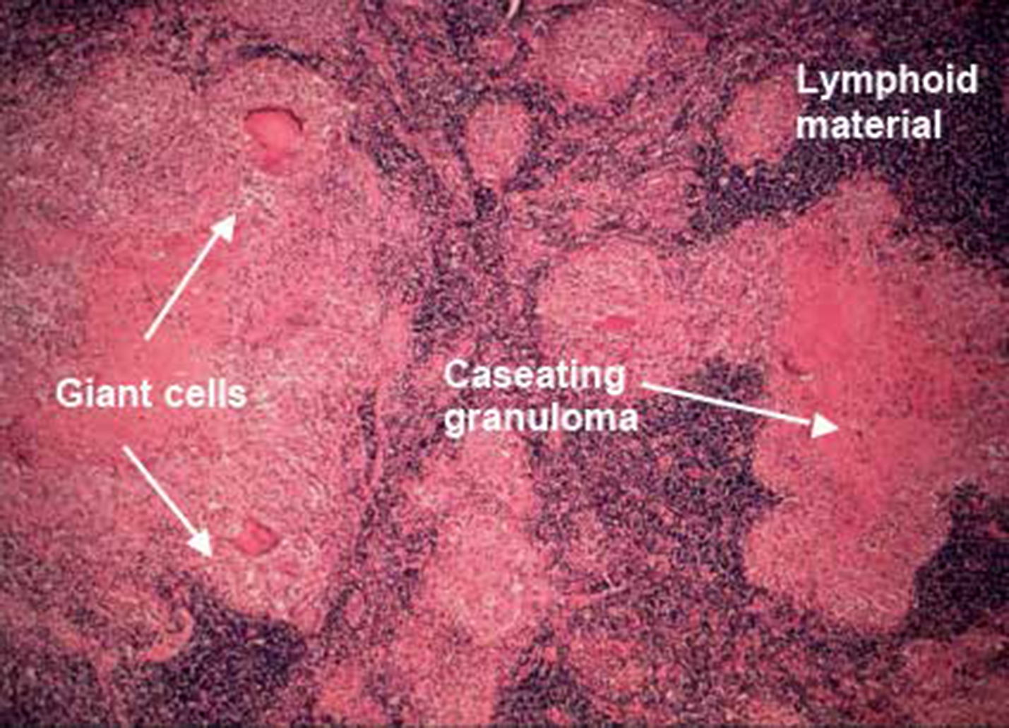
In the case of pulmonary tuberculosis, sputum smears may show acid-fast bacilli. In all cases, culture material (sputum, lymph node aspirates, etc) should be obtained whenever possible; not only to support the diagnosis but also to detect resistant disease. Currently, in the United Kingdom, some 6% of patients have isoniazid resistant disease and about 1.5% have multiple drug resistance. In non-pulmonary tuberculosis, nodal sampling may not demonstrate any bacteria and diagnosis may be clinical, supported by a number of other special investigations. Biopsy or fine needle aspiration showing caseating granuloma with Langhans giant cells (Figure 1) is suggestive of tuberculosis. This appearance may not be present if the patient is immunocompromised. In these cases, acid fast bacilli are more likely to be present.7 It is vital that any biopsy material (nodes or fine needle aspirate) should not be placed in formalin to allow culture, sensitivity and, crucially, the identification of drug resistance.
Figure 1. Lymph node with giant cells, caseating granulomas and lymphoid material.
A Mantoux test can be used but the results should be regarded with caution due to a potential false positive if there has been a previous BCG vaccination or exposure to environmental nontuberculous mycobacteria with which there is cross-reactivity in the Mantoux test; and a potential false negative if the patient is immunocompromised. The use of Interferon Gamma Release Assay tests is more specific and currently recommended for further investigation,3 in conjunction with the Mantoux test or in situations where Mantoux testing is unlikely to be reliable. These tests rely on detection of release of interferon gamma from specific T cells sensitized to the Early Secretion Antigen Target 6 (ESAT 6) and Culture Filtrate Protein 10 (CP10) antigens which are not present in BCG and are almost exclusive to the mycobacterium TB complex, with the exception of very few species of atypical mycobacteria. These tests cannot, however, distinguish between active and latent disease.
A chest X-ray can be used to help identify those patients with concurrent pulmonary tuberculosis, evidence of old pulmonary disease with the possibility of re-activation, and mediastinal lymphadenopathy.
Tuberculosis is a notifiable disease, requiring contact tracing to identify and treat those who have been infected. An untreated individual with pulmonary tuberculosis infects 10–15 people per year.3 A smear-positive individual is more infectious and contact tracing has to include work contacts and close family members.
Non-pulmonary tuberculosis is non-infectious despite its often dramatic clinical appearance. Patients should be fully informed of this as the stigma of tuberculous infection may delay presentation and compliance with treatment in certain ethnic groups. Current NICE guidelines3 still recommend contact tracing for non-pulmonary tuberculosis because this may identify an unknown pulmonary index case from whom a patient with non-pulmonary tuberculosis has contracted the disease.
The treatment for lymph node tuberculosis is the same as for pulmonary tuberculosis and is often started based on clinical suspicion without definitive cultures. Once the diagnosis of tuberculosis has been made, patients should only be treated by a respiratory or infectious diseases physician with expertise in tuberculosis and the patient should be supported by a key worker, normally a specialist nurse, to ensure compliance and manage any complications (such as resistant disease) effectively. Treatment lasts for 6 months and then stops, even if there is evidence of residual nodes, new nodes or sinus formation, which occurs in 10–15% of cases owing to immune response to tuberculoproteins from disrupted macrophages. An individual with pulmonary tuberculosis is considered to be non-infectious after 2 weeks of treatment.7
Case report
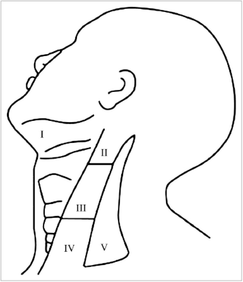
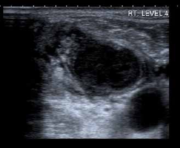
A 79-year-old Caucasian male was referred with a four-week history of a neck mass overlying the sternocleidomastoid muscle. There was no history of systemic upset or dysphagia and no relevant medical or infectious history. Clinical assessment found a fixed, firm and non-tender 3 cm lump on the right of the neck at level IV (Figure 2). On examination, it was thought that malignancy was the most likely diagnosis. Flexible nasendoscopy was unremarkable. An ultrasound examination demonstrated abnormal lymph nodes in the right neck at levels II–V. There was breach of the nodal capsule in the largest 18 mm node which also showed intra-nodal cystic necrosis with surrounding oedema of the tissue (periadenitis) (Figure 3).
Figure 2. Anatomical levels of the neck.Figure 3. A 2 cm lymph node mass at right level 4 demonstrating extracapsular breach, intra-nodal cystic necrosis and surrounding tissue oedema.
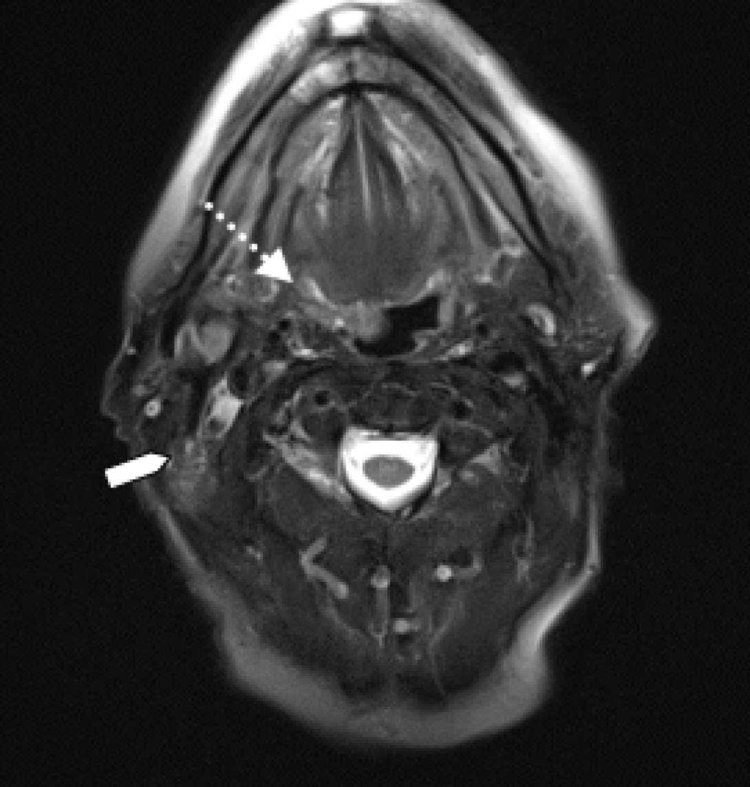
Multiple fine needle aspiration biopsies only demonstrated acellular necrotic debris. An MRI highlighted asymmetry of the tonsils, with some infilling of the right glossotonsillar sulcus (Figure 4) and the right tonsil demonstrated a small focus of high signal. The initial radiological impression was that of a primary tonsillar tumour with ipsilateral metastatic neck disease. A CT scan of the chest revealed multiple bilateral pulmonary nodules which were consistent with metastases.
Figure 4. Axial T2 image demonstrating some infilling of the right glossotonsillar sulcus (dotted arrow). The arrow demonstrates the most cranial aspect of the nodal mass in the posterior triangle.
A second attempt at fine needle aspiration revealed necrosis and acute inflammation with a focal granulomatous component. The head and neck multidisciplinary team scheduled the patient for tonsillectomy and lymph node excision, aiming for definitive pathological diagnosis. The histology reported the tonsil to show no evidence of malignancy. The lymph node was consistent with a caseating granulomatous process which extended into surrounding skeletal muscle. A diagnosis of pulmonary and non-pulmonary tuberculosis was given.
The patient was referred to the tuberculosis clinic and successfully treated with the standard 4 drug regimen of 6 months rifampicin and isoniazid plus pyrazinamide and ethambutol in the initial 2-month phase. The Health Protection Agency for Enhanced Tuberculosis Surveillance was notified.
Discussion
This case highlights the difficulties in diagnosing tuberculosis. This patient had no history of known tuberculosis contact and no associated symptoms (Table 1).
Cough for more than 2 weeks
Haemoptysis
Chest pain
Weakness
Weight loss
Fatigue
Fever
Night sweats
The differential diagnosis for neck masses is large (Table 2) but a clinical examination and history should enable those likely to have tuberculosis to be identified.
Midline neck masses
Upper
Skin and subcutaneous lesions
Cysts (epidermoid, dermoid, teratoid)
Lipoma
Lymph nodes
Infectious - EBV, TB, toxoplasmosis among others
Inflammatory - sarcoidosis, rheumatoid disease
Neoplastic
- Metastatic
- Lymphoma
- Leukaemia
Thyroglossal cysts
Lower
Thyroid
Solitary nodules
Multinodular goitre
Diffusely enlarged
Lateral neck masses
Skin and subcutaneous lesions
Cysts (epidermoid, dermoid, teratoid)
Lipoma
Developmental masses
Lymphangioma
Branchial cysts
Lymph nodes
Infectious - EBV, TB, toxoplasmosis among others
Inflammatory - sarcoidosis, rheumatoid disease
Neoplastic
- Metastatic
- Lymphoma
- Leukaemia
Neurogenic tumours
Neurofibromas
Schwannomas
Vascular tumours
Carotid body tumours
Salivary glands
It is advisable that all patients with a likely diagnosis of tuberculosis are referred directly to the respiratory tuberculosis team. In the case presented here the patient had been referred to the Neck Lump Clinic which was set up following national guidelines to improve head and neck cancer outcomes. These clinics are staffed by a consultant surgeon, a consultant radiologist and a consultant pathologist and are well placed to diagnose and refer patients that have tuberculosis.
In the dental setting emergency patients with tuberculosis can be treated as long as simple measures are in place to protect the dental team. All members of the dental team should be appropriately vaccinated, rubber dam should be used where possible and ultrasonic scaling should be avoided. Other patients should be protected by minimizing the time the patient spends in the waiting room. It should be recognized that prolonged contact is necessary to contract tuberculosis and so the risk for clinicians and nurses, even with smear-positive individuals, is considered to be negligible.
Conclusion
Tuberculosis is still relatively common in urban areas and dentists need to have an increased suspicion for infection with tuberculosis in patients presenting with cervical lymphadenopathy, especially in patients from ethnic minority groups. Tuberculosis can be hard to diagnose, even by specialists, and may require a multidisciplinary approach. The rapid and appropriate referral of patients with tuberculosis is part of the national strategy to regain control of this disease.